. 2024 May 3;12(5):e008799.
doi: 10.1136/jitc-2024-008799.

Combining toll-like receptor agonists with immune checkpoint blockade affects antitumor vaccine efficacy

Affiliations

Combining toll-like receptor agonists with immune checkpoint blockade affects antitumor vaccine efficacy

Donghwan Jeon et al. J Immunother Cancer. .

Abstract

Background: T cell checkpoint receptors are expressed when T cells are activated, and modulation of the expression or signaling of these receptors can alter the function of T cells and their antitumor efficacy. We previously found that T cells activated with cognate antigen had increases in the expression of PD-1, and this was attenuated in the presence of multiple toll-like receptor (TLR) agonists, notably TLR3 plus TLR9. In the current report, we sought to investigate whether combining TLR agonists with immune checkpoint blockade can further augment vaccine-mediated T cell antitumor immunity in murine tumor models.

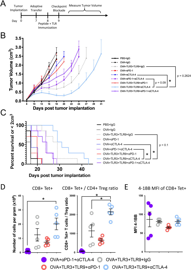
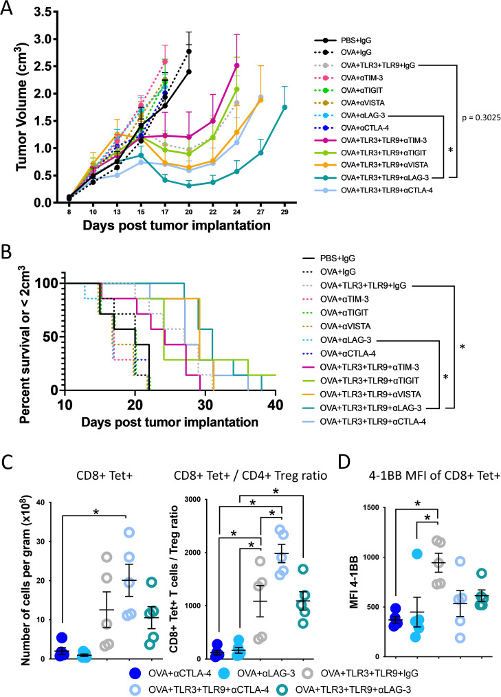
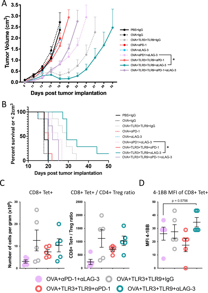
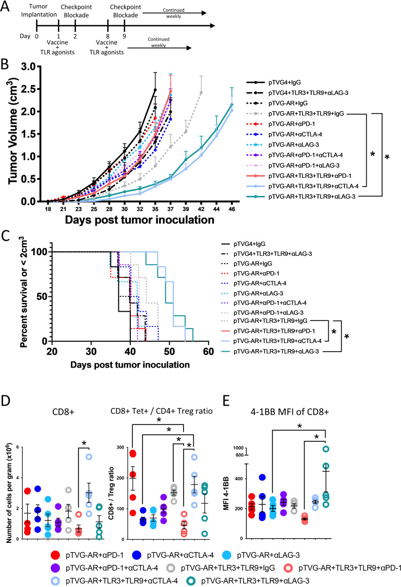
Methods: TLR agonists (TLR3 plus TLR9) and immune checkpoint inhibitors (antibodies targeting PD-1, CTLA-4, LAG-3, TIM-3 or VISTA) were combined and delivered with vaccines or vaccine-activated CD8+T cells to E.G7-OVA or MyC-CaP tumor-bearing mice. Tumors were assessed for growth and then collected and analyzed by flow cytometry.

Results: Immunization of E.G7-OVA tumor-bearing mice with SIINFEKL peptide vaccine, coadministered with TLR agonists and αCTLA-4, demonstrated greater antitumor efficacy than immunization with TLR agonists or αCTLA-4 alone. Conversely, the antitumor efficacy was abrogated when vaccine and TLR agonists were combined with αPD-1. TLR agonists suppressed PD-1 expression on regulatory T cells (Tregs) and activated this population. Depletion of Tregs in tumor-bearing mice led to greater antitumor efficacy of this combination therapy, even in the presence of αPD-1. Combining vaccination with TLR agonists and αCTLA-4 or αLAG-3 showed greater antitumor than with combinations with αTIM-3 or αVISTA.

Conclusion: The combination of TLR agonists and αCTLA-4 or αLAG-3 can further improve the efficacy of a cancer vaccine, an effect not observed using αPD-1 due to activation of Tregs when αPD-1 was combined with TLR3 and TLR9 agonists. These data suggest that optimal combinations of TLR agonists and immune checkpoint blockade may improve the efficacy of human anticancer vaccines.

Keywords: Immune Checkpoint Inhibitor; Prostate Cancer; T regulatory cell - Treg; Toll-like receptor - TLR; Vaccine.


Conflict of interest statement

Competing interests: DGM has ownership interest, has received research support and serves as consultant to Madison Vaccines, which has licensed the pTVG-AR vaccine described in this manuscript. The other authors have no relevant potential conflicts of interest.

Figures

Figure 1
Figure 1
Combining TLR agonists with αCTLA-4, but not αPD-1, improved the efficacy of peptide-activated OT-I CD8+T cells in suppressing E.G7 tumor growth. (A) Ovalbumin-expressing E.G7 (eg,7-OVA-PD-L1high) cells were implanted in C57BL/6 mice and permitted to grow until tumors were palpable (7 days). OT-I splenocytes were then adoptively transferred and mice were immunized subcutaneously the following day with SIINFEKL (OVA) peptide alone, or with TLR agonists [TLR3 (Poly I:C) and TLR9 (ODN1826)]. Mice were further treated with αPD-1 and/or αCTLA-4 the day following immunization. (B) Shown are the tumor growth curves (mean+SE, n=7 animals per group). (C) Survival plots using the time to death or when tumors reached 2 cm3 in size, whichever occurred first. Data shown are from one of two independent studies (see online supplemental figure 2). (D) In a parallel study, animals were treated as in A, but tumors were collected at day 15 and evaluated for the frequency of infiltrating CD3+CD8+ tetramer+ T cells per gram of tumor and the number of Tregs (CD3+CD4+ CD25+ FoxP3+). (E) Tumor-infiltrating CD3+CD8+ tetramer+T cells were further evaluated for 4-1BB expression by flow cytometry. Asterisks indicate p<0.05 assessed by the mixed-effects model with Geisser-Greenhouse correction and Tukey’s multiple comparisons test with individual variances (B), by log-rank test (C), or by the one-way ANOVA with Tukey’s multiple comparisons test (D, E). Error bars represent SEM.
Figure 2
Figure 2
TLR agonists activated Tregs by lowering PD-1 expression, and these Tregs were more functionally suppressive of CD8+T cell proliferation and antitumor efficacy. Splenocytes were collected from the spleens of OT-II mice and stimulated in vitro for 3 days with class II OVA peptide (ISQAVHAAHAEINEAGR, OVA-II) or media alone (untreated) in the presence or absence of TLR agonists (TLR3+TLR9). (A) The median fluorescence intensity (MFI) of PD-1 was determined by flow cytometry in activated CD4+T cells (CD3+CD4+ CD25+ FoxP3-) and Tregs (CD3+CD4+ CD25+ FoxP3+). (B) Purified Tregs were coincubated with naïve OT-I CD3+CD8+ T cells labeled with CFSE, in a 1:1 ratio, in the presence of IL-2 and anti-CD3/anti-CD28 beads. Proliferation of CD3+CD8+ T cell was analyzed via flow cytometry by CFSE loss, and the % of cells with CFSE loss is shown. Upper panel shows representative flow cytometry data, and lower panel shows quantification (n=3 per condition). (C) Purified Tregs were coincubated with CD3+CD8+ cells from wild type mice in a 1:1 ratio for 48 hours, and analyzed for the expression of IFNγ, perforin, Granzyme B, and TNFα. Shown are the MFI of expression of these in CD3+CD8+ cells. (D) Treatment of mice with E.G7-OVA-PD-L1high tumors was conducted as described in (A). The day following peptide immunization (with or without TLR agonists), mice were treated with immune checkpoint blockade (or control IgG), using αPD-1, αCTLA-4, or a CTLA-4-targeting antibody that depletes Treg (αTreg). Animals receiving αTreg or αCTLA-4 received additional antibody treatment on days 11 and 13. Shown are the tumor growth curves (mean+SE, n=7 animals per group). Asterisks indicate p<0.05 assessed by the one-way ANOVA with Tukey’s multiple comparisons test (A and B) or by the mixed-effects model with Geisser-Greenhouse correction and Tukey’s multiple comparisons test with individual variances (C). Error bars represent SEM.
Figure 3
Figure 3
Combination of TLR agonists and αLAG-3 with peptide-activated CD8+T cells elicited prolonged suppression of E.G7-OVA-PDL1high tumor growth. Ovalbumin-expressing E.G7 cells (eg,7-OVA-PD-L1high) were implanted in C57BL/6 mice and permitted to grow until tumors were palpable (7 days). As in figure 1A, OT-I splenocytes were then adoptively transferred and mice were immunized subcutaneously the following day with SIINFEKL (OVA) peptide, with or without TLR agonists. Mice were then treated with immune checkpoint blockade, using αTIM-3, αTIGIT, αVISTA, αLAG-3, αCTLA-4, or IgG control, the day following immunization. (A) Shown are the tumor growth curves (mean+SE, n=7 animals per group). (B) Survival plots using the time to death or when tumors reached 2 cm3 in size, whichever occurred first. (C) In a parallel study, animals were treated as in A but tumors were collected at day 16 and evaluated for the frequency of infiltrating CD3+CD8+ tetramer+ T cells per gram of tumor and the number of Tregs (CD3+CD4+ CD25+ FoxP3+). (D) Tumor-infiltrating CD3+CD8+ tetramer+ T cells were further evaluated for 4-1BB expression by flow cytometry. Asterisks indicate p<0.05 assessed by the mixed-effects model with Geisser-Greenhouse correction and Tukey’s multiple comparisons test with individual variances (A), by log-rank test (B), or by one-way ANOVA with Tukey’s multiple comparisons test (C, D). Error bars represent SEM. Results are from one experiment and are representative of data from three independent experiments (shown in online supplemental figure 5 and 6). ANOVA, analysis of variance; MFI, median fluorescence intensity; TLR, toll-like receptor.
Figure 4
Figure 4
Peptide-activated CD8+T cells had greater antitumor efficacy against E.G7 tumors when delivered with TLR agonists and αLAG-3 than with αPD-1 and αLAG-3. Ovalbumin-expressing E.G7 cells were implanted in C57BL/6 mice and permitted to grow until tumors were palpable (7 days). As in figures 1 and 3, OT-I splenocytes were then adoptively transferred and mice were immunized subcutaneously the following day with SIINFEKL (OVA) peptide, with or without TLR agonists. Mice were then treated with αPD-1 and/or αLAG-3 (or IgG control), the day following immunization. (A) Shown are the tumor growth curves (mean+SEM, n=7 animals per group). (B) Survival plots using the time to death or when tumors reached 2 cm3 in size, whichever occurred first. Data shown are representative of two independent studies (see online supplemental figure 8). (C) In a parallel study, animals were treated as in A but tumors were collected at day 16 and evaluated for the frequency of infiltrating CD3+CD8+ tetramer+T cells per gram of tumor and the number of Tregs. (D) Tumor-infiltrating CD3+CD8+ tetramer+T cells were further evaluated for 4-1BB and expression by flow cytometry. Asterisks indicate p<0.05 assessed by the mixed-effects model with Geisser-Greenhouse correction and Tukey’s multiple comparisons test with individual variances (A), by log-rank test (B), or by the one-way ANOVA with Tukey’s multiple comparisons test (C, D). Error bars represent SEM. ANOVA, analysis of variance; MFI, median fluorescence intensity; TLR, toll-like receptor.
Figure 5
Figure 5
Combination of TLR agonists and immune checkpoint blockade improves the antitumor efficacy of a DNA vaccine in a murine prostate tumor model. Male FVB mice were implanted subcutaneously with 106 MyC-CaP tumor cells. (A) Mice were immunized intradermally weekly beginning on day 1 with control vector (pTVG4) or DNA encoding AR ligand-binding domain (pTVG-AR), and delivered alone or with TLR agonists. Mice were then treated with αPD-1, αCTLA-4, αLAG-3, combinations of antibodies, or IgG control the day after each vaccination. (B) Shown are the growth curves for each group (mean+SEM, n=6–7 animals per group). (C) Survival plots using the time to death or when tumors reached 2 cm3 in size, whichever occurred first. Data shown are representative of two independent studies (see online supplemental figure 11). (D) In a parallel study, animals were treated as in A, but tumors were collected at day 33 and evaluated for the frequency of infiltrating CD3+CD8+ T cells per gram of tumor and the number of Tregs (CD3+CD4+ CD25+FoxP3+). (E) Tumor-infiltrating CD3+CD8+ T cells were further evaluated for 4-1BB expression by flow cytometry. Asterisks indicate p<0.05 assessed by the mixed-effects model with Geisser-Greenhouse correction and Tukey’s multiple comparisons test with individual variances (B), by log-rank test (C), or by one-way ANOVA with Tukey’s multiple comparisons test (D, E). Error bars represent SEM. ANOVA, analysis of variance; TLR, toll-like receptor.

Similar articles

Cited by

References

    1. Janeway CA. The immune system evolved to discriminate infectious nonself from noninfectious self. Immunol Today 1992;13:11–6. 10.1016/0167-5699(92)90198-G - DOI - PubMed
    1. Kawai T, Akira S. Toll-like receptors and their crosstalk with other innate receptors in infection and immunity. Immunity 2011;34:637–50. 10.1016/j.immuni.2011.05.006 - DOI - PubMed
    1. Takeda K, Kaisho T, Akira S. Toll-like receptors. Annu Rev Immunol 2003;21:335–76. 10.1146/annurev.immunol.21.120601.141126 - DOI - PubMed
    1. Takeda K, Akira S. Toll-like receptors. Curr Protoc Immunol 2015;109:14. 10.1002/0471142735.im1412s109 - DOI - PubMed
    1. Tomai MA, Vasilakos JP. TLR agonists as vaccine adjuvants. In: Baschieri S, ed. Innovation in Vaccinology: from design, through to delivery and testing. Dordrecht: Springer Netherlands, 2012: 205–28.

Publication types

Substances