. 2024 May 3;15(1):3738.
doi: 10.1038/s41467-024-47450-x.

Adjuvant-dependent impact of inactivated SARS-CoV-2 vaccines during heterologous infection by a SARS-related coronavirus

Affiliations

Adjuvant-dependent impact of inactivated SARS-CoV-2 vaccines during heterologous infection by a SARS-related coronavirus

Jacob A Dillard et al. Nat Commun. .

Abstract

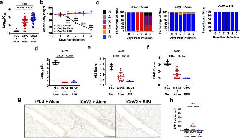
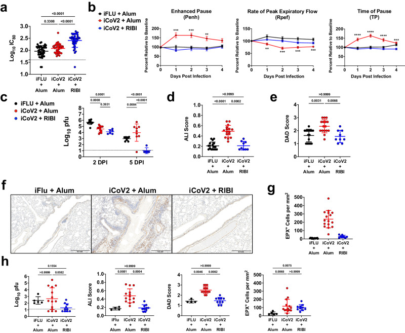
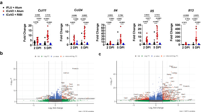
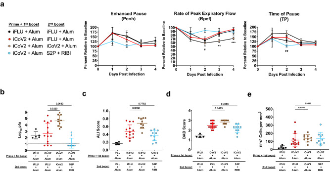
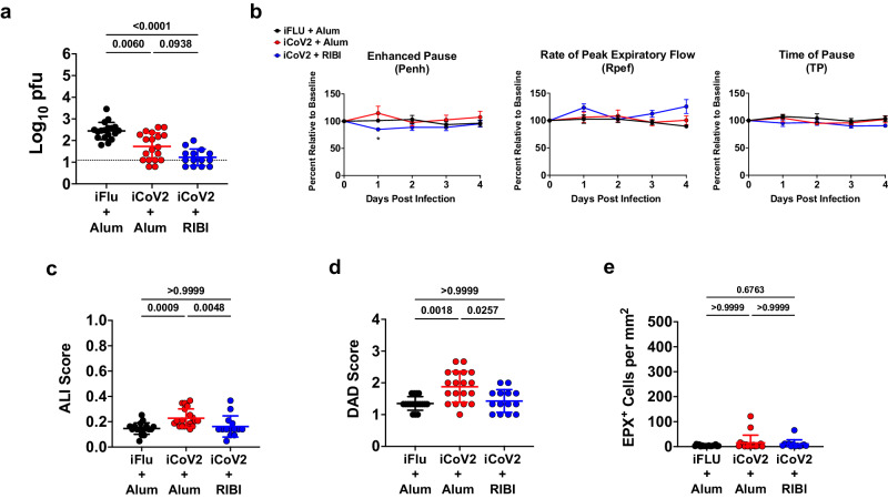
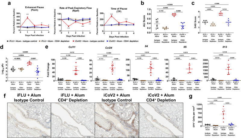
Whole virus-based inactivated SARS-CoV-2 vaccines adjuvanted with aluminum hydroxide have been critical to the COVID-19 pandemic response. Although these vaccines are protective against homologous coronavirus infection, the emergence of novel variants and the presence of large zoonotic reservoirs harboring novel heterologous coronaviruses provide significant opportunities for vaccine breakthrough, which raises the risk of adverse outcomes like vaccine-associated enhanced respiratory disease. Here, we use a female mouse model of coronavirus disease to evaluate inactivated vaccine performance against either homologous challenge with SARS-CoV-2 or heterologous challenge with a bat-derived coronavirus that represents a potential emerging disease threat. We show that inactivated SARS-CoV-2 vaccines adjuvanted with aluminum hydroxide can cause enhanced respiratory disease during heterologous infection, while use of an alternative adjuvant does not drive disease and promotes heterologous viral clearance. In this work, we highlight the impact of adjuvant selection on inactivated vaccine safety and efficacy against heterologous coronavirus infection.


Conflict of interest statement

R.S.B. has served on the Scientific Advisory Boards for Takeda vaccines, VaxArt and Invivyd Therapeutics, and has collaborations with Gilead, Janssen Pharmaceuticals, Pardas Biosciences, and Chimerix. M.T.H. has collaborations with Moderna, Inc. and Chimerix. R.S.B., K.H.D. III, and S.R.L. are listed as inventors on patents pertaining to the mouse-adapted SARS-CoV-2 viruses (MA10 and MA10-B.1.351; Patent number 11,225,508) and the SARS-CoV-2 nanoLuciferase viruses (SARS-CoV-2-nLuc and B.1.351-nLuc; Patent number 11,492,379) used in this study. The remaining authors declare no competing interests.

Figures

Fig. 1
Fig. 1. Inactivated vaccine protects mice against SARS-CoV-2 but causes adjuvant-dependent type 2 inflammation.
a Post-boost serum-neutralizing antibody titers against SARS-CoV-2 (D614G) in mice vaccinated with inactivated influenza virus adjuvanted with aluminum hydroxide (iFLU + Alum, n = 49), inactivated SARS-CoV-2 adjuvanted with aluminum hydroxide (iCoV2 + Alum, n = 45), or inactivated SARS-CoV-2 adjuvanted with Sigma Adjuvant System adjuvant (iCoV2 + RIBI, n = 50); IC80 = 80% inhibitory concentration. b, c Body weight change (b) and clinical scores (c) in iFLU + Alum (n = 12), iCoV2 + Alum (n = 19), or iCoV2 + RIBI (n = 4) vaccinated mice following challenge with SARS-CoV-2-MA10. d Pulmonary viral titers at 5 DPI following SARS-CoV-2-MA10 infection in iFLU + Alum (n = 5), iCoV2 + Alum (n = 8), or iCoV2 + RIBI (n = 4) vaccinated mice; pfu plaque-forming units. e, f Acute lung injury (ALI) (e) and diffuse alveolar damage (DAD) (f) scores in hematoxylin and eosin (H&E)-stained lungs at 5 DPI following SARS-CoV-2-MA10 infection in iFLU + Alum (n = 5), iCoV2 + Alum (n = 12), or iCoV2 + RIBI (n = 4) vaccinated mice. g, h Representative photomicrographs (g) (scale bar = 0.5 mm) and quantification (h) of pulmonary eosinophils immunohistochemically labeled for eosinophil peroxidase (EPX, brown cells) at 5 DPI following SARS-CoV-2-MA10 infection in iFLU + Alum (n = 3), iCoV2 + Alum (n = 8), or iCoV2 + RIBI (n = 4) vaccinated mice. a, d–f, h Individual data points represent independent biological replicates; solid horizontal lines and error bars represent group means ± standard deviation (SD); data analyzed by Kruskal–Wallis test with Dunn’s multiple comparisons; solid horizontal lines above data represent pairwise comparisons with p values; a, d Dotted line represents assay limit of detection. b Results reported as mean ± SD; ****p < 0.0001, iFLU + Alum versus iCoV2 + Alum, ###p < 0.001, ####p < 0.0001, iFLU + Alum versus iCoV2 + RIBI, two-way ANOVA with Tukey’s multiple comparisons. c Clinical scoring system: 0 = normal (blue), 1 = piloerection (orange), 2 = piloerection + kyphosis (red), 3 = piloerection, kyphosis, and reduced movement (purple), 4 = markedly reduced movement and/or labored breathing (gray), and 5 = moribund, dead or euthanized (black). Data presented as combined results from one (b, c), two (e, f, h), three (d), or four (a) independent animal experiments.
Fig. 2
Fig. 2. Inactivated SARS-CoV-2 vaccine causes enhanced disease during infection by a SARS-related coronavirus.
a Post-boost serum-neutralizing antibody titers against Rs-SHC014-CoV in mice vaccinated with inactivated influenza virus adjuvanted with aluminum hydroxide (iFLU + Alum, n = 43), inactivated SARS-CoV-2 adjuvanted with aluminum hydroxide (iCoV2 + Alum, n = 36), or inactivated SARS-CoV-2 adjuvanted with Sigma Adjuvant System adjuvant (iCoV2 + RIBI, n = 39); IC80 = 80% inhibitory concentration. b Pulmonary function measured by whole-body plethysmography in iFLU + Alum (n = 12), iCoV2 + Alum (n = 24), or iCoV2 + RIBI (n = 12) vaccinated mice following challenge with Rs-SHC014-CoV 4 weeks post-boost; results reported as mean ± standard error of the mean; **p < 0.01, **p < 0.001, ****p < 0.0001, iFLU + Alum versus iCoV2 + Alum, two-way ANOVA with Geisser-Greenhouse correction and Tukey’s multiple comparisons. c Pulmonary viral titers following Rs-SHC014-CoV infection in iFLU + Alum (2 DPI n = 12, 5 DPI n = 10), iCoV2 + Alum (2 DPI n = 12, 5 DPI n = 9), or iCoV2 + RIBI (2 DPI n = 6, 5 DPI n = 5) -vaccinated mice; pfu plaque-forming units. d, e Acute lung injury (ALI) (d) and diffuse alveolar damage (DAD) (e) scores in hematoxylin and eosin (H&E)-stained lungs at 5 DPI following Rs-SHC014-CoV infection in iFLU + Alum (ALI n = 17, DAD n = 16), iCoV2 + Alum (n = 16), or iCoV2 + RIBI (n = 9) vaccinated mice. f, g Representative photomicrographs (f) (scale bar = 0.5 mm) and quantification (g) of pulmonary eosinophils immunohistochemically labeled for eosinophil peroxidase (EPX, brown cells) at 5 DPI following Rs-SHC014-CoV infection in iFLU + Alum (n = 17), iCoV2 + Alum (n = 16), or iCoV2 + RIBI (n = 8) vaccinated mice. h Viral lung titers (left panel), ALI scores (second from left panel), DAD scores (second from right panel), and EPX+ cells (right panel) at 5 DPI in mice vaccinated with iFLU + Alum (titer and EPX n = 5, ALI and DAD n = 4), iCoV2 + Alum (n = 14), or iCoV2 + RIBI (n = 10) and challenged with Rs-SHC014-CoV 10.5 months post-boost. a, ce, g, h Individual data points represent independent biological replicates taken from discrete samples; horizontal lines and error bars represent group means ± standard deviation; data analyzed by Kruskal–Wallis test with Dunn’s multiple comparisons. a, c, h Dotted line represents the assay limit of detection. Data presented as combined results from one (c, h), two (b), three (d, e, g), or four (a) independent animal experiments.
Fig. 3
Fig. 3. Vaccine adjuvants promote divergent immune gene expression patterns during heterologous infection.
a Type 2 cytokine gene expression (Ccl11 C-C motif chemokine 11, Ccl24 C-C motif chemokine 24, Il4 interleukin 4, Il5 interleukin 5, Il13 interleukin 13) normalized to Gapdh expression at 2- and 5 days post-infection (DPI) with Rs-SHC014-CoV in mice vaccinated with inactivated influenza virus adjuvanted with aluminum hydroxide (iFLU + Alum), inactivated SARS-CoV-2 adjuvanted with aluminum hydroxide (iCoV2 + Alum), or inactivated SARS-CoV-2 adjuvanted with Sigma Adjuvant System adjuvant (iCoV2 + RIBI); Ccl11, Ccl24 2 DPI – iFLU + Alum (n = 12), iCoV2 + Alum (n = 12), iCoV2 + RIBI (n = 6); Ccl11, Ccl24 5 DPI – iFLU + Alum (n = 14), iCoV2 + Alum (n = 13), iCoV2 + RIBI (n = 9). Il4, Il5, Il13 2 DPI – iFLU + Alum (n = 12), iCoV2 + Alum (n = 12), iCoV2 + RIBI (n = 6); Il4, Il5, Il13 5 DPI – iFLU + Alum (n = 13), iCoV2 + Alum (n = 13), iCoV2 + RIBI (n = 9); individual data points represent independent biological replicates; results presented from one animal experiment and analyzed by Kruskal–Wallis test with Dunn’s multiple comparisons correction; solid horizontal lines and error bars overlaying data represent group means ± standard deviation; solid horizontal lines above data represent pairwise comparisons with p values. b, c Volcano plots showing differential expression by RNA-Seq between iCoV2 + Alum relative to iCoV2 + RIBI at 2 DPI (b) and 5 DPI (c) following infection with Rs-SHC014-CoV. Fold change (FC) is shown along the X-axis (with 1.5 log2 FC thresholds represented by vertical dashed lines) and significance along the Y-axis (with false discovery rate-adjusted q < 0.05 thresholds represented by horizontal dashed lines). Key genes are highlighted in the upper right (iCoV2 + Alum expression > iCoV2 + RIBI) and upper left (iCoV2 + Alum expression < iCoV2 + RIBI) quadrants. 2 DPI (n = 12) and 5 DPI (n = 9) for iCoV2 + Alum, and 2 DPI (n = 6) and 5 DPI (n = 5) for iCoV2 + RIBI; results presented from one animal experiment and analysis described in detail in RNA Sequencing (RNA-Seq) in Methods. Source data are provided as a Source Data file. RNA Sequencing raw .fastq data files are submitted to the Sequence Read Archive (SRA) database under BioProject ID PRJNA1022427.
Fig. 4
Fig. 4. Alternative boost vaccination partially reduces vaccine-enhanced disease.
a Pulmonary function measured by whole-body plethysmography following Rs-SHC014-CoV challenge in mice vaccinated with inactivated influenza virus adjuvanted with aluminum hydroxide (iFLU + Alum) initially followed by a second boost vaccination with iFLU + Alum 9.5 months post-first boost (n = 5) or vaccinated with inactivated SARS-CoV-2 adjuvanted with aluminum hydroxide (iCoV2 + Alum) initially followed by a second boost vaccination with iFLU + Alum (n = 14), iCoV2 + Alum (n = 10), or stabilized spike protein adjuvanted with Sigma Adjuvant System adjuvant (S2P + RIBI, n = 9) 9.5 months post-first boost; results represented by one experiment and reported as group mean ± standard error of the mean; **p < 0.01, ***p < 0.001, iCoV2 + Alum – iFLU + Alum versus iCoV2 + Alum – iCoV2 + Alum, #p < 0.05, ##p < 0.01, iCoV2 + Alum – iFLU + Alum versus iCoV2 + Alum – S2P + RIBI, two-way ANOVA with Geisser-Greenhouse correction and Dunnett’s multiple comparisons correction. b Pulmonary viral titers at 5 days post-infection (DPI) following Rs-SHC014-CoV infection; dotted line represents assay limit of detection; pfu plaque forming units. c, d Acute lung injury (ALI) (c) and diffuse alveolar damage (DAD) (d) scores in hematoxylin and eosin (H&E)-stained lungs at 5 DPI following Rs-SHC014-CoV infection. e Quantification of pulmonary eosinophils immunohistochemically labeled for eosinophil peroxidase (EPX, brown cells) at 5 DPI following Rs-SHC014-CoV infection. be Individual data points represent independent biological replicates; solid horizontal lines and error bars overlaying data represent group mean ± standard deviation; data analyzed by Kruskal–Wallis test with Dunn’s multiple comparisons correction; solid horizontal lines above data represent pairwise comparisons with p values; iFLU + Alum – iFLU + Alum (n = 5), iCoV2 + Alum – iFLU + Alum (n = 14), iCoV2 + Alum – iCoV2 + Alum (n = 10), iCoV2 + Alum – S2P + RIBI (n = 10). iCoV2 + Alum – iFLU + Alum control group data are repeated from Fig. 2 and served as the iCoV2 + Alum group for the final 10.5-month post-boost vaccination (Fig. 2h) and the control group for comparison to later secondary boost vaccination (Fig. 4).
Fig. 5
Fig. 5. Vaccine immune serum promotes cross-protection with modest pathology during heterologous infection.
a Pulmonary viral titers at 5 days post-infection (DPI) following Rs-SHC014-CoV infection in naïve mice that received a passive serum transfer from mice vaccinated with inactivated influenza virus adjuvanted with aluminum hydroxide (iFLU + Alum, n = 18), inactivated SARS-CoV-2 adjuvanted with aluminum hydroxide (iCoV2 + Alum, n = 19), or inactivated SARS-CoV-2 adjuvanted with Sigma Adjuvant System adjuvant (iCoV2 + RIBI, n = 14); dotted line represents assay limit of detection; pfu plaque forming units. b Pulmonary function measured by whole-body plethysmography following challenge with Rs-SHC014-CoV in naïve mice that received a passive serum transfer from mice vaccinated with iFLU + Alum (n = 6), iCoV2 + Alum (n = 6), or iCoV2 + RIBI (n = 6); results reported as group mean ± standard error of the mean; *p < 0.05, iFLU + Alum versus iCoV2 + Alum, two-way ANOVA with Geisser-Greenhouse correction and Tukey’s multiple comparisons correction. c, d Acute lung injury (ALI) (c) and diffuse alveolar damage (DAD) (d) scores in hematoxylin and eosin (H&E)-stained lungs at 5 DPI following Rs-SHC014-CoV infection in naïve mice that received a passive serum transfer from mice vaccinated with iFLU + Alum (n = 18), iCoV2 + Alum (n = 19), or iCoV2 + RIBI (n = 14). e Quantification of pulmonary eosinophils immunohistochemically labeled for eosinophil peroxidase (EPX, brown cells) at 5 DPI following Rs-SHC014-CoV infection in naïve mice that received a passive serum transfer from mice vaccinated with iFLU + Alum (n = 18), iCoV2 + Alum (n = 19), or iCoV2 + RIBI (n = 13). Individual data points represent independent biological replicates; solid lines and error bars represent group mean ± standard deviation; data analyzed by Kruskal–Wallis test with Dunn’s multiple comparisons correction; solid horizontal lines above data represent pairwise comparisons with p values; data presented as combined results from one (b) or two (a, c, d, e) independent animal experiments.
Fig. 6
Fig. 6. CD4+ T helper cells promote vaccine-enhanced disease during heterologous infection.
a Pulmonary function measured by whole-body plethysmography following Rs-SHC014-CoV challenge in mice vaccinated with inactivated influenza virus adjuvanted with aluminum hydroxide (iFLU + Alum) or inactivated SARS-CoV-2 adjuvanted with aluminum hydroxide (iCoV2 + Alum) and administered anti-CD4 antibody (iFLU + Alum – CD4+ depletion, n = 12; iCoV2 + Alum – CD4+ depletion, n = 14) or isotype control antibody (iFLU + Alum – isotype control, n = 12; iCoV2 + Alum – isotype control, n = 13) prior to Rs-SHC014-CoV challenge; results reported as group mean ± standard error of the mean; ***p < 0.001, ****p < 0.0001, iCoV2 + Alum – isotype control versus iCoV2 + Alum – CD4+ depletion, two-way ANOVA with Geisser-Greenhouse correction and Dunnett’s multiple comparisons correction. be Acute lung injury (ALI) scores (b), diffuse alveolar damage (DAD) scores (c), viral titers (pfu plaque forming units) (d), and type 2 cytokine gene expression (Ccl11 C-C motif chemokine 11, Ccl24 C-C motif chemokine 24, Il4 interleukin 4, Il5 interleukin 5, Il13 interleukin 13) (e) in lungs at 5 DPI following Rs-SHC014-CoV infection in mice vaccinated with iFLU + Alum or iCoV2 + Alum and administered anti-CD4 antibody or isotype control antibody prior to Rs-SHC014-CoV challenge. f, g Representative photomicrographs (f) (scale bar = 0.5 mm) and quantification (g) of pulmonary eosinophils immunohistochemically labeled for eosinophil peroxidase (EPX, brown cells) at 5 DPI following Rs-SHC014-CoV infection in mice vaccinated with iFLU + Alum or iCoV2 + Alum and administered anti-CD4 antibody or isotype control antibody prior to Rs-SHC014-CoV challenge. bg iFLU + Alum – isotype control (n = 8), iFLU + Alum – CD4+ depletion (n = 8), iCoV2 + Alum – isotype control (n = 7), iCoV2 + Alum – CD4+ depletion (n = 8). be, g Individual data points represent independent biological replicates; solid lines and error bars represent group mean ± standard deviation; data analyzed by Kruskal–Wallis test with Dunn’s multiple comparisons correction; solid horizontal lines above data represent pairwise comparisons with p values. d Dotted line represents the assay limit of detection. Data presented as combined results from one (bg) or two (a) independent animal experiments.

Update of

References

    1. Johns Hopkins Coronavirus Resource Center. COVID-19 Dashboard. (Center for Systems Science and Engineering at Johns Hopkins University, 2023).
    1. Yeyati, E. L. & Filippini, F. Social and economic impact of COVID-19. Brookings Global Working Paper #158. The Brookings Institution Global Economy and Development Program. (2021).
    1. Cui J, Li F, Shi ZL. Origin and evolution of pathogenic coronaviruses. Nat. Rev. Microbiol. 2019;17:181–192. doi: 10.1038/s41579-018-0118-9. - DOI - PMC - PubMed
    1. Menachery VD, et al. A SARS-like cluster of circulating bat coronaviruses shows potential for human emergence. Nat. Med. 2015;21:1508–1513. doi: 10.1038/nm.3985. - DOI - PMC - PubMed
    1. Menachery VD, et al. SARS-like WIV1-CoV poised for human emergence. Proc. Natl Acad. Sci. USA. 2016;113:3048–3053. doi: 10.1073/pnas.1517719113. - DOI - PMC - PubMed

Publication types

MeSH terms