. 2024 May 3;14(1):10196.
doi: 10.1038/s41598-024-59144-x.

Gentamicin loaded niosomes against intracellular uropathogenic Escherichia coli strains

Affiliations

Gentamicin loaded niosomes against intracellular uropathogenic Escherichia coli strains

Jacopo Forte et al. Sci Rep. .

Abstract

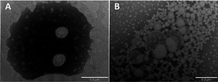
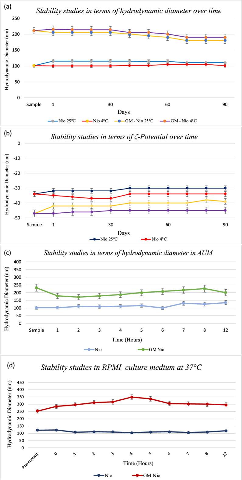
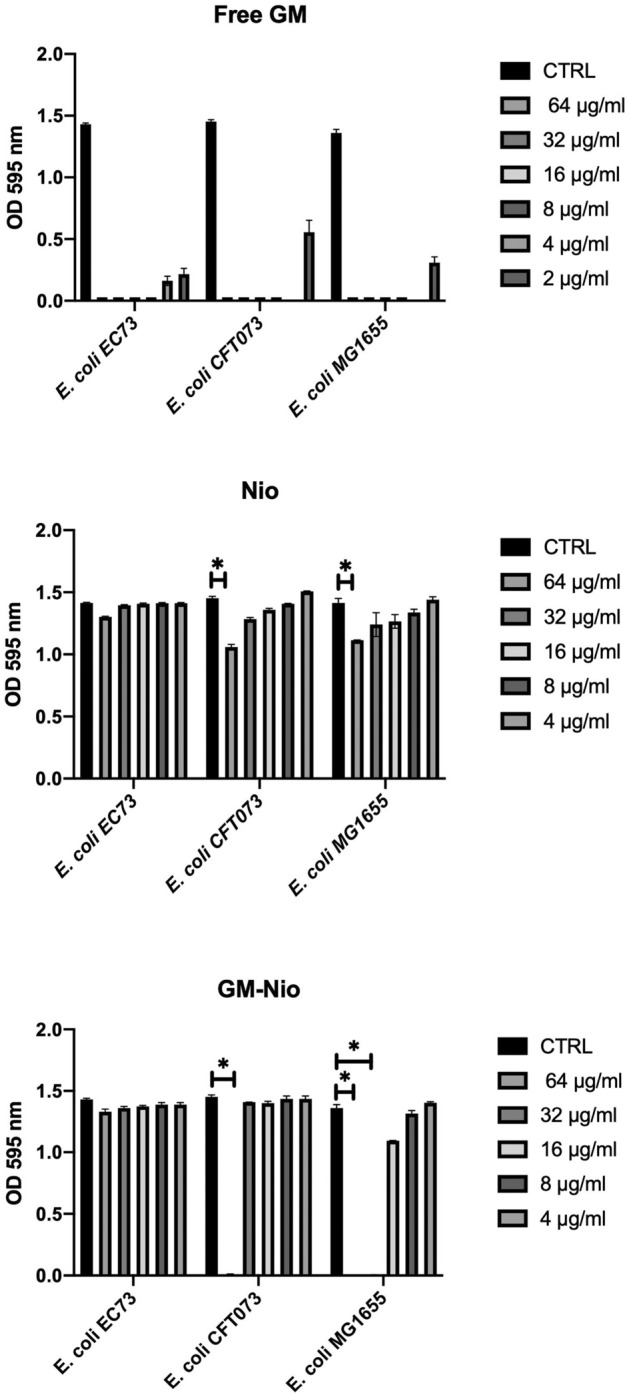
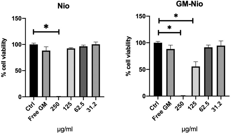
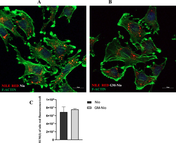
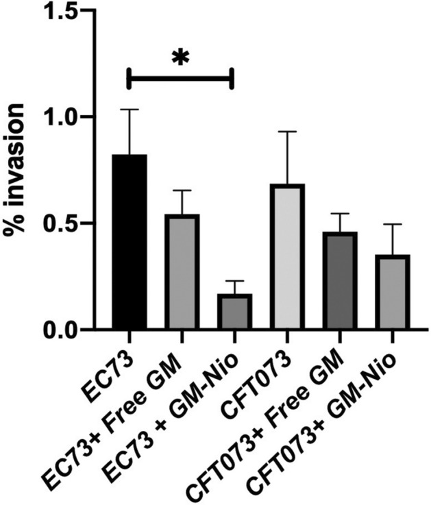
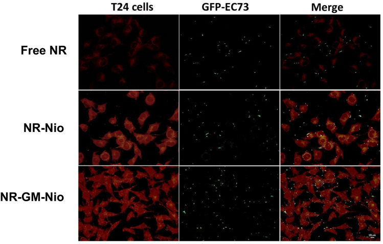
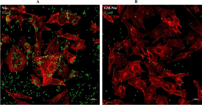
Urinary tract infections (UTIs) are the most common bacterial infections and uropathogenic Escherichia coli (UPEC) is the main etiological agent of UTIs. UPEC can persist in bladder cells protected by immunological defenses and antibiotics and intracellular behavior leads to difficulty in eradicating the infection. The aim of this paper is to design, prepare and characterize surfactant-based nanocarriers (niosomes) able to entrap antimicrobial drug and potentially to delivery and release antibiotics into UPEC-infected cells. In order to validate the proposed drug delivery system, gentamicin, was chosen as "active model drug" due to its poor cellular penetration. The niosomes physical-chemical characterization was performed combining different techniques: Dynamic Light Scattering Fluorescence Spectroscopy, Transmission Electron Microscopy. Empty and loaded niosomes were characterized in terms of size, ζ-potential, bilayer features and stability. Moreover, Gentamicin entrapped amount was evaluated, and the release study was also carried out. In addition, the effect of empty and loaded niosomes was studied on the invasion ability of UPEC strains in T24 bladder cell monolayers by Gentamicin Protection Assay and Confocal Microscopy. The observed decrease in UPEC invasion rate leads us to hypothesize a release of antibiotic from niosomes inside the cells. The optimization of the proposed drug delivery system could represent a promising strategy to significatively enhance the internalization of antimicrobial drugs.


Conflict of interest statement

The authors declare no competing interests.

Figures

Figure 1
Figure 1
Transmission electron microscopy images of empty (A) and GM-loaded niosomes (B).
Figure 2
Figure 2
Result of investigation on physicochemical stability over time. Effect of storage temperature (25 °C and 4 °C) hydrodynamic diameter and ζ-potential (A, B) and stability studies in terms of hydrodynamic diameter in AUM (C) and in RPMI culture medium (D). Data was obtained as the means of three independent experiments.
Figure 3
Figure 3
GM release profile over 12 h. Data was obtained as the means of three independent experiments.
Figure 4
Figure 4
Susceptibility test with Free-GM, empty Nio and GM loaded Nio. Data were expressed as mean ± SD. All considered conditions were compared to untreated control. *p value ≤ 0.05. Data are the averages of triplicate samples from three identical experiments and the error bars represent standard deviations. Statistically significant differences compared with the negative control (untreated cells) are indicated by asterisks (*, p < 0.05).
Figure 5
Figure 5
Cytotoxicity activity of empty Nio and GM loaded Nio on T24 cells. Results were expressed as % cell viability compared to untreated control. Free GM was assayed at 250 µg/ml. Data are the averages of duplicate samples from three identical experiments and the error bars represent standard deviations. Statistically significant differences compared with the negative control (untreated cells) are indicated by asterisks (*, p < 0.05).
Figure 6
Figure 6
Representative confocal images of NR (A) and NR-GM-co-loaded niosomes (B) treated cells. Under these conditions nile red fluorescence is appreciable within the cell with a similar distribution pattern. Scale bars 20 µm. (C) The graph represents the quantitative analysis of red fluorescence intensity (Sum(I)/cell) in T24 cells treated with NR empty niosome and NR GM-loaded niosomes (means S.E.M.; Student’s t-test, p = n.s., n = 354 vs. 328 cells respectively) FITC phalloidin for F-actin cytoskeletal visualization was used.
Figure 7
Figure 7
Bacterial counts and invasion percentages of T24 cells challenged with EC73 and CFT073 strains in absence or presence of GM-Nio and free GM. Results were expressed as percentage of cell invasion compared to untreated control (without niosomes). Data are the averages of duplicate samples from three identical experiments and the error bars represent standard deviations. Statistically significant differences compared with the negative control (untreated cells) are indicated by asterisks (*, p < 0.05).
Figure 8
Figure 8
Localization of GFP-labeled E. coli EC73 on T24 cells during adhesion assay (1 h infection) in presence of free NR, NR- and NR-GM-niosomes.
Figure 9
Figure 9
Immunofluorescence analysis of the bacteria infected cells treated with Nio (A) and with GM-Nio (B). It is evident the presence of intracellular bacteria and a clear reduction in the number of bacteria in the samples treated with GM Nio. Rodamine phalloidin for F-actin cytoskeletal visualization was used.

References

    1. Foxman B. The epidemiology of urinary tract infection. Nat. Rev. Urol. 2010;7:653–660. doi: 10.1038/nrurol.2010.190. - DOI - PubMed
    1. Totsika M, et al. Uropathogenic Escherichia coli mediated urinary tract infection. Current Drug Targets. 2012;13:1386–1399. doi: 10.2174/138945012803530206. - DOI - PubMed
    1. Martinez JJ, Mulvey MA, Schilling JD, Pinkner JS, Hultgren SJ. Type 1 pilus-mediated bacterial invasion of bladder epithelial cells. EMBO J. 2000;19:2803–2812. doi: 10.1093/emboj/19.12.2803. - DOI - PMC - PubMed
    1. Klein RD, Hultgren SJ. Urinary tract infections: microbial pathogenesis, host–pathogen interactions and new treatment strategies. Nat. Rev. Microbiol. 2020;18:211–226. doi: 10.1038/s41579-020-0324-0. - DOI - PMC - PubMed
    1. Miao Y, Li G, Zhang X, Xu H, Abraham SN. A TRP channel senses lysosome neutralization by pathogens to trigger their expulsion. Cell. 2015;161:1306–1319. doi: 10.1016/j.cell.2015.05.009. - DOI - PMC - PubMed

Publication types

MeSH terms