. 2024 Jul;16(7):1200-1208.
doi: 10.1038/s41557-024-01490-4. Epub 2024 May 3.

Selection of a promiscuous minimalist cAMP phosphodiesterase from a library of de novo designed proteins

Affiliations

Selection of a promiscuous minimalist cAMP phosphodiesterase from a library of de novo designed proteins

J David Schnettler et al. Nat Chem. 2024 Jul.

Abstract

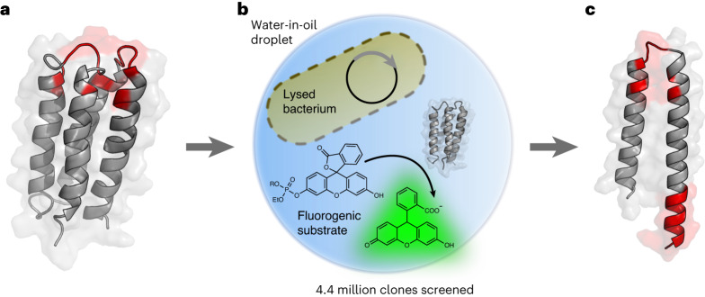
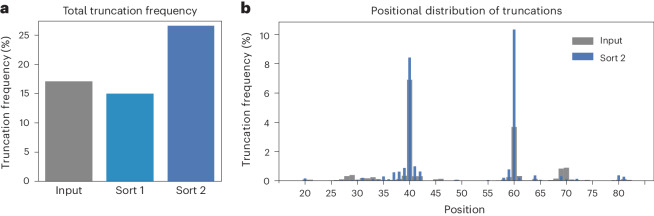
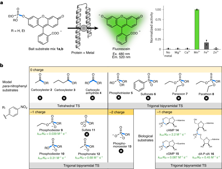
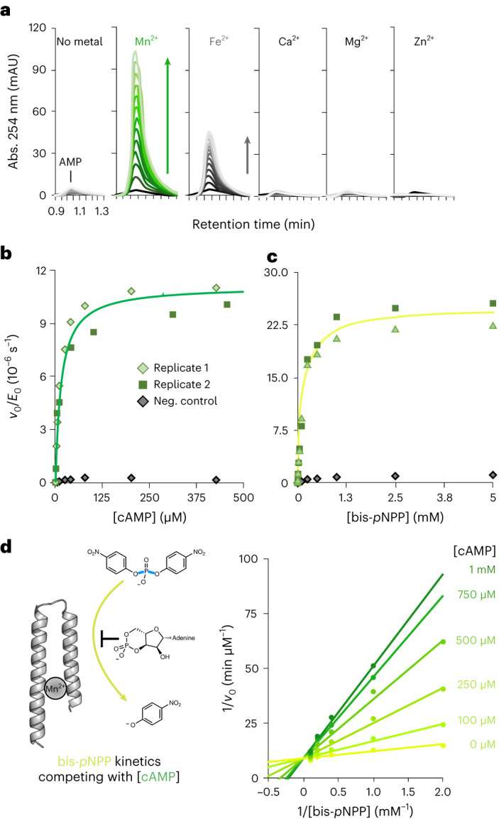
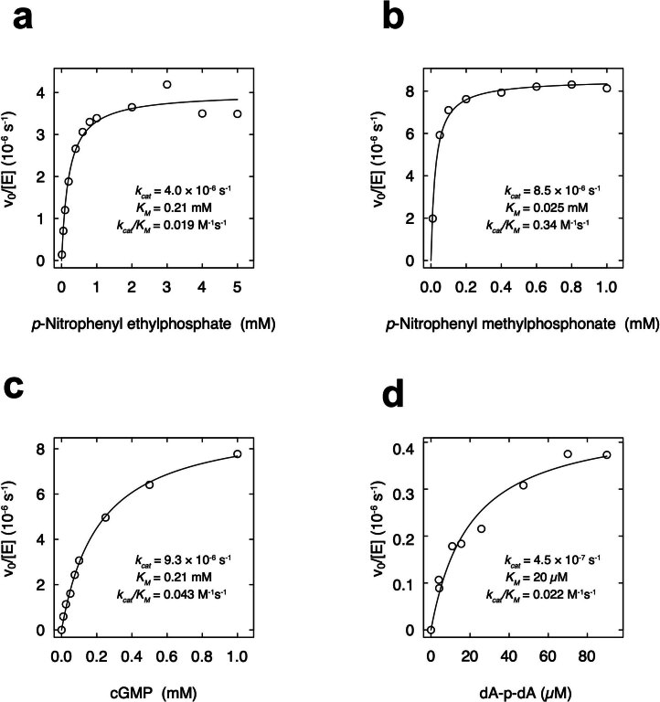
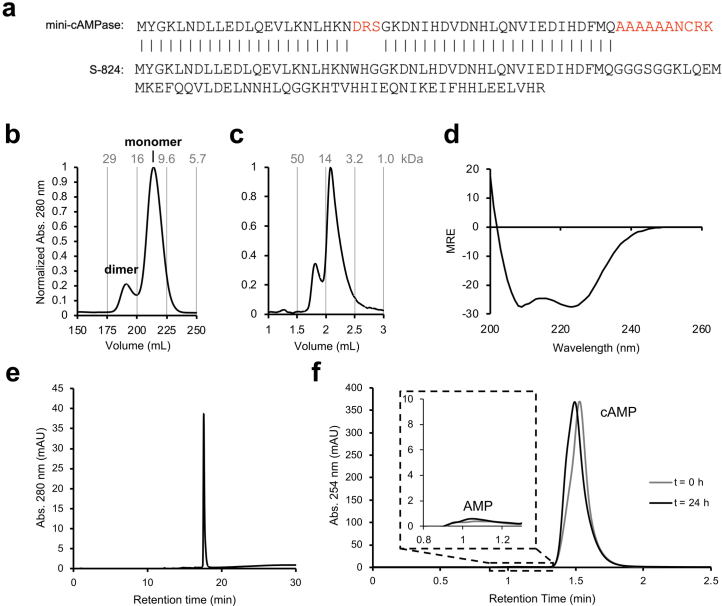
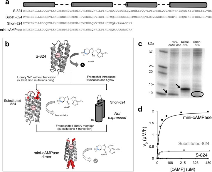
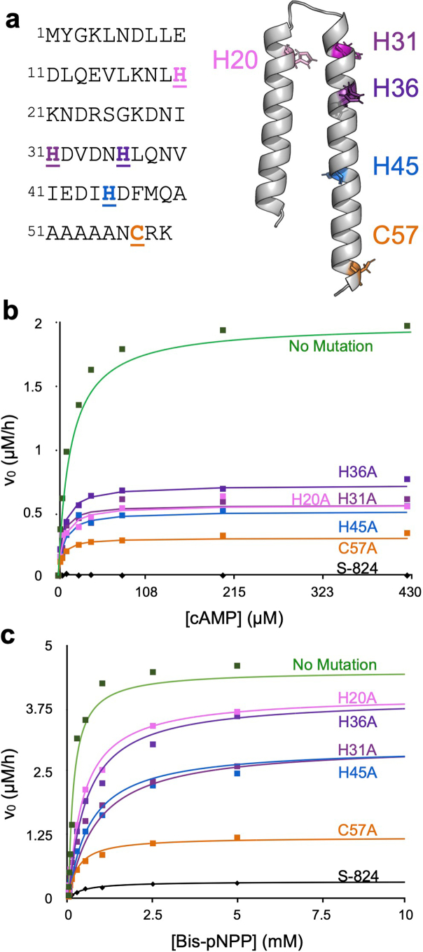
The ability of unevolved amino acid sequences to become biological catalysts was key to the emergence of life on Earth. However, billions of years of evolution separate complex modern enzymes from their simpler early ancestors. To probe how unevolved sequences can develop new functions, we use ultrahigh-throughput droplet microfluidics to screen for phosphoesterase activity amidst a library of more than one million sequences based on a de novo designed 4-helix bundle. Characterization of hits revealed that acquisition of function involved a large jump in sequence space enriching for truncations that removed >40% of the protein chain. Biophysical characterization of a catalytically active truncated protein revealed that it dimerizes into an α-helical structure, with the gain of function accompanied by increased structural dynamics. The identified phosphodiesterase is a manganese-dependent metalloenzyme that hydrolyses a range of phosphodiesters. It is most active towards cyclic AMP, with a rate acceleration of ~109 and a catalytic proficiency of >1014 M-1, comparable to larger enzymes shaped by billions of years of evolution.


Conflict of interest statement

The authors declare no competing interests.

Figures

Fig. 1
Fig. 1. Droplet screening of a library of de novo designed 4-helix bundles enriches truncated sequences with phosphoesterase activity.
a, De novo designed 4-helix bundle library. A library containing ~1.7 million variants based on the stably folded de novo designed 4-helix bundle protein S-824 was screened for phosphoesterase activity. The diversified residues of S-824 are shown in red (degenerate codons used: NDT/VRC/RRC). b, Ultrahigh-throughput microdroplet screening. The library was subjected to FADS on a microfluidic chip, and the 0.1–0.2% most fluorescent droplets (of a total of 4.4 million screened) were selected. c, Truncated peptide with phosphoesterase activity. The selection yielded catalytically active, truncated peptides consisting of a helix-turn-helix motif of ~60 amino acids (mutated sites are shown in red), illustrated here with a structural model of mini-cAMPase generated with AlphaFold2/ColabFold,. Panel b adapted with permission from ref. under a Creative Commons licence CC BY 4.0.
Fig. 2
Fig. 2. NGS analysis reveals enrichment of truncated proteins across the library.
a, Relative frequency (percentage of truncated sequences among the total number of sequences) of truncated reads in the input library (grey; 17%), after sorting 1 (light blue; 15%) and after sorting 2 (dark blue; 27%) reveals 1.6-fold enrichment of premature stop codons after sort 2, albeit close to 0% would be expected. b, The frequency of truncations at every sequenced position in the input library (grey, broad bars) and after sorting 2 (blue, narrow bars) shows 1.2- and 2.8-fold enrichments of truncations at position 40 and 60, respectively, after sorting 2. Source data
Fig. 3
Fig. 3. Metal requirement and substrate scope.
a, Metal dependence of the enzymatic reaction for the mixture of fluorogenic bait substrates: 100 µM mini-cAMPase was incubated with ~200 µM substrate mixture and 200 µM Ca2+, Mg2+, Mn2+, Fe2+ or Zn2+. The histogram shows that mini-cAMPase requires Mn2+ for its catalytic activity. Data are presented as mean values of biological duplicates (n = 2), with error bars representing ±1 s.d. b, Activity was tested against substrates sampling different ground-state charges (from 0 to −2) and transition-state (TS) geometries (tetrahedral and trigonal–bipyramidal). The mini-cAMPase hydrolyses p-nitrophenyl phosphodiesters and phosphonates. The highest activity was observed for cAMP hydrolysis. The scissile bond is highlighted in blue. Cross symbols indicate no detectable activity. R indicates the p-nitrophenol leaving group. Source data
Fig. 4
Fig. 4. Kinetic characterization of enzymatic activity.
a, Metal dependence of the mini-cAMPase activity. Spectral scans are shown that report on the appearance of AMP on reverse-phase (RP) HPLC (100 µM mini-cAMPase incubated with 100 µM respective divalent metal and 250 µM cAMP). Each successive trace is 1.5 h apart, with a total time of 18 h. b, Michaelis–Menten kinetics for cAMPase activity in biological duplicates with 50 µM protein and 200 µM MnCl2, measured at 25 °C in PBS (pH 7.4). The purification control (grey) shows the background activity for the same preparation steps on the protein without the His6-tag. Rates are the mean of three independent repeat datasets. Each colour shows a biological replicate. The purification control is shown in grey. c, Michaelis–Menten plot for the phosphodiesterase activity of 50 µM mini-cAMPase and 200 µM MnCl2 with the model substrate bis(p-nitrophenyl) phosphate in biological duplicates. Each colour shows a biological replicate. As before, the purification control is shown in grey. d, Competition between bis-pNPP and cAMP hydrolysis (left) shown in a Lineweaver–Burke plot (right). Lines connect kinetics for a single cAMP concentration, with higher cAMP concentrations shown in darkening hues of green. The shared y intercept indicates competitive inhibition with Ki = 70 ± 8 µM cAMP. Michaelis–Menten kinetics were assayed for bis(p-nitrophenyl) phosphate hydrolysis by 100 µM protein with 200 µM MnCl2 in increasing cAMP concentrations. Source data
Fig. 5
Fig. 5. Mini-cAMPase is a dynamic α-helical dimer.
Across all panels, yellow represents the disulfide-bonded dimer, green is the reduced protein, and black is the parental protein (S-824). a,b, Preparation-scale (a) and analytical-scale (b) SEC shows that reduced mini-cAMPase elutes at double its monomeric molecular mass (that is, it is a noncovalent dimer). c, RP-HPLC traces show reduced (green), partly oxidized (yellow–green, 125 µM H2O2) and fully oxidized (yellow, 250 µM H2O2) protein. d, Activity under a gradient of increasingly oxidizing conditions to promote disulfide formation. Non-reducing SDS–PAGE reveals the relative abundance of disulfides (single measurement). Below each gel lane we show the HPLC traces indicating the corresponding activity of 50 µM mini-cAMPase incubated with 200 µM Mn2+ and 250 µM cAMP. The HPLC traces monitor the appearance of AMP (assayed by RP-HPLC) measured every 2 h for 24 h. The mutant C57A is included for comparison. e, 1H NMR showing the amide region for S-824 (black) and mini-cAMPase (reduced, green; oxidized, yellow). The broad and poorly resolved peaks indicate that the protein does not form a well-ordered structure and remains dynamic, even after oxidation. f, CD spectra showing that mini-cAMPase (green, yellow) is helical, but less so than the parent 4-helix bundle (black). There is no discernible shift in secondary structure caused by oxidation of C57. MRE, mean residue ellipticity. g, Melting curves measuring CD at 222 nm show that oxidized mini-cAMPase (yellow, TM > 90 °C) has a melting curve similar to S-824 (black, TM > 90 °C), with both proteins more thermostable than the reduced mini-cAMPase (green, TM ≈ 72 °C). Source data
Extended Data Fig. 1
Extended Data Fig. 1. Enrichment of single-nucleotide deletions causing frameshifts after sorting 2.
(a) Relative frequency (percentage of sequences including one single nucleotide deletion of total number of sequences) of single nucleotide deletions in input library (grey, 12%), after sorting 1 (light blue, 10%) and after sorting 2 (dark blue, 19%) reveals 1.6-fold enrichment of single nucleotide deletions after sorting 2. (b) Frequency of single nucleotide deletions at every sequenced position in input library (grey, broad bars) and after sorting 2 (blue, narrow bars). Deletions between position 55 and 117 cause a stop codon at codon 40 while deletions between position 121 and 177 cause a stop codon at codon 60. Deletions at position 147 (as found in the variant mini-cAMPase) are enriched 8.7-fold (0.18 vs 1.56%). Source data
Extended Data Fig. 2
Extended Data Fig. 2. Metal binding effects on structure and thermal stability by Circular Dichroism (CD).
(a) Full CD spectra of 40 µM mini-cAMPase in TBS without added Mn2+ (green) and with added 200 µM Mn2+ (pink). (b) Melting curves of the same samples as in (a), with and without manganese. Source data
Extended Data Fig. 3
Extended Data Fig. 3. Controls for purification of mini-cAMPase.
(a) Purification of (His)6-tagged mini-cAMPase on the nickel-NTA column, with the taken fractions boxed in yellow. These are then put on (b) the preparation-scale sizing column, where the fractions containing the protein are taken (again in yellow). To control for any endogenous proteins that come from this purification steps, in the same cellular background, the un-tagged protein was expressed alongside with the same fractions taken from the nickel-NTA column (c) and sizing column (d) here shown as grey boxes. (e) The samples with (green) and without (grey) the (His)6-tag are compared by HPLC, with the main peak being the protein mini-cAMPase. Inset is the magnified baseline. (f) The raw data for a single data point of the purification background control in Fig. 4b, c. The inset is the appearance of AMP. 250 µM cAMP was incubated with 200 µM Mn2+ in the background solution. This plot lacks concentration units but is diluted only slightly by the addition of cAMP and Mn2+. Note that, in contrast to the purified protein fraction, the control fraction was not diluted for this assay. Therefore, we estimate that the background activity represents an overestimate as any potential endogenous contaminants in the control fraction would be 2-fold to 5-fold more concentrated in the control fraction as compared to the protein fraction. These are the same conditions used for cAMPase activity characterization in Fig. 6. The very low observed activity in the control fraction indicates that the observed activity of mini-cAMPase cannot be attributed to an endogenous contaminant. Source data
Extended Data Fig. 4
Extended Data Fig. 4. Michaelis-Menten plots for kinetics of mini-cAMPase.
Steady-state kinetics were measured with (a) p-nitrophenyl ethylphosphate (b) p-nitrophenyl methylphosphonate, (c) cGMP, and (d) dA-P-dA. Protein was lyophilized after HPLC, and resuspended in 50 mM HEPES-NaOH, 150 mM NaCl, 5 mM DTT, pH 8.0 at 25 °C. Curves with cAMP and bis-pNPP are shown in Fig. 4b, c. Source data
Extended Data Fig. 5
Extended Data Fig. 5. S-824 purification and lack of activity.
(a) Sequence comparison of characterized hit protein and S-824 with mismatches highlighted in red. (b) Preparation-scale and (c) analytical-scale size-exclusion chromatography shows that S-824 exists predominantly as a monomer, but a dimer is partially present. (d) Circular Dichroism spectra of S-824 in TBS shows that it is helical. (e) Reverse-phase HPLC of S-824 shows that it is of high purity (>99%). (f) The raw data for a single data point in Extended Data Fig. 7b, sampled before and after 24 h for 50 µM S-824 incubated with 200 µM Mn2+ and 250 µM cAMP (the same conditions used for cAMPase activity characterization in Extended Data Fig. 7b), showing that S-824 lacks cAMPase activity. Source data
Extended Data Fig. 6
Extended Data Fig. 6. Substitutions and truncations contribute to phosphodiesterase activity.
(a) Protein sequences showing the changes that bridge S-824 and mini-cAMPase. (b) Scheme of the relationship between the sequences and the effect of the truncation on function. (c) SDS-PAGE of whole cell extracts shows that Short-824 is poorly expressed (single measurement). (d) Michaelis-Menten kinetics comparing S-824, mini-cAMPase, and Substituted-824, showing that Substituted-824 is ≈ 6-fold less active (in kcat/KM) than the truncated mini-cAMPase protein. Source data
Extended Data Fig. 7
Extended Data Fig. 7. Impact of alanine mutations on the activity of mini-cAMPase.
(a) Protein sequence and predicted structure with hydrophobic residues in yellow. Sites of mutated residues are emphasized and colour-coded. (b) cAMPase kinetics of alanine point mutants targeting the potential metal binding residues, alongside the inactive ancestor S-824 (black diamonds along baseline, with example raw data in Extended Data Fig. 5). (c) Bis-pNPP kinetics of alanine point mutants, alongside the inactive ancestor S-824. Note that panels (b) and (c) are plotted on different scales. Source data
Extended Data Fig. 8
Extended Data Fig. 8. Molecular dynamics simulations with mini-cAMPase.
(a) The dynamics of S-824 in either of the two observed topologies, as well as mini-cAMPase were analyzed by 100 ns molecular dynamics simulations. (b) Molecular dynamics simulations indicate that the mini-cAMPase helical-bundle core becomes slightly more rigid. In contrast, the C-terminal tails is highly dynamic, which could potentially explain the experimental observations from 1H NMR and CD spectroscopy. (c) RMSD plots indicate that the simulations are stable and converged.
Extended Data Fig. 9
Extended Data Fig. 9. Structure prediction of S-824 and the mini-cAMPase dimer.
(a) AlphaFold2 reproduced the NMR model of S-824. In contrast, the mini-cAMPase dimer adopts a different topological isomer in which the overall topology is mirrored. EMSfold predicted that S-824 populates that other topoisomer. (b) MultiSFold was used to dissect the topological dynamism further. For S-824, the topology observed in NMR was predicted exclusively. For mini-cAMPase, both topologies were observed in a ratio of 40:60. In addition to the more flexible C-terminus, the variability in topology could be another explanation for the experimental observation from 1H NMR and CD spectroscopy.

Similar articles

Cited by

References

    1. Yeh AH-W, et al. De novo design of luciferases using deep learning. Nature. 2023;614:774–780. - PMC - PubMed
    1. Tiessen A, Pérez-Rodríguez P, Delaye-Arredondo LJ. Mathematical modeling and comparison of protein size distribution in different plant, animal, fungal and microbial species reveals a negative correlation between protein size and protein number, thus providing insight into the evolution of proteomes. BMC Res. Notes. 2012;5:85. - PMC - PubMed
    1. Mandecki W. A method for construction of long randomized open reading frames and polypeptides. Protein Eng. 1990;3:221–226. - PubMed
    1. Prijambada ID, et al. Solubility of artificial proteins with random sequences. FEBS Lett. 1996;382:21–25. - PubMed
    1. Keefe AD, Szostak JW. Functional proteins from a random-sequence library. Nature. 2001;410:715–718. - PMC - PubMed

Publication types

MeSH terms

Substances

LinkOut - more resources