. 2024 Sep;261(5):1073-1092.
doi: 10.1007/s00709-024-01953-y. Epub 2024 May 4.

Viable protoplast isolation, organelle visualization and transformation of the globally distributed plant pathogen Phytophthora cinnamomi

Affiliations

Viable protoplast isolation, organelle visualization and transformation of the globally distributed plant pathogen Phytophthora cinnamomi

Aayushree Kharel et al. Protoplasma. 2024 Sep.

Abstract

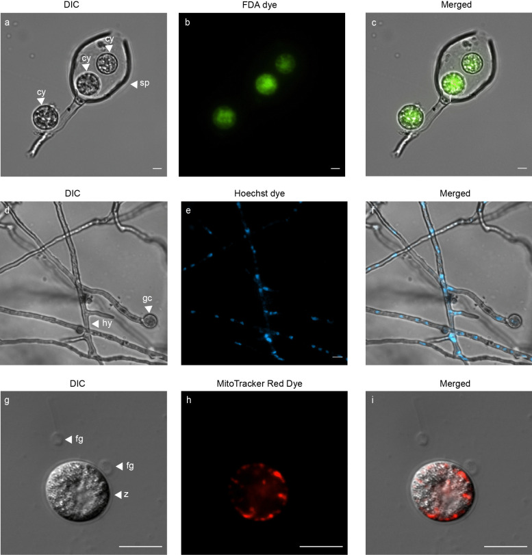
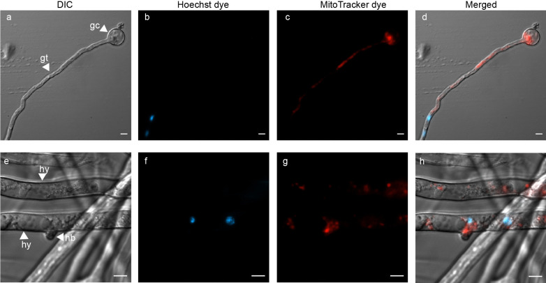
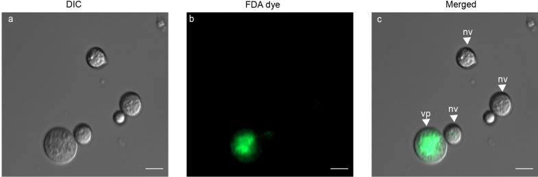
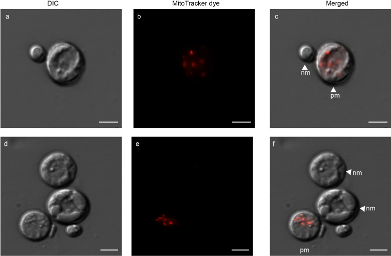
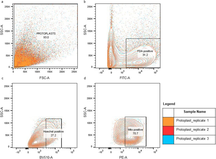
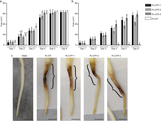
Phytophthora cinnamomi is an oomycete plant pathogen with a host range of almost 5000 plant species worldwide and therefore poses a serious threat to biodiversity. Omics technology has provided significant progress in our understanding of oomycete biology, however, transformation studies of Phytophthora for gene functionalisation are still in their infancy. Only a limited number of Phytophthora species have been successfully transformed and gene edited to elucidate the role of particular genes. There is a need to escalate our efforts to understand molecular processes, gene regulation and infection mechanisms of the pathogen to enable us to develop new disease management strategies. The primary obstacle hindering the advancement of transformation studies in Phytophthora is their challenging and unique nature, coupled with our limited comprehension of why they remain such an intractable system to work with. In this study, we have identified some of the key factors associated with the recalcitrant nature of P. cinnamomi. We have incorporated fluorescence microscopy and flow cytometry along with the organelle-specific dyes, fluorescein diacetate, Hoechst 33342 and MitoTracker™ Red CMXRos, to assess P. cinnamomi-derived protoplast populations. This approach has also provided valuable insights into the broader cell biology of Phytophthora. Furthermore, we have optimized the crucial steps that allow transformation of P. cinnamomi and have generated transformed isolates that express a cyan fluorescent protein, with a transformation efficiency of 19.5%. We therefore provide a platform for these methodologies to be applied for the transformation of other Phytophthora species and pave the way for future gene functionalisation studies.

Keywords: Phytophthora cinnamomi; Flow cytometry; Fluorescence microscopy; PEG/CaCl2 transformation; Protoplast.


Conflict of interest statement

The authors have no competing interests to declare that are relevant to the content of this article.

Figures

Fig. 1
Fig. 1
Light microscope images of P. cinnamomi stained with fluorescent dyes a P. cinnamomi sporangia (sp) with encysted zoospores (cy). b Each cyst is emitting green fluorescence when stained with FDA. d Germinated cyst (gc) and hyphae (hy) of wild type P. cinnamomi (Pc-WT), with DIC microscopy. e Nuclei within the hyphae is distributed throughout the coenocytic hyphae, stained with Hoechst 33342 (blue). g Zoospore at the point of encysting (z), with the flagella (fg) detached. h The mitochondria, stained in red with MitoTracker™ Red CMXRos, is spread along the periphery of the encysting zoospore, underlying the cell membrane. c, f and i are images with the individual channels merged. (Scale = 10 μm)
Fig. 2
Fig. 2
Use of dual fluorescent dyes to visualize organelles in asexual structures of P. cinnamomi a Germinated cyst (gc), with a growing germ tube (gt). b and c germ tube harbouring two nuclei (blue) and multiple mitochondria (red), respectively e A high resolution image that shows mature hyphae (hy), with hyphal branching (hb). f and g shows two nuclei and multiple mitochondria spread along the cytoplasm of the hyphae, respectively. d and h are images with the individual channels merged. (Scale = 5 μm)
Fig. 3
Fig. 3
Comparison of the effect of starting material and growth media on P. cinnamomi protoplast yields. Three different growth media (10% cV8, pea and rich pea), without (-) or with (+) β-sitosterol, were used for the growth of the starting material in order to isolate protoplasts. The starting material used was: a Mycelia harvested from plugs incubated in media for 18 hours. b Mycelia harvested from plugs incubated in media for 40 hours. c Germinated cysts harvested from media after 18 hours of incubation. Different letters above each bar diagram represents a significant difference calculated using 2-way ANOVA and Tukey multiple comparison
Fig. 4
Fig. 4
Summary of percentage of regeneration of protoplasts in pea broth. Protoplasts isolated from different starting materials (mature hyphae of 18 or 40 hours, or germinated cysts of 18 hours) grown in pea broth with β-sitosterol were regenerated in pea broth with 0.5 M mannitol. Evidently, protoplasts generated from germinated cysts had the highest percentage of regeneration. Different letters above the error bars indicate significant difference calculated using 2-way ANOVA and Tukey multiple comparison
Fig. 5
Fig. 5
Fluorescein diacetate (FDA)-stained protoplasts of P. cinnamomi. a Protoplasts, of varying sizes, with DIC microscopy. b Protoplast emitting bright, green fluorescence suggesting the protoplast is intact and contains fluorescein esterase within the cell (viable). Protoplasts that lack the fluorescence are non-viable. Image with the individual channels merged. Viable protoplasts are labelled vp, while non-viable protoplasts are labelled as nv (Scale = 10 μm)
Fig. 6
Fig. 6
Hoechst 33342 stained nuclei of P. cinnamomi protoplasts a A high-resolution image of protoplast with DIC microscopy. b Two nuclei within the protoplast visualized with Hoechst 33342 stain d Protoplasts of varying sizes. e Only one of the protoplasts contains a nucleus, while the other protoplast is anucleated. c and f are images with the individual channels merged. Protoplasts with nucleus (np) and without (anucleated protoplast - ap) are labelled in the image. (Scale = 5 μm)
Fig. 7
Fig. 7
Novel use of MitoTracker™ Red CMXRos to stain mitochondria within P. cinnamomi protoplasts a Protoplasts of varying sizes with DIC microscopy. b Mitochondria present within one of the protoplasts is visible, with MitoTracker™ Red CMXRos stain. d Multiple protoplasts clustered together. e Only one of the protoplasts harbours mitochondria, while the others have non-functional mitochondria or lack them completely. The mitochondria are randomly spread within the protoplast. c and f are images with the individual channels merged. Indicated are the protoplasts with mitochondria (pm) and protoplasts which either lack mitochondria or, where present, are non-functional (nm). (Scale = 5 μm)
Fig. 8
Fig. 8
Dual staining of P. cinnamomi protoplasts to visualize the nucleus and mitochondria and High resolution image of protoplasts, with DIC microscopy. b Hoechst 33342 stained two nuclei present within one of the protoplasts, while the other protoplast is anucleated. c Mitochondria within the protoplasts are fluorescing red as they were stained with MitoTracker™ Red CMXRos. f and g Protoplast with a single nucleus and multiple mitochondria, respectively. d and h are images with the individual channels merged. Indicated are protoplasts that contain both nuclei and mitochondria (nmp), and an anucleated protoplast with mitochondria (amp). The vacuole (v) is labelled as well. (Scale = 5 μm)
Fig. 9
Fig. 9
Flow cytometric analysis of the protoplasts isolated from germinated cysts (18 hours) of P. cinnamomi. a Approximately 93% of events were gated for protoplast. The total number of protoplasts analyzed was 10,000. Given that P. cinnamomi protoplasts had a wide variety of sizes, almost all data points obtained were gated with the forward scatter channel (FSC-A) and the side scatter channel (SSC-A). b 31.2% of FDA positive protoplasts were identified with the FITC-A channel, suggesting that in this protoplast population only 3120 protoplasts were viable. c From the viable protoplast population, protoplasts with at least one nucleus were identified with Hoechst 33342 stain, under BV510-A channel (27.2%). d Within the Hoechst 33342 positive population, protoplasts containing mitochondria were identified with MitoTracker™ Red CMXRos stain, under PE-A channel (70.7%)
Fig. 10
Fig. 10
Validation of P. cinnamomi transformation through PCR and microscopy a and b PCR assay of nptII and CFP gene in G418-resistant transformants with genomic DNA (gDNA) and complementary DNA (cDNA), respectively. P. cinnamomi transformants, Pc-CFP-1, Pc-CFP-2 and Pc-CFP-4, contained the nptII and CFP gene. A faint band for CFP gene in cDNA for Pc-CFP-2 and Pc-CFP-4 suggests low expression of the fluorescent gene. Wildtype, Pc-WT, does not contain the nptII and CFP gene. Actin was used as an internal control for P. cinnamomi. Microscopic visualization of the cyan fluorescent protein localised within the hyphae (hy) and zoospores (z) present within the sporangia (sp) of P. cinnamomi transformants. (Scale = 20 μm)
Fig. 11
Fig. 11
in vitro comparison of growth of P. cinnamomi transformants and wildtype, and in planta development of lesions in roots inoculated with the wildtype or transformants a and b compares the growth area of the mycelia of the transformants and wildtype in the absence and presence of G418, respectively. The x-axis represents days elapsed since the transfer of plugs. Different letters above each bar diagram represents a significant difference calculated using 2-way ANOVA and Tukey multiple comparison. c Lupin roots showing visible lesions (black brackets) developed following inoculation with wildtype P. cinnamomi, Pc-WT, and the transformants, Pc-CFP-1, Pc-CFP-2 and Pc-CFP-4, 7 days after inoculation. (Scale = 1 cm)

Similar articles

Cited by

References

    1. Ah-Fong AM, Judelson HS (2011) Vectors for fluorescent protein tagging in Phytophthora: tools for functional genomics and cell biology. Fungal Biol 115(9):882–890. 10.1016/j.funbio.2011.07.001 10.1016/j.funbio.2011.07.001 - DOI - PubMed
    1. Allardyce JA, Rookes JE, Cahill DM (2012) Defining plant resistance to Phytophthora cinnamomi: a standardized approach to assessment. J Phytopathol 160(6):269–276. 10.1111/j.1439-0434.2012.01895.x 10.1111/j.1439-0434.2012.01895.x - DOI
    1. Bailey AM, Mena GL, Herrera-Estrella L (1993) Transformation of four pathogenic Phytophthora spp by microprojectile bombardment on intact mycelia. Current Genet 23:42–46. 10.1007/BF00336748 10.1007/BF00336748 - DOI - PubMed
    1. Bargmann BO, Birnbaum KD (2009) Positive fluorescent selection permits precise, rapid, and in-depth overexpression analysis in plant protoplasts. Plant Physiol 149(3):1231–1239. 10.1104/pp.108.133975 10.1104/pp.108.133975 - DOI - PMC - PubMed
    1. Bartnicki-Garcia S (1966) Chemistry of hyphal walls of Phytophthora. J Gen Microbiol 42:1. 10.1099/00221287-42-1-57 10.1099/00221287-42-1-57 - DOI - PubMed

LinkOut - more resources