Review
. 2024 Jun;20(6):673-688.
doi: 10.1038/s41589-024-01593-6. Epub 2024 May 3.

Structures, mechanisms and applications of RNA-centric CRISPR-Cas13

Affiliations
Review

Structures, mechanisms and applications of RNA-centric CRISPR-Cas13

Hui Yang et al. Nat Chem Biol. 2024 Jun.

Abstract

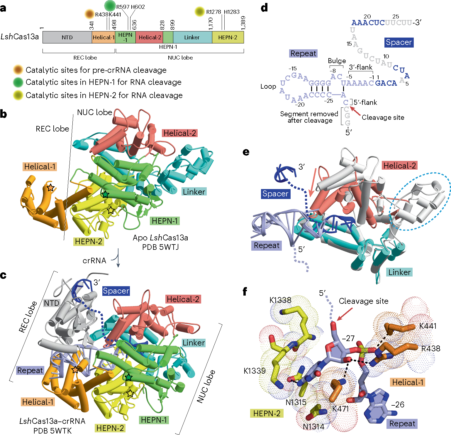
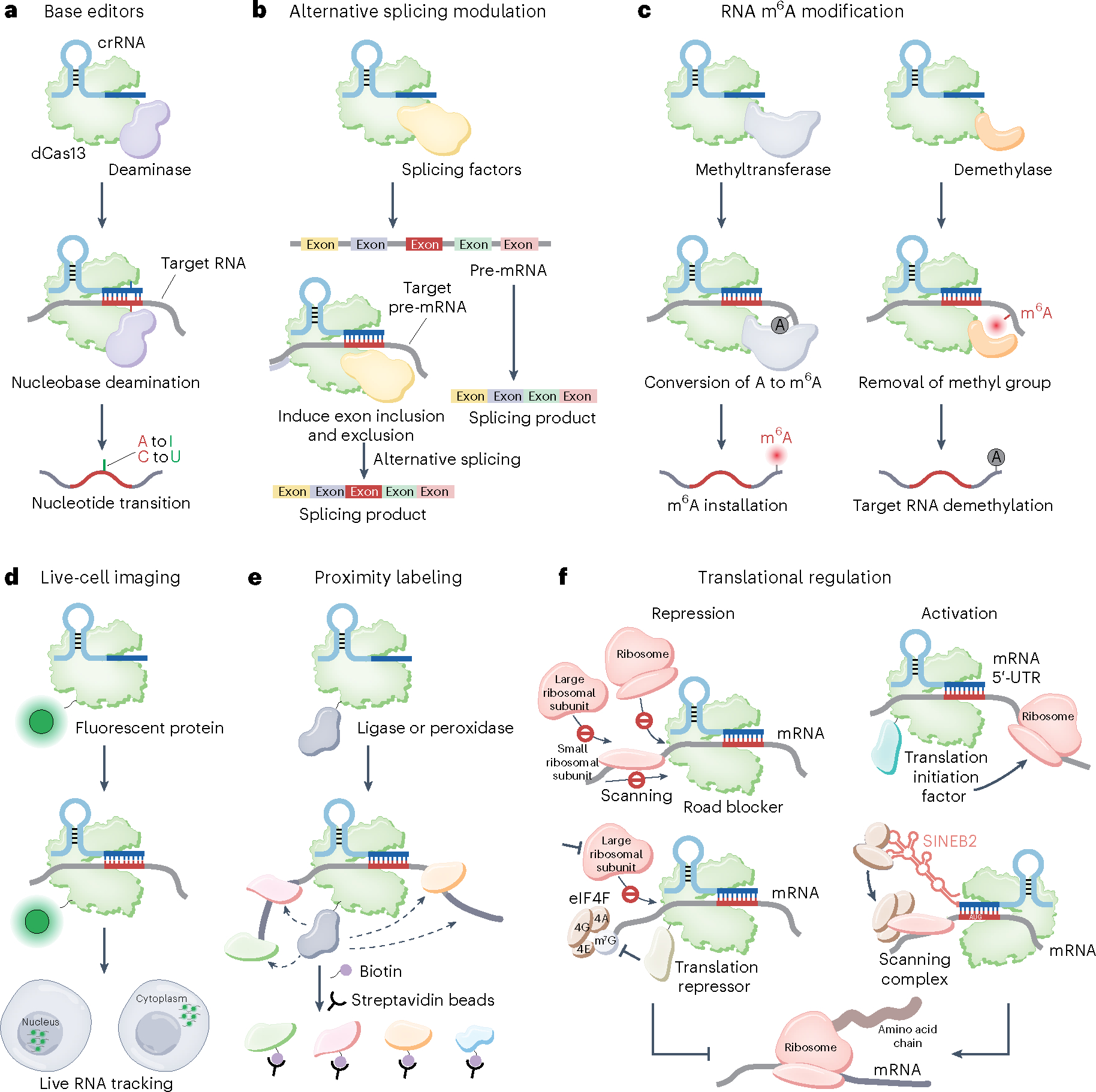
Prokaryotes are equipped with a variety of resistance strategies to survive frequent viral attacks or invading mobile genetic elements. Among these, CRISPR-Cas surveillance systems are abundant and have been studied extensively. This Review focuses on CRISPR-Cas type VI Cas13 systems that use single-subunit RNA-guided Cas endonucleases for targeting and subsequent degradation of foreign RNA, thereby providing adaptive immunity. Notably, distinct from single-subunit DNA-cleaving Cas9 and Cas12 systems, Cas13 exhibits target RNA-activated substrate RNase activity. This Review outlines structural, biochemical and cell biological studies toward elucidation of the unique structural and mechanistic principles underlying surveillance effector complex formation, precursor CRISPR RNA (pre-crRNA) processing, self-discrimination and RNA degradation in Cas13 systems as well as insights into suppression by bacteriophage-encoded anti-CRISPR proteins and regulation by endogenous accessory proteins. Owing to its programmable ability for RNA recognition and cleavage, Cas13 provides powerful RNA targeting, editing, detection and imaging platforms with emerging biotechnological and therapeutic applications.


Conflict of interest statement

Competing interests

The authors declare no competing interests.

Figures

Fig. 1 |
Fig. 1 |. Three stages of CRISPR–Cas13 adaptive immunity: adaptation, expression and interference.
CRISPR–Cas13 loci consist of a CRISPR array and neighboring cas genes. In the adaption stage, the adaptation machinery acquires fragments of invading mobile genetic elements and integrates them into the CRISPR array as new spacers, which generates heritable immunological memory. CRISPR repeats are represented as cyan rectangles, and spacers are represented as differently colored diamonds. The cas1 and cas2 genes, which encode adaptation modules, are missing in most Cas13 loci. How these Cas13 systems acquire new spacers remains unclear. During the CRISPR expression stage, the CRISPR array is transcribed as pre-crRNA and is processed into mature crRNA by Cas13, such that each crRNA carries a single spacer flanked with a part of the direct repeat on one side. The topologies of pre-crRNA in types VI-A, VI-B and VI-D systems, highlighting the conserved sequences and secondary structures, as well as the differences among different subtypes are shown in the zoomed-in box. Pre-crRNA processing sites are indicated by red arrows. The mature crRNA assembles with its effector protein Cas13 to form a surveillance complex that recognizes and degrades foreign genetic elements complementary to the crRNA spacer during interference. At the interference stage, guide–target base-pairing requirements enable sequence-specific targeting and lead to activation of Cas13 for the cleavage of target RNA at promiscuous positions outside the pairing region. Additionally, upon target RNA recognition, Cas13 causes collateral RNase activity against environmental substrate RNA, which induces cell dormancy. Acrs encoded by bacteriophages can directly inactivate Cas13 and prevent target cleavage.
Fig. 2 |
Fig. 2 |. Functional module organization of CRISPR type VI Cas13 loci.
The functional modules involved in adaptation, expression and interference for each subtype are illustrated in the inserted panels. The approximate positions of the two HEPN domains are indicated in dark green within each effector Cas13a (C2c2), Cas13b (C2c6), Cas13c (C2c7), Cas13d (CasRx), Cas13bt (further classified as Cas13X and Cas13Y) and Cas13ct proteins. The average sizes of Cas13 proteins are indicated under the corresponding genes, among which Cas13d (~930 amino acids (aa)) and Cas13bt and Cas13ct (~800 amino acids) proteins are relatively smaller than their Cas13a–Cas13c protein counterparts (larger than 1,100 amino acids). Genes encoding Cas1, Cas2, Csx27, Csx28 and WYL domain proteins are found only in some species in a set of indicated subtypes and are marked by dashed outlines. The dendrogram on the left of the Cas13 systems is adapted from refs. ,, and shows the potential evolutionary relationships. The branch lengths are not scaled and do not indicate evolutionary distances. HTH, helix–turn–helix. TM, transmembrane.
Fig. 3 |
Fig. 3 |. Mechanisms of crRNA recognition and maturation in type VI-A systems.
a, Schematic representation of the domain architecture of LshCas13a. The catalytic sites for pre-crRNA cleavage and target RNA cleavage are labeled. b,c, Overall structures of LshCas13a in apo (b; PDB 5WTJ) and crRNA-bound (c; PDB 5WTK) states. The line indicates the boundary between the REC lobe and the NUC lobe. The NTD domain in the REC lobe, which cannot be traced in the apo form, is clearly visible in the crRNA-bound form. The bound crRNA is embedded in the Cas13 scaffold with the entire repeat segment traceable, but only some segments of the spacer are detectable in the structure of the complex. The direct repeat and the spacer are colored in light blue and dark blue, while disordered regions are indicated as dashed lines. The catalytic residues for pre-crRNA cleavage are indicated by orange stars, while the R-X4–6-H motifs in the two HEPN domains for target RNA cleavage are indicated by green and yellow stars, respectively. d, Topology showing crRNA in the Cas13a-bound state on binary complex formation. Loop, flank and bulge elements are labeled. Nucleotides not observed in the structure are colored in gray. The pre-crRNA processing site is indicated by a red arrow. e, Domain movements induced by crRNA loading. The structure of Cas13a in the apo state (in silver) is superimposed with the structure of Cas13a in the crRNA-bound state (in color). The major domain movements occur in the helical-2 domain that shifts toward the linker domain. A portion of the linker domain is disordered in the crRNA-bound state, which is indicated as a cyan dashed circle. f, Detailed interaction between the pre-crRNA cleavage site nucleotide C(−27) and Cas13a.
Fig. 4 |
Fig. 4 |. Mechanisms of target RNA recognition in type VI-A systems.
a, Schematic representation of the domain architecture of LbuCas13a. The catalytic sites in the pair of HEPN domains for target RNA cleavage are indicated. b,c, Overall structures of the LbuCas13a–crRNA binary complex in the absence (b, pre-target bound; PDB 5XWY) and presence (c, target bound; PDB 5XWP) of target RNA. The REC and NUC lobes are labeled. The R-X4–6-H motifs in the two HEPN domains for target cleavage are indicated by colored stars. d,e, Topology of crRNA (d) and crRNA–target RNA (e) in the LbuCas13a binary and ternary complexes. The direct repeat, spacer and target RNA are colored in light blue, dark blue and red, respectively. The loop, flank and bulge elements of crRNA are labeled. Nucleotides not observed in the structure are colored in gray. The conformational change in the 3′-flank of the crRNA direct repeat is indicated by a red arrow. The 3′-flank region (−3 to −1) of the direct repeat of LbuCas13a–crRNA moves toward the stem loop as indicated by red arrows. f, Domain movements in the Cas13a–crRNA binary complex induced by bound target RNA on ternary complex formation. The structure of LbuCas13a in the pre-target RNA-bound state (in silver) is superimposed with that in the target RNA-bound state (in color). The major domain movements occur in the helical-2 and HEPN-1 domains and are indicated by colored arrows. g, Conformational changes in catalytic residues of the pair of HEPN domains on proceeding from the Cas13a–crRNA binary complex (in silver) to the ternary complex with added target RNA (in color). Target RNA loading induces the formation of a composite catalytic pocket involving a pair of HEPN domains (in color). Lbu, LbuCas13a.
Fig. 5 |
Fig. 5 |. Mechanism of self–non-self-discrimination in type VI-A systems.
a, Schematic representation of LshCas13a domain architecture. b, Overall structure of the LshCas13a–crRNA binary complex bound to target RNA (PDB 7DMQ) in which the target RNA anti-tag segment pairs with the crRNA tag segment. The NTD domain is disordered as indicated as the dashed box in a. c, Topology showing pairing alignment between crRNA and anti-tag containing target RNA. The 8-nucleotide tag segment is highlighted with a black box. The 8-nucleotide anti-tag and 28-nucleotide target regions of target RNA are colored in black and red, respectively. The conformational changes of the tag segment are indicated by a red arrow. d, Comparison of catalytic residues of the pair of HEPN domains on proceeding from the Cas13a–crRNA binary complex (in silver) to the ternary complex (in color) with added anti-tag target RNA. The catalytic residues remain apart from each other, indicating that Cas13a is inactive after binding with anti-tag target RNA. Lsh, LshCas13a.
Fig. 6 |
Fig. 6 |. Modulation mechanisms of type VI systems involving anti-CRISPRs and accessory proteins.
a, Proposed modulation mechanisms mediated by anti-CRISPRs and accessory proteins. b, AlphaFold2-predicted overall structure (left) and electrostatic surface (right) of BzCsx27. The hydrophobic surface might be associated with membrane. c, Overall structure of the PbCsx28 octamer (PDB 8GI1). Transmembrane regions are indicated. The predicted key residues R152 and H157 in the R-X4–6-H motif are shown as a stick model in the zoomed-in box. d, Overall domain alignment and structure of the RspWYL1 dimer (top; PDB 6OAW). Monomer A (Mol A) is shown in a ribbon mode, and monomer B (Mol B) is shown as a surface mode. e, Schematic representation of the domains of LseCas13a and AcrVIA1. f, Two views of a ribbon diagram showing Acr protein AcrVIA1 (in surface) bound to LseCas13–crRNA (in ribbon) (PDB 6VRB). AcrVIA1 directly binds to and interacts with distinct domains of Cas13, thereby blocking access to target RNA.
Fig. 7 |
Fig. 7 |. Active Cas13-based RNA-targeting technologies.
a, DNA or RNA samples are amplified by isothermal amplification and then transcribed into ssRNA. b, Binding of the crRNA to the complementary target sequence activates Cas13 collateral cleavage of quenched fluorescent ssRNA reporters, thereby releasing fluorescence signal. c, Schematic of Cas13–Csm6-based nucleic acid detection.
Fig. 8 |
Fig. 8 |. dCas13-based RNA manipulation technologies.
a, RNA base editing. The dCas13–deaminase fusion can be used for site-specific nucleotide conversion. b, Programmable alternative splicing modulation. dCas13 fused to splicing factors can be used for pre-mRNA alternative splicing modulation to generate a customized splicing product. c, Programmable RNA m6A modification regulation. dCas13 fused to methyltransferase complex (left) or demethylase (right) can mediate the m6A methylation levels of target RNA. d, Endogenous RNA imaging and tracking. dCas13 fused to a fluorescent protein enables the visualization of RNA in live cells. By fusing different localization signals with Cas13, target RNA can be transported to the desired cellular location, such as the nucleus and the cytoplasm. e, Cas13-based proximity labeling. dCas13 fused with ligase or peroxidase can be applied to label proximal proteins with biotin and facilitate the discovery of new proteins interacting with target RNA. f, dCas13 recruited to mRNA may physically abrogate ribosome binding and movement, thereby impeding translation initiation and elongation. dCas13 fused with a translational repressor provides an alternative suppression mechanism upon m7G cap binding, increasing translational repression efficiency. dCas13 fused with a translational initiation factor targeting the 5′-UTR of mRNA enables the recruitment of ribosomal subunits and thus presumably increases translation initiation. Through ribosome recruitment by the SINEB2 element, the dCasRx–SINEB2 system allows for translation activation by targeting the initiation region around the AUG start codon. eIF4, eukaryotic initiation factor 4.

References

    1. Jia N & Patel DJ Structure-based functional mechanisms and biotechnology applications of anti-CRISPR proteins. Nat. Rev. Mol. Cell Biol. 22, 563–579 (2021). - PubMed
    1. Borges AL, Davidson AR & Bondy-Denomy J The discovery, mechanisms, and evolutionary impact of anti-CRISPRs. Annu. Rev. Virol. 4, 37–59 (2017). - PMC - PubMed
    1. Davidson AR et al. Anti-CRISPRs: protein inhibitors of CRISPR–Cas systems. Annu. Rev. Biochem. 89, 309–332 (2020). - PMC - PubMed
    1. Li Y & Bondy-Denomy J Anti-CRISPRs go viral: the infection biology of CRISPR–Cas inhibitors. Cell Host Microbe 29, 704–714 (2021). - PMC - PubMed
    1. Pawluk A, Davidson AR & Maxwell KL Anti-CRISPR: discovery, mechanism and function. Nat. Rev. Microbiol. 16, 12–17 (2018). - PubMed

Publication types

MeSH terms

LinkOut - more resources