. 2024 Jun;11(6):1478-1491.
doi: 10.1002/acn3.52062. Epub 2024 May 4.

Primary mitochondrial disorders and mimics: Insights from a large French cohort

Affiliations

Primary mitochondrial disorders and mimics: Insights from a large French cohort

Cécile Rouzier et al. Ann Clin Transl Neurol. 2024 Jun.

Abstract

Objective: The objective of this study was to evaluate the implementation of NGS within the French mitochondrial network, MitoDiag, from targeted gene panels to whole exome sequencing (WES) or whole genome sequencing (WGS) focusing on mitochondrial nuclear-encoded genes.

Methods: Over 2000 patients suspected of Primary Mitochondrial Diseases (PMD) were sequenced by either targeted gene panels, WES or WGS within MitoDiag. We described the clinical, biochemical, and molecular data of 397 genetically confirmed patients, comprising 294 children and 103 adults, carrying pathogenic or likely pathogenic variants in nuclear-encoded genes.

Results: The cohort exhibited a large genetic heterogeneity, with the identification of 172 distinct genes and 253 novel variants. Among children, a notable prevalence of pathogenic variants in genes associated with oxidative phosphorylation (OXPHOS) functions and mitochondrial translation was observed. In adults, pathogenic variants were primarily identified in genes linked to mtDNA maintenance. Additionally, a substantial proportion of patients (54% (42/78) and 48% (13/27) in children and adults, respectively), undergoing WES or WGS testing displayed PMD mimics, representing pathologies that clinically resemble mitochondrial diseases.

Interpretation: We reported the largest French cohort of patients suspected of PMD with pathogenic variants in nuclear genes. We have emphasized the clinical complexity of PMD and the challenges associated with recognizing and distinguishing them from other pathologies, particularly neuromuscular disorders. We confirmed that WES/WGS, instead of panel approach, was more valuable to identify the genetic basis in patients with "possible" PMD and we provided a genetic testing flowchart to guide physicians in their diagnostic strategy.


Conflict of interest statement

The authors declare no competing financial interest.

Figures
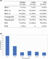
Figure 1
Figure 1
(A) Overview of patients according to MDC score, consanguinity and molecular testing (targeted panels, WES, WGS). Patients were classified according to the MDC score. Patients with a PMDC score of 1 had either an OXPHOS deficiency or multiple mtDNA deletions. N, number. (B) Age distribution of individuals at the time of enrolment.
Figure 2
Figure 2
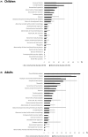
Percentage of patients for clinical symptoms following the Human Phenotype Ontology (HPO) classification. Children clinical features are represented in the left panel (A), in black for children with primary PMD (N = 235) and in gray for children with PMD mimics (N = 59). Adults' clinical features are represented in the right panel (B), in black for adults with primary PMD (N = 87) and in gray for adults with PMD MIMICS (N = 16). OA, optic atrophy; RP, retinitis pigmentosa.
Figure 3
Figure 3
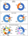
Analysis of pathogenic variants identified in the 294 children (left panels) and the 103 adults (right panels). (A) Mode of inheritance, autosomal dominant (AD), autosomal recessive (AR), X‐linked (XL) are indicated with the percentage. (B) Molecular classification according to the type of variants (Missense; indels:Insertion/deletion; nonsense; splice variant; CNV: Copy number variation. (C) The most frequently mutated genes are listed if pathogenic variants were identified at least 3 times in adults, and 6 times in children. In brackets: absolute numbers/frequencies. The mitochondrial functions are represented by colors: blue: mtDNA maintenance, green: lipids metabolism, orange: carbohydrate metabolism, brown: assembly factors, yellow: mitochondrial translation, pink: mitochondrial protein metabolism, purple: mitochondrial dynamics, gray: non‐mitochondrial gene.
Figure 4
Figure 4
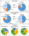
Histology and biochemical characterization of the patient cohort according to age, children and adults with mitochondrial (MD) and PMD mimics (PMD mimics). (A) Histology analysis. Pie charts showing the patient number (n) and percentages with normal (light blue and light orange) or abnormal, including ragged‐red fibers, COX negative fibers, mitochondrial proliferation or excessive lipids accumulation, (dark blue and dark orange) muscle histology in children and adults (B) Pie charts depicting the number (n) of patients with normal (light blue and light orange) or abnormal (dark blue and dark orange) respiratory chain (RC) activities. (C) Summary of the outcome of respiratory chain activity with the percentages of complexes of the respiratory chain; CI, complex I; CII, complex II; CIII, complex III; CIV, complex IV; CV, complex V; or combined or quinone deficiencies.
Figure 5
Figure 5
Diagnostic approach for mitochondrial disorders. Score 2–4: possible PMD, score ≥5 probable PMD.

References

    1. Schaefer AM, Taylor RW, Turnbull DM, Chinnery PF. The epidemiology of mitochondrial disorders—past, present and future. Biochim Biophys Acta. 2004;1659(2–3):115‐120. - PubMed
    1. Tan J, Wagner M, Stenton SL, et al. Lifetime risk of autosomal recessive mitochondrial disorders calculated from genetic databases. EBioMedicine. 2020;54:102730. - PMC - PubMed
    1. Morava E, van den Heuvel L, Hol F, et al. Mitochondrial disease criteria: diagnostic applications in children. Neurology. 2006;67(10):1823‐1826. - PubMed
    1. Niyazov DM, Kahler SG, Frye RE. Primary mitochondrial disease and secondary mitochondrial dysfunction: importance of distinction for diagnosis and treatment. Mol Syndromol. 2016;7(3):122‐137. - PMC - PubMed
    1. Bannwarth S, Procaccio V, Lebre AS, et al. Prevalence of rare mitochondrial DNA mutations in mitochondrial disorders. J Med Genet. 2013;50(10):704‐714. - PMC - PubMed

Substances