The impact of temperature on vascular function in connection with vascular laser treatment
- PMID: 38703271
- PMCID: PMC11069475
- DOI: 10.1007/s10103-024-04070-7
The impact of temperature on vascular function in connection with vascular laser treatment
Abstract
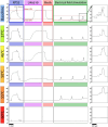
Pulsed dye lasers are used effectively in the treatment of psoriasis with long remission time and limited side effects. It is, however, not completely understood which biological processes underlie its favorable outcome. Pulsed dye laser treatment at 585-595 nm targets hemoglobin in the blood, inducing local hyperthermia in surrounding blood vessels and adjacent tissues. While the impact of destructive temperatures on blood vessels has been well studied, the effects of lower temperatures on the function of several cell types within the blood vessel wall and its periphery are not known. The aim of our study is to assess the functionality of isolated blood vessels after exposure to moderate hyperthermia (45 to 60°C) by evaluating the function of endothelial cells, smooth muscle cells, and vascular nerves. We measured blood vessel functionality of rat mesenteric arteries (n=19) by measuring vascular contraction and relaxation before and after heating vessels in a wire myograph. To this end, we elicited vascular contraction by addition of either high potassium solution or the thromboxane analogue U46619 to stimulate smooth muscle cells, and electrical field stimulation (EFS) to stimulate nerves. For measurement of endothelium-dependent relaxation, we used methacholine. Each vessel was exposed to one temperature in the range of 45-60°C for 30 seconds and a relative change in functional response after hyperthermia was determined by comparison with the response per stimulus before heating. Non-linear regression was used to fit our dataset to obtain the temperature needed to reduce blood vessel function by 50% (Half maximal effective temperature, ET50). Our findings demonstrate a substantial decrease in relative functional response for all three cell types following exposure to 55°C-60°C. There was no significant difference between the ET50 values of the different cell types, which was between 55.9°C and 56.9°C (P>0.05). Our data show that blood vessel functionality decreases significantly when exposed to temperatures between 55°C-60°C for 30 seconds. The results show functionality of endothelial cells, smooth muscle cells, and vascular nerves is similarly impaired. These results help to understand the biological effects of hyperthermia and may aid in tailoring laser and light strategies for selective photothermolysis that contribute to disease modification of psoriasis after pulsed dye laser treatment.
Keywords: Blood vessels; Endothelial cells; Hyperthermia; Smooth muscle cells; Vascular nerves; Wire myography.
© 2024. The Author(s).
Conflict of interest statement
All authors certify that they have no affiliations with or involvement in any organization or entity with any financial interest or non-financial interest in the subject matter or materials discussed in this manuscript.
Figures


References
MeSH terms
Grants and funding
LinkOut - more resources
Full Text Sources

