. 2024 Dec;61(12):10159-10181.
doi: 10.1007/s12035-024-04196-3. Epub 2024 May 4.

Neuroprotective Role of Selenium Nanoparticles Against Behavioral, Neurobiochemical and Histological Alterations in Rats Subjected to Chronic Restraint Stress

Affiliations

Neuroprotective Role of Selenium Nanoparticles Against Behavioral, Neurobiochemical and Histological Alterations in Rats Subjected to Chronic Restraint Stress

Sarah A Elfakharany et al. Mol Neurobiol. 2024 Dec.

Abstract

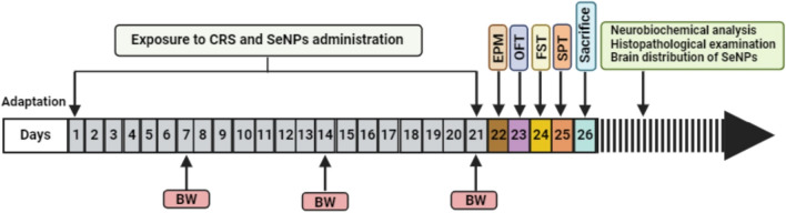
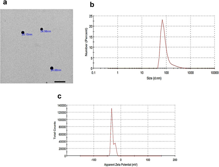
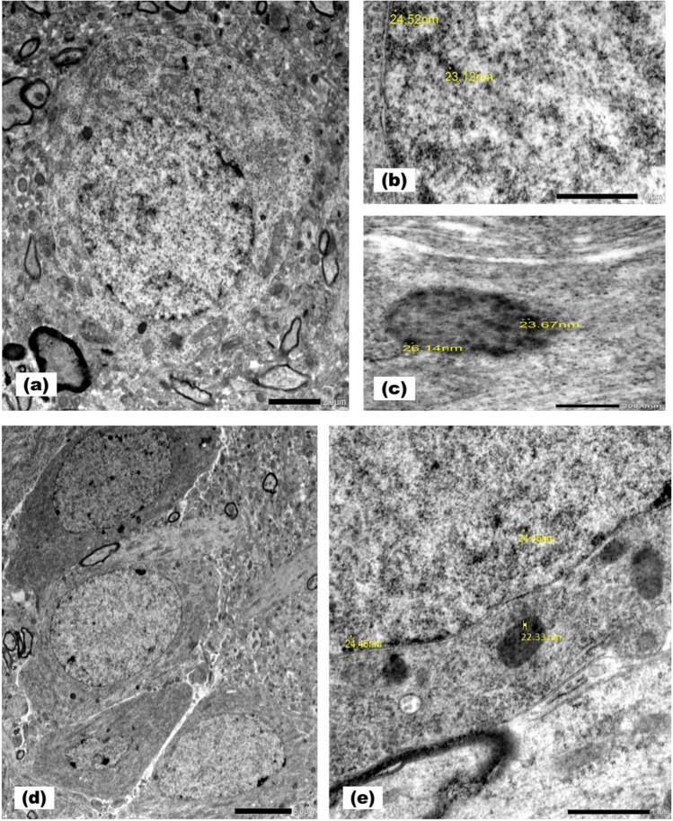
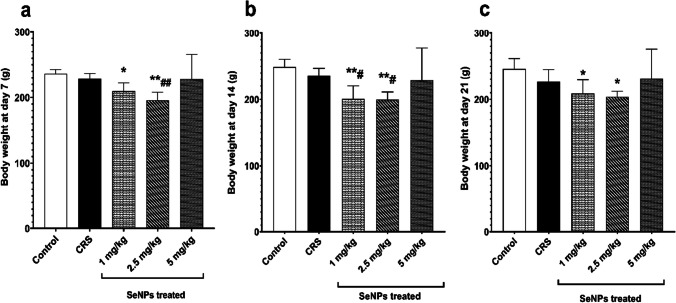
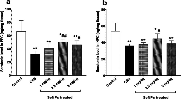
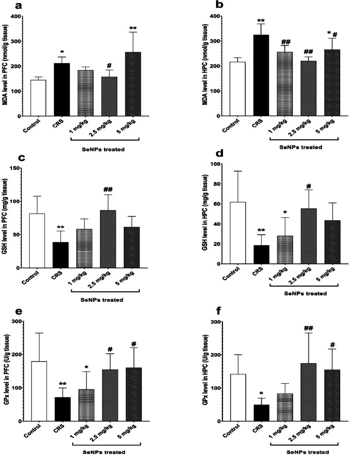
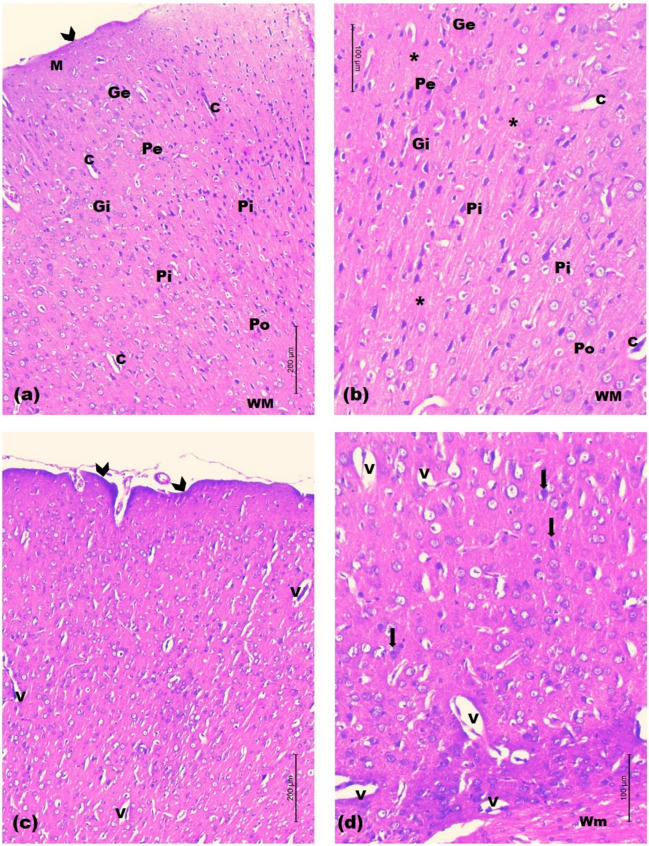
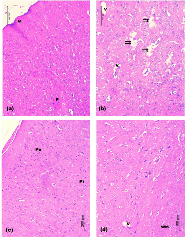
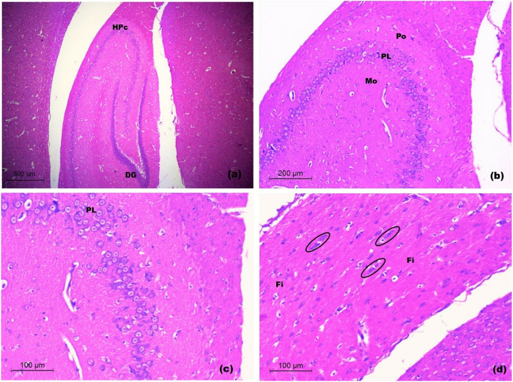
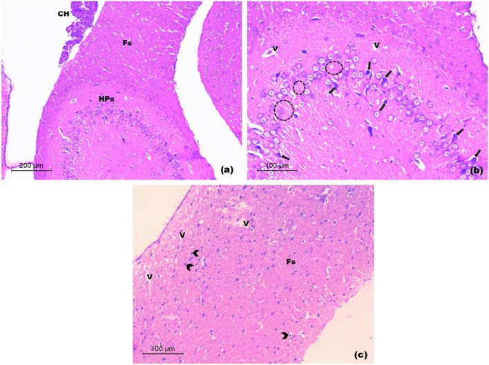
Chronic stress induces changes in the prefrontal cortex and hippocampus. Selenium nanoparticles (SeNPs) showed promising results in several neurological animal models. The implementation of SeNPs in chronic restraint stress (CRS) remains to be elucidated. This study was done to determine the possible protective effects of selenium nanoparticles on behavioral changes and brain oxidative stress markers in a rat model of CRS. 50 rats were divided into three groups; control group (n = 10), untreated CRS group (n = 10) and CRS-SeNPs treated group (n = 30). Restraint stress was performed 6 h./day for 21 days. Rats of CRS-SeNPs treated group received 1, 2.5 or 5 mg/kg SeNPs (10 rats each) by oral gavage for 21 days. Rats were subjected to behavioral assessments and then sacrificed for biochemical and histological analysis of the prefrontal cortex and hippocampus. Prefrontal cortical and hippocampal serotonin levels, oxidative stress markers including malondialdehyde (MDA), reduced glutathione (GSH) and glutathione peroxidase (GPx), tumor necrosis factor alpha (TNF-α) and caspase-3 were assessed. Accordingly, different doses of SeNPs showed variable effectiveness in ameliorating disease parameters, with 2.5 mg/kg dose of SeNPs showing the best improving results in all studied parameters. The present study exhibited the neuroprotective role of SeNPs in rats subjected to CRS and proposed their antioxidant, anti-inflammatory and anti-apoptotic effects as the possible mechanism for increased prefrontal cortical and hippocampal serotonin level, ameliorated anxiety-like and depressive-like behaviors and improved prefrontal cortical and hippocampal histological architecture.

Keywords: Caspase-3; Chronic restraint stress; Oxidative stress; Selenium nanoparticles; Serotonin; TNF-α.


Conflict of interest statement

Declarations. Ethics Approval: The study protocol was approved by the institutional Medical Ethics Committee, Faculty of Medicine (IRB NO: 00012098; FWA NO: 00018699; approval NO: 0106414). Consent to Participate: Not applicable. Consent for Publication: Not applicable. Competing Interests: The authors declare no competing interests.

Figures

Fig. 1
Fig. 1
Schematic representation of the experimental design. CRS: chronic restraint stress, SeNPs: selenium nanoparticles, BW: body weight, EPM: elevated plus maze test, OFT: open field test, FST: forced swim test, SPT: sucrose preference test
Fig. 2
Fig. 2
Characterization of SeNPs. Transmission electron micrograph of selenium nanoparticles (scale bar 100 nm) showing size of 20–30 nm in (a). Size distribution of 100% peak at a size of 92.01 nm is shown in (b). Zeta potential of -30.5 mV is revealed in (c)
Fig. 3
Fig. 3
Transmission electron micrographs of prefrontal cortex a-c and hippocampus d-e of selenium nanoparticles-treated rats. A prefrontal cortical neuron a harboring selenium nanoparticles in the nucleus b and the lysosomes c. Selenium nanoparticles are revealed in the nucleoplasm and lysosomes of a pyramidal neuron in the hippocampus proper d-e (Mic. Mag. a × 2500, b × 8000, c × 25000, d × 1200 and e × 8000)
Fig. 4
Fig. 4
Body weight of rats in the different studied groups at day 7 (a), at day 14 (b) and at day 21 (c). Data is expressed as Mean ± SD and analyzed by (one-way ANOVA) separately at days 7, 14 and 21, followed when significant by Post Hoc Test (Tukey).* p < 0.05, **p ≤ 0.001 significant difference compared to control group. #p < 0.05, ##p ≤ 0.001 significant difference compared to untreated chronic restraint stress group. CRS: chronic restraint stress, SeNPs: selenium nanoparticles, g: grams
Fig. 5
Fig. 5
Neurobehavioral parameters of rats in the different studied groups a-h. a Number of entries in closed arms. b Time spent in closed arms. c Number of entries in open arms. d Time spent in open arms. e Number of squares crossed. f Number of rears. g Immobility time. h Percentage of sucrose preference. Data is expressed as median (intra-quartile range) and analyzed by non-parametric ANOVA (Kruskal Wallis, KW) in a, c, d & g. Pair wise comparisons are analyzed using Mann Whitney U test. Data is expressed as Mean ± SD and analyzed by (one-way ANOVA), followed when significant by Post Hoc Test (Tukey) in b, e, f & h. * p < 0.05, **p ≤ 0.001 significant difference compared to control group. #p < 0.05, ##p ≤ 0.001 significant difference compared to untreated chronic restraint stress group. CRS: chronic restraint stress, SeNPs: selenium nanoparticles, sec: seconds
Fig. 6
Fig. 6
Serotonin level (in ng/mg tissue) in the prefrontal cortex a and hippocampus b of rats in the different studied groups. Data is expressed as Mean ± SD and analyzed by (one-way ANOVA), followed when significant by Post Hoc Test (Tukey). * p < 0.05, **p ≤ 0.001 significant difference compared to control group. #p < 0.05, ##p ≤ 0.001 significant difference compared to untreated chronic restraint stress group. CRS: chronic restraint stress, SeNPs: selenium nanoparticles, PFC: prefrontal cortex, HPC: hippocampus
Fig. 7
Fig. 7
Oxidative stress markers of rats in the different studied groups (a-f). Malondialdehyde level (in nmol/g tissue) in the prefrontal cortex (a) and hippocampus (b). Reduced glutathione level (in mg/g tissue) in the prefrontal cortex (c) and hippocampus (d). Glutathione peroxidase level (in U/g tissue) in the prefrontal cortex (e) and hippocampus (f). Data is expressed as Mean ± SD and analyzed by (one-way ANOVA), followed when significant by Post Hoc Test (Tukey). * p < 0.05, **p ≤ 0.001 significant difference compared to control group. #p < 0.05, ##p ≤ 0.001 significant difference compared to untreated chronic restraint stress group. CRS: chronic restraint stress, SeNPs: selenium nanoparticles, MDA: malondialdehyde, GSH: reduced glutathione, GPx: glutathione peroxidase, PFC: prefrontal cortex, HPC: hippocampus
Fig. 8
Fig. 8
TNF-α level (in ng/g protein) in the prefrontal cortex a and hippocampus b, and caspase-3 level (in pg/g protein) in the prefrontal cortex c and hippocampus d of rats in the different studied groups. Data is expressed as Mean ± SD and analyzed by (one-way ANOVA), followed when significant by Post Hoc Test (Tukey). * p < 0.05, **p ≤ 0.001 significant difference compared to control group. #p < 0.05, ##p ≤ 0.001 significant difference compared to untreated chronic restraint stress group. CRS: chronic restraint stress, SeNPs: selenium nanoparticles, TNF-α: tumor necrosis factor alpha, PFC: prefrontal cortex, HPC: hippocampus
Fig. 9
Fig. 9
Light H&E-stained micrographs of the prefrontal cortices of control and untreated chronic restraint stress rats. Histological sections of control rats in (a) and (b). The normal architecture with the molecular layer (M) beneath the pia mater (arrow head) is seen. The external granular (Ge), the external pyramidal (Pe) and the internal granular (Gi) layers are observed. The internal pyramidal cell layer (Pi) is seen with its large pyramidal neuronal cell bodies. The polymorphic layer of the cortex (Po) is seen deeper to the internal pyramidal cell layer. The neuropil (asterisk) is revealed among the cell layers with numerous blood capillaries (C). White matter (WM). Histological sections of untreated chronic restraint stress rats in (c) and (d). The irregular cortical surface is seen (arrow heads). The disturbed neuronal layers are seen within a lightly-stained spongy-like neuropil with vacuoles (V). Occasional pyknotic nuclei are seen (arrows). Notice, the disorganized white matter (Wm) (Mic. Mag. a × 100, b × 200, c × 100 and d × 200)
Fig. 10
Fig. 10
Light H&E-stained micrographs of the prefrontal cortices of rats treated with 1 and 2.5 mg/kg of SeNPs. Sections of rats treated with 1 mg/kg SeNPs in a and b show restoration of the laminar architecture of cortical neurons (M, P). Few vacuoles (V) are seen with sporadic areas of mouth-eaten neuropil (double arrows). Sections of rats treated with 2.5 mg/kg SeNPs in c and d reveal the normal architecture of cortical neurons and the neuropil. Few small vacuoles are seen (V). Note, the regularly arranged nerve fiber bundles of the white matter (WM). External (Pe) and internal pyramidal (Pi) cell layers (Mic. Mag. a × 100, b × 200, c × 100 and d × 200)
Fig. 11
Fig. 11
Light H&E-stained micrographs of the prefrontal cortices of rats treated with 5 mg/kg of SeNPs in a and b show congested blood vessels of the subarachnoid space (stars). Vacuolar necrotic areas are revealed with structureless fibrillar material (NF). Note, disorganized cortical layers (Mic. Mag. a × 100, b × 200)
Fig. 12
Fig. 12
Light H&E-stained micrographs of hippocampus of control rats. Sections of control rats in a-d. The normal architecture of hippocampus proper (HPc) and dentate gurus (DG) is seen. The hippocampus proper appears with the molecular layer (Mo), the prominent pyramidal cell layer (PL) and the polymorphic layer (Po). Note, the well-organized fimbria (Fi) and the rows of neuroglia (Oval) (Mic. Mag. a × 40, b × 100, c × 200 and d × 200)
Fig. 13
Fig. 13
Light H&E-stained micrographs of hippocampus untreated CRS rats a-c. The disorganized pyramidal layer of hippocampus proper (HPs in a) is seen with evident neuronal loss (dotted circles in b) and focal vacuoles of the polymorphic layer (V in b). Many apoptotic pyramidal neurons are revealed (arrows in b). Note, the disorganized fimbria (Fs in a and c) with vacuoles (V in c) and cellular infiltration (arrow heads in c). The choroid plexus of inferior horn of lateral ventricle (CH in a) is seen (Mic. Mag. a × 100, b × 200 and c × 200)
Fig. 14
Fig. 14
Light H&E-stained micrographs of hippocampus of rats treated with 1 and 2.5 mg/kg of SeNPs. Sections a and b show the almost normal architecture of hippocampus proper and fimbria with prominent capillaries and larger blood vessels (BV). A number of pyramidal neurons (PL) are seen extending into the polymorphic layer. Note, very few apoptotic pyramidal neurons (arrows) (Mic. Mag. a × 100, b × 200)
Fig. 15
Fig. 15
Light H&E-stained micrographs of hippocampus of rats treated with 5 mg/kg of SeNPs a-b. Evident thinning out of the pyramidal cell layer with neuronal loss (dotted circles in a and b) is seen. Some apoptotic pyramidal cells (arrows in b). Note, few vacuoles (V in b) of the polymorphic layer of hippocampus proper (Mic. Mag. a × 100, b × 200)

References

    1. Becerril-Chávez H, Colín-González AL, Villeda-Hernández J, Galván-Arzate S, Chavarría A, de Lima ME, Túnez I, Santamaría A (2017) Protective effects of S-allyl cysteine on behavioral, morphological and biochemical alterations in rats subjected to chronic restraint stress: antioxidant and anxiolytic effects. J Funct Foods 35:105–114. 10.1016/j.jff.2017.05.034
    1. Hegab II, Baarhoma RA, Abd El-Latif RN, El-Esawy R (2019) The prospective protective effect of selenium against chronic restraint stress-induced memory impairment in male albino rats. Med J Cairo Univ 87:1563–1572. 10.21608/mjcu.2019.53576
    1. Alkadhi K (2013) Brain physiology and pathophysiology in mental stress. ISRN 2013. 10.1155/2013/806104
    1. Chu B, Marwaha K, Sanvictores T, Ayers D (2021) Physiology, stress reaction. StatPearls Publishing Treasure Island (FL). https://www.ncbi.nlm.nih.gov/books/NBK541120/. Accessed 30 September 2021 - PubMed
    1. Phyu P, Dumenigo R, Oms JD, Gunaseelan L, Foroughi G, Rizvi SAA (2017) The influence of chronic stress on the development of psychiatric conditions. Consultant 57:76–83. 10.1017/S1092852918000585

MeSH terms