. 2024 Oct 3;147(10):3547-3561.
doi: 10.1093/brain/awae140.

Hippocampal aggregation signatures of pathogenic UBQLN2 in amyotrophic lateral sclerosis and frontotemporal dementia

Affiliations

Hippocampal aggregation signatures of pathogenic UBQLN2 in amyotrophic lateral sclerosis and frontotemporal dementia

Kyrah M Thumbadoo et al. Brain. .

Abstract

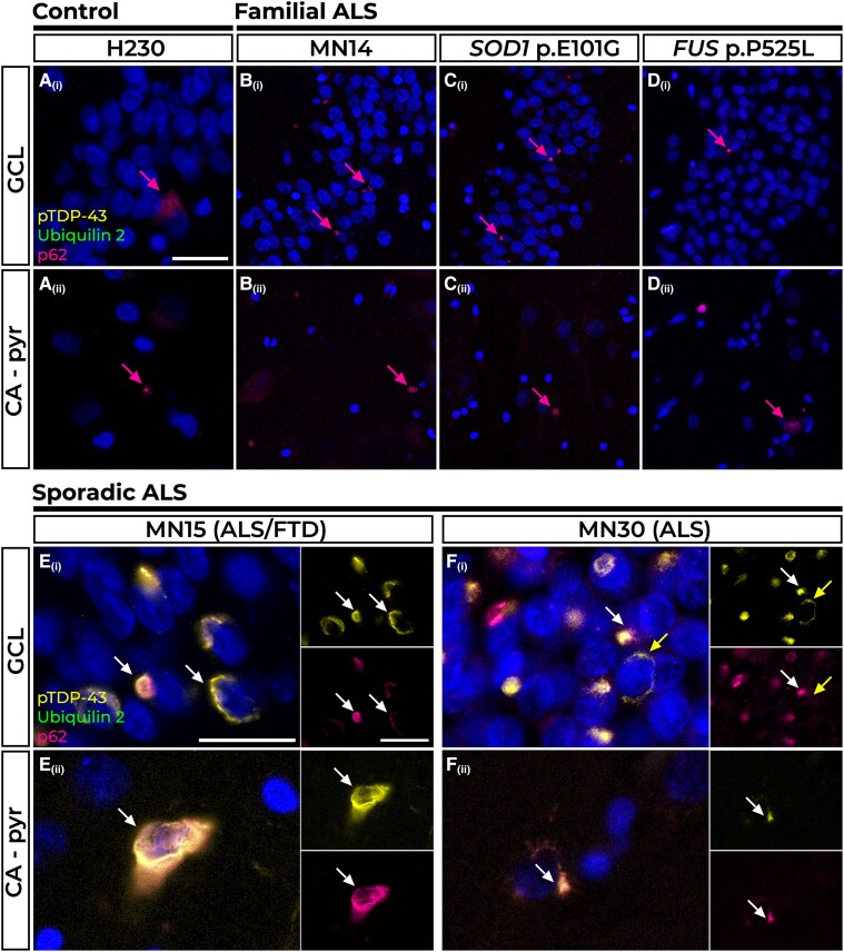
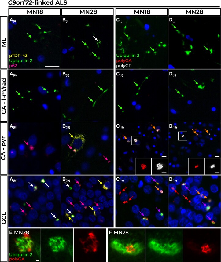
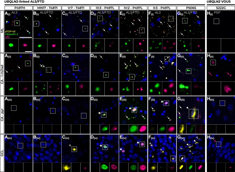
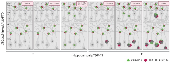
Pathogenic variants in the UBQLN2 gene cause X-linked dominant amyotrophic lateral sclerosis and/or frontotemporal dementia characterized by ubiquilin 2 aggregates in neurons of the motor cortex, hippocampus and spinal cord. However, ubiquilin 2 neuropathology is also seen in sporadic and familial amyotrophic lateral sclerosis and/or frontotemporal dementia cases not caused by UBQLN2 pathogenic variants, particularly C9orf72-linked cases. This makes the mechanistic role of mutant ubiquilin 2 protein and the value of ubiquilin 2 pathology for predicting genotype unclear. Here we examine a cohort of 44 genotypically diverse amyotrophic lateral sclerosis cases with or without frontotemporal dementia, including eight cases with UBQLN2 variants [resulting in p.S222G, p.P497H, p.P506S, p.T487I (two cases) and p.P497L (three cases)]. Using multiplexed (five-label) fluorescent immunohistochemistry, we mapped the co-localization of ubiquilin 2 with phosphorylated TDP-43, dipeptide repeat aggregates and p62 in the hippocampus of controls (n = 6), or amyotrophic lateral sclerosis with or without frontotemporal dementia in sporadic (n = 20), unknown familial (n = 3), SOD1-linked (n = 1), FUS-linked (n = 1), C9orf72-linked (n = 5) and UBQLN2-linked (n = 8) cases. We differentiate between (i) ubiquilin 2 aggregation together with phosphorylated TDP-43 or dipeptide repeat proteins; and (ii) ubiquilin 2 self-aggregation promoted by UBQLN2 pathogenic variants that cause amyotrophic lateral sclerosis and/or frontotemporal dementia. Overall, we describe a hippocampal protein aggregation signature that fully distinguishes mutant from wild-type ubiquilin 2 in amyotrophic lateral sclerosis with or without frontotemporal dementia, whereby mutant ubiquilin 2 is more prone than wild-type to aggregate independently of driving factors. This neuropathological signature can be used to assess the pathogenicity of UBQLN2 gene variants and to understand the mechanisms of UBQLN2-linked disease.

Keywords: UBQLN2; amyotrophic lateral sclerosis (ALS); frontotemporal dementia (FTD); hippocampus; neuropathology; ubiquilin 2.


Conflict of interest statement

The authors report no competing interests.

Figures

Figure 1
Figure 1
Pathology in the hippocampal dentate gyrus and cornu ammonis regions of control, familial and sporadic cases. The majority of control tissue had no pTDP-43 or ubiquilin 2 pathology, but one case had sparse p62 immunoreactivity in the granule cell layer (GCL) and cornu ammonis – pyramidal cells (CA – pyr) layers (A Control H230, pink arrows). Such p62-positive inclusions were also found in the GCL [B(i)–D(i)] and CA – pyr layers [B(ii)–D(ii)] in Case MN14, an amyotrophic lateral sclerosis (ALS) case of unknown genotypic cause (B), Case MN24, a SOD1 p.E101G case (C) and Case P525L, a FUS p.P525L case (D). P62-positive perinuclear pTDP-43 aggregates negative for ubiquilin 2 were found in the GCL and CA – pyr regions of n = 6 sporadic cases represented here by ALS+FTD Case MN15 [E(i and ii), white arrows] and ALS Case MN30 [F(i and ii), white arrows]. Rare pTDP-43 aggregates negative for p62 are indicated by the yellow arrow in F(ii). Scale bars = 50 µm (AD); and 25 µm (E and F). FTD = frontotemporal dementia.
Figure 2
Figure 2
Pathology in the hippocampal dentate gyrus and cornu ammonis regions of C9orf72-linked cases. All C9orf72-linked amyotrophic lateral sclerosis/frontotemporal dementia (ALS/FTD) cases showed wispy ubiquilin 2 aggregates in the molecular layer (ML) and cornu ammonis – lacunosum-molecular and radiatum (CA – l-m/rad) layers that were predominantly p62 negative [A(i and ii) and B(i and ii), green arrows], with larger aggregates sometimes co-labelling with p62 in the ML [B(i), white arrow]. In Case MN18, representative of n = 4 C9orf72 cases (80%), the CA – pyr (pyramidal) cells harboured stellate p62 aggregates [A(iii), pink arrow] while the GCL showed stellate p62 aggregates either with ubiquilin 2 [A(iv), white arrows] or without [A(iv), pink arrow]. Case MN28 shared this pattern of p62 and ubiquilin 2 pathology with the additional presence of perinuclear pTDP-43 inclusions, often found without other co-labelling in the CA – pyr region [B(iii), yellow arrow] or granule cell layer (GCL) [B(iv), yellow arrow]. Ubiquilin 2 aggregates in the ML and CA –l-m/rad layers were negative for dipeptide repeat proteins in all C9orf72 cases [C(i and ii) and D(i and ii), green arrows]. The dominant pathology in CA – pyr regions was the presence of polyGA and polyGP inclusions negative for ubiquilin 2 [C(iii) and D(iii), orange arrowheads]. All GCL ubiquilin 2 was co-localized with at least one dipeptide repeat protein [C(iv) and D(iv), purple arrows]. Ubiquilin 2-negative aggregates were comprised of both dipeptide repeat proteins [C(iv) and D(iv), orange arrows] or most commonly, polyGA without other co-labelling [C(iv) and D(iv), red arrows]. Super-resolution STED microscopy of ubiquilin 2-positive polyGA aggregates in the GCL of Case MN28 demonstrated polyGA enmeshed with and encircling ubiquilin 2 (E) or ubiquilin 2 labelling around a core of polyGA (F). Scale bar in main images AD = 50 µm; magnifications in C(iv) and D(iv), 25 µm; and 1 µm in E and F.
Figure 3
Figure 3
Pathology in the hippocampal dentate gyrus and cornu ammonis regions of UBQLN2-linked cases. All UBQLN2-linked amyotrophic lateral sclerosis/frontotemporal dementia (ALS/FTD) cases showed numerous p62-positive ubiquilin 2 aggregates, indicated by white arrows, in the molecular layer (ML) [A(i)–G(i)] and cornu ammonis – lacunosum-molecular and radiatum (CA – l-m/rad) layers [A(ii)–G(ii)]. Cases P497H and MN17-T487I showed no aggregate labelling in the CA – pyr (pyramidal) cells [A(iii) and B(iii)] or granule cell layer (GCL) [A(iv) and B(iv)]. ALS+FTD Case V:7 – T487I showed no pathology in the CA – pyr cells [C(iii)] and very rare p62-positive pTDP-43 aggregates negative for ubiquilin 2 in the GCL [C(iv), hollow white arrowhead]. All three p.P497L mutant cases had rare intranuclear p62-positive ubiquilin 2 inclusions negative for pTDP-43 in the CA – pyr cells [D(iii)–F(iii)] and Cases III:3 – P497L and IV:2 – P497L also showed these in the GCL [D(iv) and E(iv), all indicated by asterisk]. In Case II:3 – P497L no such intranuclear inclusions were observed in the GCL but frequent p62-positive pTDP-43 aggregates were seen instead [F(iv), hollow arrowheads]. Case P506S had aggregates positive for p62, pTDP-43 and ubiquilin 2 in the CA – pyr cells [G(iii), filled white arrowhead] and the GCL [G(iv), filled white arrowheads] and numerous compact p62-positive but ubiquilin 2-negative pTDP-43 aggregates in the GCL [G(iv), hollow white arrowheads]. A progressive supranuclear palsy (PSP) case harbouring a UBQLN2 p.S222G variant of uncertain significance (VOUS) (H) had no pathology in the ML [H(i)] or CA – pyr cells [H(iii)], but showed sparse p62 positivity (pink arrows) in the CA – l-m/rad layers [H(ii)] and granule cell layer (GCL) [H(iv)]. Scale bar in main images = 25 µm; magnifications, 5 µm.
Figure 4
Figure 4
Schematic representation of the hippocampal neuropathological signature defining UBQLN2-linked ALS/FTD. In all UBQLN2-linked amyotrophic lateral sclerosis/frontotemporal dementia (ALS/FTD) cases, mutant ubiquilin 2 forms punctate, p62-positive aggregates in the cornu ammonis – lacunosum-molecular and radiatum (CA – l-m/rad) layers and molecular layer (ML), comprising a unique neuropathological signature. The neuropathology in the CA – pyr layer and granule cell layer (GCL) is variable between cases. The CA – pyr may show intranuclear p62-positive ubiquilin 2 aggregates (all three P497L cases) or aggregates positive for p62, pTDP-43 and ubiquilin 2 (Case P506S). The GCL may show intranuclear p62-positive ubiquilin 2 aggregates (Cases III:3 and IV:2 – P497L) or pTDP-43 aggregates that are frequent and ubiquilin 2-labelled (Cases p.P506S and II:3 – P497L) and/or rare and ubiquilin 2-negative (Cases p.P506S, II:3 – P497L, and V7 – p.T487I), suggesting a pathological cascade in which granule cell layer pTDP-43 aggregates provide a scaffold around which mutant ubiquilin 2 can aggregate. Image created in Biorender.com.

References

    1. Deng HXX, Chen W, Hong STT, et al. Mutations in UBQLN2 cause dominant X-linked juvenile and adult-onset ALS and ALS/dementia. Nature. 2011;477:211–215. - PMC - PubMed
    1. Marín I. The ubiquilin gene family: Evolutionary patterns and functional insights. BMC Evol Biol. 2014;14:63. - PMC - PubMed
    1. Hjerpe R, Bett JS, Keuss MJ, et al. UBQLN2 mediates autophagy-independent protein Aggregate clearance by the proteasome. Cell. 2016;166:935–949. - PMC - PubMed
    1. Itakura E, Zavodszky E, Shao S, Wohlever ML, Keenan RJ, Hegde RS. Ubiquilins chaperone and triage mitochondrial membrane proteins for degradation. Mol Cell. 2016;63:21–33. - PMC - PubMed
    1. Protter DSW, Parker R. Principles and properties of stress granules. Trends Cell Biol. 2016;26:668–679. - PMC - PubMed

MeSH terms