. 2025 Apr:70:515-529.
doi: 10.1016/j.jare.2024.04.027. Epub 2024 May 3.

Tetrahydroxy stilbene glucoside rejuvenates aging hematopoietic stem cells with predilection for lymphoid differentiation via AMPK and Tet2

Affiliations

Tetrahydroxy stilbene glucoside rejuvenates aging hematopoietic stem cells with predilection for lymphoid differentiation via AMPK and Tet2

Dan Gao et al. J Adv Res. 2025 Apr.

Abstract

Introduction: Aging of hematopoietic stem cells (HSCs) has emerged as an important challenge to human health. Recent advances have raised the prospect of rejuvenating aging HSCs via specific medical interventions, including pharmacological treatments. Nonetheless, efforts to develop such drugs are still in infancy until now.

Objectives: We aimed to screen the prospective agents that can rejuvenate aging HSCs and explore the potential mechanisms.

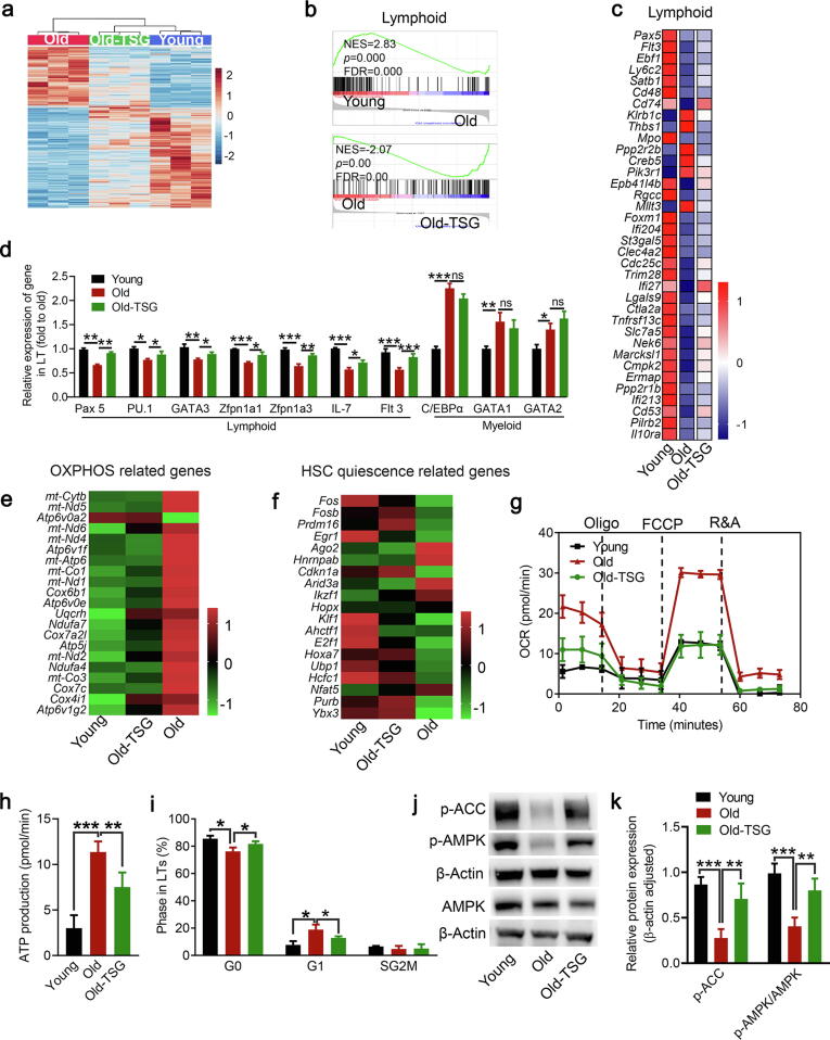
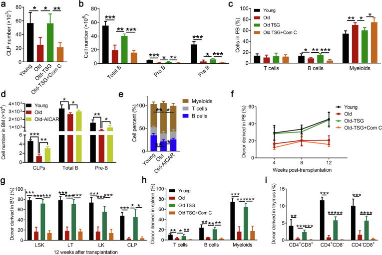
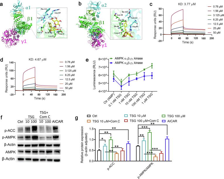
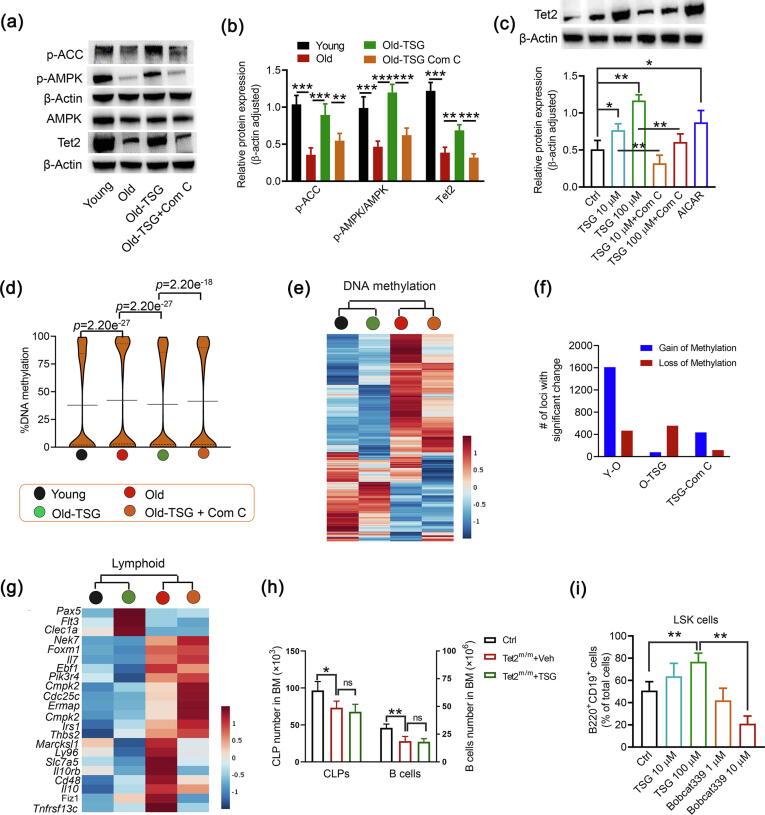
Methods: We screened a set of natural anti-aging compounds through oral administration to sub-lethally irradiated mice, and identified 2,3,5,4'-tetrahydroxystilbene-2-O-β-D-glucoside (TSG) as a potent rejuvenating agent for aging HSCs. Then naturally aged mice were used for the follow-up assessment to determine the HSC rejuvenating potential of TSG. Finally, based on the transcriptome and DNA methylation analysis, we validated the role of the AMP-activated protein kinase (AMPK)-ten-eleven-translocation 2 (Tet2) axis (the AMPK-Tet2 axis) as the underlying mechanisms of TSG for ameliorating HSCs aging.

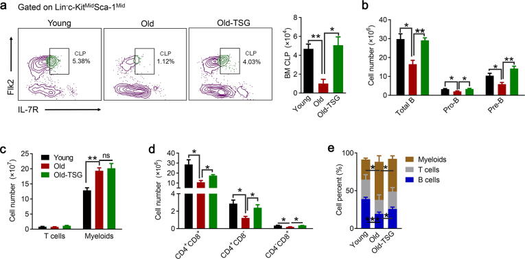
Results: TSG treatment not only significantly increased the absolute number of common lymphoid progenitors (CLPs) along with B lymphocytes, but also boosted the HSCs/CLPs repopulation potential of aging mice. Further elaborated mechanism research demonstrated that TSG supplementation restored the stemness of aging HSCs, as well as promoted an epigenetic reprograming that was associated with an improved regenerative capacity and an increased rate of lymphopoiesis. Such effects were diminished when the mice were co-treated with an AMPK inhibitor, or when it was performed in Tet2 knockout mice as well as senescent cells assay.

Conclusion: TSG is effective in rejuvenating aging HSCs by modulating the AMPK- Tet2 axis and thus represents a potential candidate for developing effective HSC rejuvenating therapies.

Keywords: AMPK-Tet2 axis; Aging; Hematopoietic stem cells; Lymphoid differentiation; Tetrahydroxy stilbene glucoside.


Conflict of interest statement

Declaration of competing interest The authors declare that they have no known competing financial interests or personal relationships that could have appeared to influence the work reported in this paper.

Figures

None
Graphical abstract
Fig. 1
Fig. 1
A approach for screening agents with hematopoietic stem cell (HSC) rejuvenation potential. (a) A schematic illustration of the screening process. (b-d) A small number of candidate compounds are first hand-picked from highly acclaimed TCM antiaging herbs for hematopoiesis and the mice dosages are calculated based on the human dose. The primary experimental screen of nine selected agents is then first carried out with total body irradiated (TBI) mice to identify agents that have a significant positive effect on the systematic analysis of the hematopoietic system. When a positive agent is identified, it is then subjected to a secondary experimental screen in naturally aging mice based on the same criteria. When an agent passes this secondary screen, it is then subjected to a series of analyses for a full assessment of its efficacy in HSC rejuvenation in naturally aging mice. (d) Tetrahydroxy stilbene glucoside (TSG), the sole agent that was found to significantly improve the number of CLPs based on this test. (e) Specifically, the representative FACS plots and quantification of the total number of B cells and pre-B cells in the bone marrow (BM) of the untreated normal 8-week-old mice (Ctrl), TBI mice treated with vehicle (TBI), and TBI mice treated with tetrahydroxy stilbene glucoside (TBI-TSG) are shown. Data is presented as mean ± S.D. (n = 4). *: p < 0.05; **: p < 0.01; ***: p < 0.001.
Fig. 2
Fig. 2
Effects of tetrahydroxy stilbene glucoside (TSG) on key attributes of HSCs of naturally aging (80-week-old) mice. Cohorts of aging mice were treated with either vehicle (Old) or TSG (Old-TSG) for 60 days and analyzed. A cohort of 8-week-old mice was included as a control for young mice (Young). (a) Analysis of common lymphoid progenitors (CLPs). Representative FACS plots (The left panel) and quantification of CLPs (the right panel) from the bone marrow of different cohorts. (b) Absolute numbers of various sub-types of B cells in the bone marrow of different cohorts. (c) Absolute numbers of T and myeloid cells in the bone marrow of different cohorts. (d) Absolute numbers of different sub-types of T cells per thymus of different cohorts. (e) Mean percentages of peripheral B, T, and myeloid cells of different cohorts. Throughout, data is represented by mean ± S.D. (n = 4). *: p < 0.05; **: p < 0.01; ns: no significance.
Fig. 3
Fig. 3
Effects of TSG on the repopulation potential of HSCs of naturally aging mice. (a) The experimental scheme for the competitive transplantation experiment. Long-term hematopoietic stem cells (LTs) from donor mice were co-transplanted with bone marrow cells from competitor mice into recipient mice, which were analyzed at various time points after the competitive LTs transplantation. (b) Percentages of donor-derived cells in peripheral blood (PB) at various time points after transplantation. (c) Percentages of various subtypes of donor-derived bone marrow (BM) stem/progenitor cells at 12 weeks after transplantation (n = 5). (d) The experimental scheme for the common lymphoid progenitor (CLP) transplantation experiment. For each transplantation, 103 CLPs were transplanted intravenously into irradiated (4 Gy) congenic 8-week-old C57BL/6J CD45.1 mice. (e-f) Percentages of donor derived total cells, B cells, and T cells in the peripheral blood (PB) and bone marrow (BM) from recipient mice at 3 weeks after transplantation. Data is presented as mean ± S.D. (n = 5). *: p < 0.05; **: p < 0.01; ***: p < 0.001.
Fig. 4
Fig. 4
Analyses of transcriptome and metabolic status related to HSC aging. Cohorts of aging mice were treated with either vehicle (Old) or TSG (Old-TSG) for four weeks. A cohort of 8-week-old mice was included as a control for young mice (Young). (a) Clustered heatmap of the differentially expressed genes determined by RNA-seq in isolated long-term hematopoietic stem cells (LTs) from the bone marrow. Rows represent genes while columns represent LTs with 3 replicates in each group. (b) Gene set enrichment analysis (GSEA) for comparing lymphoid gene term between Young and Old as well as Old and Old-SG groups according to the results of RNA-seq. NES, normalized enrichment score; FDR, false discovery rate. (c) Heatmap for the mean expression level of genes identified as lymphoid-specific in LTs from three groups based on RNA-seq analysis. (d) Validation of lymphoid-associated and myeloid-associated genes expression by qRT-PCR in LTs isolated from the bone marrow of mice from different groups. (n = 3). (e-f) Heatmap for the mean expression level of OXPHOS related genes and HSCs quiescence related genes, respectively. (g-h) OXPHOS levels and ATP production measured by Seahorse Mito Stress analysis in the linagec-kit+Sca1+ hematopoietic stem cells (LSKs) isolated from bone marrow (n = 4). (i) Cell cycle distribution analysis of isolated long-term hematopoietic stem cells (LTs) from the bone marrow by FACS with Ki67 and DAPI staining (n = 4). (j) LSKs from the bone marrow were isolated to harvest total proteins for detection levels of p-AMPK (T172), total AMPK, and p-ACC (Ser79) by western blot (β-actin was used as loading control). (k) Semi-quantitative analysis of the blots was determined by Image J software (n = 3). Data is presented as mean ± S.D. *: p < 0.05; **: p < 0.01; ***: p < 0.001. ns: no significance.
Fig. 5
Fig. 5
Tetrahydroxy stilbene glucoside (TSG) improves hematopoietic stem cell (HSC) lymphoid differentiation and repopulation potential defects of naturally aging mice by activating AMPK. In addition to administration in mice according to the scheme shown in Fig. 1, aging mice were treated with TSG by gavage daily combined with AMP-activated protein kinase (AMPK) inhibitor compound C (Com C) by intraperitoneal injection every other day as Old-TSG + Com C group (n = 4). (a) Absolute numbers of CLPs in the bone marrow of different cohorts. (b) Absolute numbers of various sub-types of B cells in the bone marrow of different cohorts. (c) Quantification of the percentages of B cells, T cells, and myeloid cells in the peripheral blood. (d-e) The naturally aging mice were treated further with an activator of AMPK 5-aminoimidazole-4-carboxamide riboside (AICAR) for 60 days and analyzed. (n = 4). (d) The number of CLPs (the left panel in d) or the total number of B cells and pre-B cells (the right panel in d) for the 8-week-old mice (Young), the naturally aging mice treated with Vehicle (Old), and the naturally aging mice treated with AICAR (Old-AICAR) are shown. (e) Mean percentages of peripheral B, T, and myeloid cells of different cohorts. (f-i) Competitive LTs transplantation similar to experimental design of Fig. 2a (5 recipients per group). (f) Percentage of donor-derived cells in the peripheral blood of recipient mice at 4, 8, and 12 weeks after competitive LT transplants. (g) Percentages of various subtypes of donor-derived bone marrow stem/progenitor cells at 12 weeks post competitive LT transplantation. (h) Spleen analyzed for percentages of donor-derived cells at 12 weeks after transplantation. (i) Thymus analyzed for percentages of donor-derived cells at 12 weeks after transplantation. Data is expressed as mean ± SD. *P < 0.05, **P < 0.01, ***P < 0.001 compared between the indicated groups.
Fig. 6
Fig. 6
Tetrahydroxy stilbene glucoside (TSG) interacts with AMPK in vitro. (a-b) Molecular docking study of the binding affinity of TSG to AMPKα1β1γ1 and AMPKα2β1γ1 crystal structures. (c-d) SPR analyses of the interaction of TSG with AMPKα1β1γ1 and AMPKα2β1γ1. (e) Effect of TSG on the kinase activity of AMPK in a cell-free system. AMPKα1β1γ1 and AMPKα2β1γ1 kinase enzyme system treated with AMP (positive control) or different concentration of TSG were detected using ADP-GloTM kinase assay reagents for the luminescent signal (n = 4). (f) Representative western blot analysis of p-AMPK (T172), total AMPK, p-ACC (Ser79) and β-actin in senescent IMR-90 cells treated with vehicle, different concentrations of TSG (10 and 100 μM), AMPK inhibitor Comp C (10 μM) combined with TSG or AMPK activator AICAR (0.5 mM) for 48 h. (g) Semi-quantitative analysis of the blots was determined by Image J software (n = 3). Data is presented as mean ± S.D. *: p < 0.05; **: p < 0.01; ***: p < 0.001.
Fig. 7
Fig. 7
Tetrahydroxy stilbene glucoside (TSG) affects the fate of hematopoietic stem cells (HSCs) through Tet2-mediated epigenetic regulation. (a) Linagec-kit+Sca1+ hematopoietic stem cells (LSKs) from the bone marrow of different cohorts according to the experimental procedure of Fig. 5 were isolated to harvest total proteins for detection levels of p-AMPK (T172), total AMPK, p-ACC (Ser79), and Tet2 by western blot (β-actin was used as loading control). (b) Semi-quantitative analysis of the blots was determined by Image J software (n = 4). (c) Representative western blot and semi-quantitative analysis of Tet2 and β-actin in senescent IMR-90 cells treated with vehicle, different concentrations of TSG (10 and 100 μM), AMPK inhibitor Comp C (10 μM) combined with TSG or AMPK activator AICAR (0.5 mM) for 48 h (n = 3). (d-g) Long-term hematopoietic stem cells (LTs) were isolated from the bone marrow for the DNA methylation analysis. (d) Bean plots of global DNA methylation of LTs from Young, Old, Old-TSG, and Old-TSG + Com C groups. (e) Hierarchical clustering of representative 100 kb tiles of DNA methylation data of LTs from mice of four groups. (f) Number of 1 kb tiles with significant methylation differences in pairwise comparisons between LTs isolated from different groups. Total comparisons with sufficient DNA methylation data from three populations, Y-O: Young LTs to Old LTs, O-TSG: Old LTs to Old-TSG LTs, Old-TSG LTs to Old-TSG + Com C LTs. (g) Heat map of DNA methylation levels of lymphoid-specific genes in LTs. Blue denotes low methylation levels and red denotes high expression. (h) 40-week-old Tet2 mutant mice were treated with vehicle or TSG by gavage once a day continuously for 60 days, a cohort of 40-week-old littermate control mice were treated with vehicle (Ctrl). The number of CLPs (the left panel in h) and the total number of B cells (the right panel in h) in the bone marrow of the mice are shown. (i) Lymphoid differentiation potential of LSKs from the bone marrow of 8-week-old mice. 1,000 sorted LSKs were seeded onto OP9 stromal cells supplemented with cytokines needed for lymphoid differentiation and numbers of B220 and CD19 double-positive cells were calculated at the indicated intervals by FACS (n = 4). Error bars represent mean ± SD. **P < 0.01, ***P < 0.001 compared between the indicated groups. (For interpretation of the references to colour in this figure legend, the reader is referred to the web version of this article.)

References

    1. Kasbekar M., Mitchell C.A., Proven M.A., Passegué E. Hematopoietic stem cells through the ages: a lifetime of adaptation to organismal demands. Cell Stem Cell. 2023;30:1403–1420. - PMC - PubMed
    1. Pang W.W., Schrier S.L., Weissman I.L. Age-associated changes in human hematopoietic stem cells. Semin Hematol. 2017;54:39–42. - PubMed
    1. Geiger H., de Haan G., Florian M.C. The ageing haematopoietic stem cell compartment. Nat Rev Immunol. 2013;13:376–389. - PubMed
    1. de Haan G., Lazare S.S. Aging of hematopoietic stem cells. Blood. 2018;131:479–487. - PubMed
    1. Broxmeyer H.E., Liu Y., Kapur R., Orschell C.M., Aljoufi A., Ropa J.P., et al. Fate of hematopoiesis during aging. what do we really know, and what are its implications? Stem Cell Rev Rep. 2020;16:1020–1048. - PMC - PubMed

MeSH terms

LinkOut - more resources