. 2024 Jun;300(6):107338.
doi: 10.1016/j.jbc.2024.107338. Epub 2024 May 3.

MAIT cell-MR1 reactivity is highly conserved across multiple divergent species

Affiliations

MAIT cell-MR1 reactivity is highly conserved across multiple divergent species

Matthew D Edmans et al. J Biol Chem. 2024 Jun.

Abstract

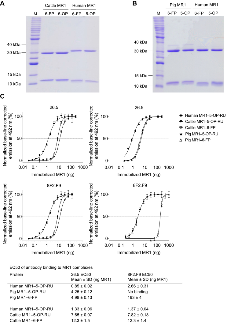
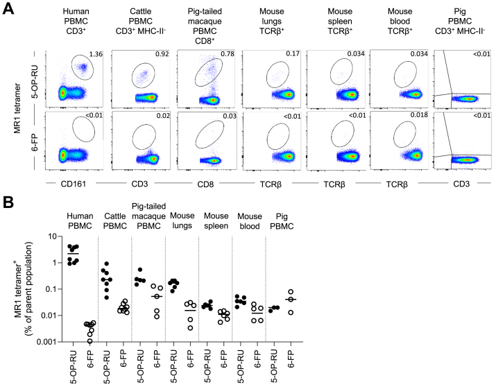
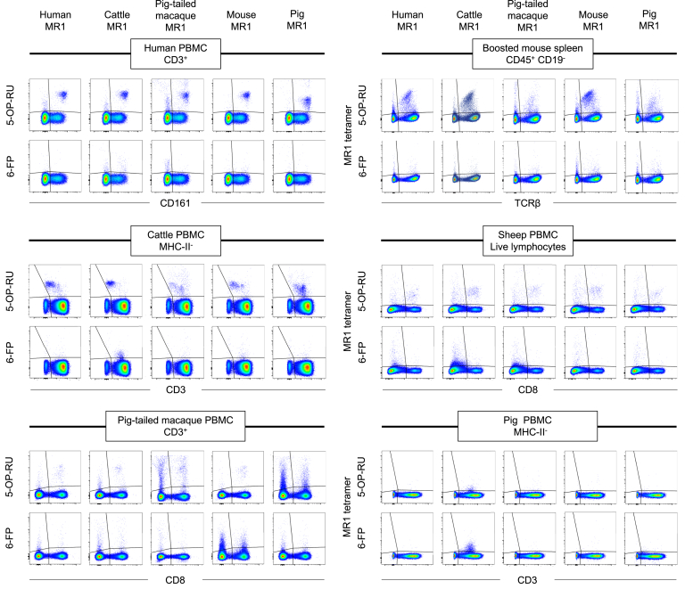
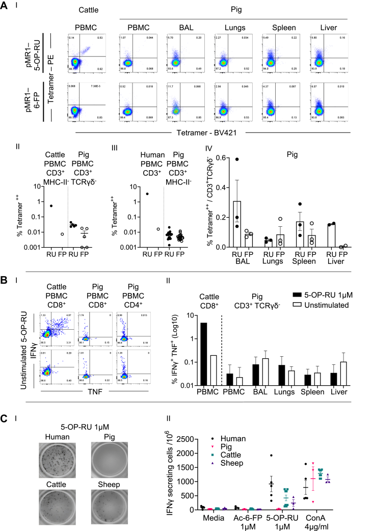
Mucosal-associated invariant T (MAIT) cells are a subset of unconventional T cells that recognize small molecule metabolites presented by major histocompatibility complex class I related protein 1 (MR1), via an αβ T cell receptor (TCR). MAIT TCRs feature an essentially invariant TCR α-chain, which is highly conserved between mammals. Similarly, MR1 is the most highly conserved major histocompatibility complex-I-like molecule. This extreme conservation, including the mode of interaction between the MAIT TCR and MR1, has been shown to allow for species-mismatched reactivities unique in T cell biology, thereby allowing the use of selected species-mismatched MR1-antigen (MR1-Ag) tetramers in comparative immunology studies. However, the pattern of cross-reactivity of species-mismatched MR1-Ag tetramers in identifying MAIT cells in diverse species has not been formally assessed. We developed novel cattle and pig MR1-Ag tetramers and utilized these alongside previously developed human, mouse, and pig-tailed macaque MR1-Ag tetramers to characterize cross-species tetramer reactivities. MR1-Ag tetramers from each species identified T cell populations in distantly related species with specificity that was comparable to species-matched MR1-Ag tetramers. However, there were subtle differences in staining characteristics with practical implications for the accurate identification of MAIT cells. Pig MR1 is sufficiently conserved across species that pig MR1-Ag tetramers identified MAIT cells from the other species. However, MAIT cells in pigs were at the limits of phenotypic detection. In the absence of sheep MR1-Ag tetramers, a MAIT cell population in sheep blood was identified phenotypically, utilizing species-mismatched MR1-Ag tetramers. Collectively, our results validate the use and define the limitations of species-mismatched MR1-Ag tetramers in comparative immunology studies.

Keywords: 5-(2-oxopropylideneamino)-6-d-ribitylaminouracil (5-OP-RU); MHC-I related protein 1 (MR1); T cell biology; T cell receptor (TCR); antigen (Ag); comparative immunology; innate-like immunity; major histocompatibility complex (MHC); mucosal-associated invariant T (MAIT) cell.


Conflict of interest statement

Conflict of interest J. Y. W. M., L. L., D. P. F., A. J. C., J. M., and S. B. G. E. are inventors on university owned patent rights (patent families WO/2015/149130 and WO/2014/005194) licensed for commercial use to Immudex and for non-profit use to the NIH Tetramer Core Facility. All other authors declare that they have no conflicts of interest with the contents of this article.

Figures

Figure 1
Figure 1
Biochemical characterization of recombinant cattle and pig MR1–5-OP-RU and MR1–6-FP monomers.A and B, 15% SDS-PAGE under non-reducing conditions of 1.5 μg purified biotinylated cattle, pig, and human MR1 in complex with β2m and loaded with 5-OP-RU (5-OP) or 6-FP in comparison to a protein ladder (M) with molecular weights of proteins indicated as relevant. Accounting for loss of 4 H atoms and 2 H atoms due to the formation of two disulphide bonds in MR1 and one disulphide bond in β2m; the molecular weights of biotinylated MR1 and β2m are as follows: human: MR1: 32,258 Da, β2m: 11,860 Da; cattle MR1: 32403 Da, β2m: 11764 Da; pig MR1: 32526 Da, β2m: 11542 Da. C, 5-OP-RU- and 6-FP-loaded cattle and pig MR1 monomers in comparison to biotinylated human MR1–5-OP-RU (assessed previously alongside other species’ MR1 molecules, (64)) in ELISA with mAbs 26.5 and 8F2.F9 showing normalized, base-line corrected dose-response curves (n = 3, mean ± SD). EC50 values, as summarized in the table, were determined based on non-linear curve fits shown in the charts. 5-OP-RU, 5-(2-oxopropylideneamino)-6-d-ribitylaminouracil; 6-FP, 6-formylpterin; β2m, β2 microglobulin; MR1, MHC-I related protein 1.
Figure 2
Figure 2
Identification of MAIT cells in multiple species using species-specific MR1–5-OP-RU and MR1–6-FP tetramers.A, representative flow cytometry plots from staining of human PBMC, cattle PBMC, pig-tailed macaque PBMC, mouse lung-, spleen- and blood-derived lymphocytes, and pig PBMC with MR1–5-OP-RU and MR1–6-FP tetramers of the corresponding species. B, frequencies of species-matched MR1–5-OP-RU and MR1–6-FP tetramer+ MAIT cells from human CD3+ PBMC (n = 4), cattle CD3+ PBMC (n = 4), pig-tailed macaque CD8+ PBMC (n = 5), mouse lungs, spleen, and whole blood-derived αβTCR+ lymphocytes (n = 6), and pig CD3+ PBMC (n = 3). Mean ± SEM is depicted. Experiments were performed once in pig-tailed macaque and mouse and twice in all other species. 5-OP-RU, 5-(2-oxopropylideneamino)-6-d-ribitylaminouracil; 6-FP, 6-formylpterin; MAIT, mucosal-associated invariant T; MR1, major histocompatibility complex-I related protein 1; PBMC, peripheral blood mononuclear cell; TCR, T cell receptor.
Figure 3
Figure 3
Flow cytometry plots of species-mismatched MR1–5-OP-RU and MR1–6-FP tetramer staining. Human, cattle, pig-tailed macaque, mouse, and pig MR1–5-OP-RU and MR1–6-FP tetramers were used to stain human PBMC, cattle PBMC, lymphocytes from MAIT cell boosted mouse spleen, pig-tailed macaque PBMC, sheep PBMC or pig PBMC. Representative final gating is shown for each species and each, MR1–5-OP-RU and MR1–6-FP tetramer. 5-OP-RU, 5-(2-oxopropylideneamino)-6-d-ribitylaminouracil; 6-FP, 6-formylpterin; MAIT, mucosal-associated invariant T; MR1, MHC-I related protein 1; PBMC, peripheral blood mononuclear cell.
Figure 4
Figure 4
Frequency and staining characteristics of MR1–5-OP-RU and MR1–6-FP tetramer+populations identified with species-matched and species-mismatched tetramer reagents. Bar charts and heatmap summaries, displaying the following: A, frequencies of species-matched (red histograms) and species-mismatched (black histograms) MR1–5-OP-RU (RU, black dots) and MR1–6-FP (FP, white dots) tetramer+ cells in human CD3+ PBMC (n = 8), cattle CD3+ PBMC (n = 8), pig-tailed macaque CD8+ PBMC (n = 5), MAIT cell boosted mouse spleen (n = 7)–derived αβTCR+ lymphocytes, and sheep CD8+ PBMC (n = 6). Histograms depict mean frequency ± SEM with each individual datapoint shown. B, species-matched (red histograms) and species-mismatched (black histograms) MR1–5-OP-RU tetramer (RU) geometric mean fluorescence intensity (gMFI) fold change over the MR1–5-OP-RU tetramer- population (background) and (C) SD of the MR1–5-OP-RU tetramer+ (RU) gMFI as described in (A). Differences between the frequencies obtained from species-matched and species-mismatched MR1–5-OP-RU staining were evaluated using a one-way ANOVA with Giesser-Greenhouse correction or, where there are missing values, a mixed effect model with repeated measures, followed by Dunnett’s multiple comparison test, comparing staining with species-matched MR1–5-OP-RU tetramer and all other species-mismatched MR1–5-OP-RU tetramers, with individual variance computed for each comparison. Only statistically significant differences (p value < 0.05) are indicated. Data are combined from either one (pig-tailed macaque), two (human, cattle, and sheep), or three (mouse) separate experiments. 5-OP-RU, 5-(2-oxopropylideneamino)-6-d-ribitylaminouracil; 6-FP, 6-formylpterin; PBMC, peripheral blood mononuclear cell; MAIT, mucosal-associated invariant T; MR1, MHC-I related protein 1; TCR, T cell receptor.
Figure 5
Figure 5
Detailed characterization of MAIT cells in pigs and sheep.A, (I) representative flow cytometry plots of cells stained with pig MR1–5-OP-RU (pMR1–5-OP-RU) or MR1–6-FP (pMR1–6-FP) tetramers conjugated to PE or BV421 among CD3+ γδTCR- pig PBMC, BAL, lungs spleen, and liver (and for comparison among CD3+ cattle PBMC), (II) the proportion of cells double positive for pig MR1–5-OP-RU (RU) or MR1–6-FP (FP) tetramers conjugated to PE or BV421 among CD3+ γδTCR- T cells in pig PBMC (n = 5) (and for comparison among CD3+ MHC-II- cattle PBMC (n = 1)), (III) the proportion of cells double positive for pig MR1–5-OP-RU (RU) or MR1–6-FP (FP) tetramers conjugated to PE or BV421 among CD3+ MHC-II- pig PBMC (n = 18) (and for comparison among CD3+ human PBMC (n = 1)), and (IV) the proportion of cells double positive for pig MR1–5-OP-RU (RU) or MR1–6-FP (FP) tetramers conjugated to PE or BV421 among CD3+ γδTCR T cells in pig (n = 2 or 3) BAL, lungs, spleen, and liver lymphocytes. B, representative flow cytometry plots (I) and quantification of the frequency (II) of the IFNγ and TNF intracellular cytokine response in cattle CD8+ PBMC (n = 1) and CD3+ γδTCR- pig (n = 3) PBMC, BAL, lungs, liver, and spleen lymphocytes, following stimulation with 1 μM 5-OP-RU for 6 h. Mean ± SEM are shown. C, representative images (I) and quantification (II) of secreted IFNγ assessed by ELISpot following coincubation of human (n = 3), pig (n = 3), cattle (n = 4), and sheep (n = 3) PBMC with medium, 1 μM Ac-6-FP, 1 μM 5-OP-RU, or 4 μg/ml ConA for 18 h. Mean SCF/106 cells ± SEM are shown. Ac-6-FP, acetyl-6-FP; 5-OP-RU, 5-(2-oxopropylideneamino)-6-d-ribitylaminouracil; 6-FP, 6-formylpterin; MAIT, mucosal-associated invariant T; MHC, major histocompatibility complex; MR1, MHC-I related protein 1; TCR, T cell receptor; PBMC, peripheral blood mononuclear cell.
Figure 6
Figure 6
Frequency and TCRα usage of putative MAIT cells in cattle, sheep and pig PBMC.A, frequencies of TRAV1+ (I) and putative MAIT TCR CDR3α sequence+ (II) of total TRA bulk sequenced cattle, sheep, and pig PBMC (n = 2). B, frequency of TRAJ33, TRAJ12, or TRAJ20 gene segment usage among TRAV1+TRA sequences from cattle, sheep, and pig PBMC. C, relative frequency and sequence of putative MAIT TCR CDR3α-loops in each animal. D, sequence logo (Seq2Logo) of the amino acid weighting of the CDR3α-loops from putative MAIT TCRs in each species (note that the Y95, highlighted in green, is conserved between all species). MAIT, mucosal-associated invariant T; PBMC, peripheral blood mononuclear cell; TCR, T cell receptor.

References

    1. Porcelli S., Yockey C.E., Brenner M.B., Balk S.P. Analysis of T cell antigen receptor (TCR) expression by human peripheral blood CD4-8- alpha/beta T cells demonstrates preferential use of several V beta genes and an invariant TCR alpha chain. J. Exp. Med. 1993;178:1–16. - PMC - PubMed
    1. Tilloy F., Treiner E., Park S.H., Garcia C., Lemonnier F., de la Salle H., et al. An invariant T cell receptor alpha chain defines a novel TAP-independent major histocompatibility complex class Ib-restricted alpha/beta T cell subpopulation in mammals. J. Exp. Med. 1999;189:1907–1921. - PMC - PubMed
    1. Treiner E., Duban L., Bahram S., Radosavljevic M., Wanner V., Tilloy F., et al. Selection of evolutionarily conserved mucosal-associated invariant T cells by MR1. Nature. 2003;422:164–169. - PubMed
    1. Hashimoto K., Hirai M., Kurosawa Y. A gene outside the human MHC related to classical HLA class I genes. Science. 1995;269:693. - PubMed
    1. Miley M.J., Truscott S.M., Yu Y.Y., Gilfillan S., Fremont D.H., Hansen T.H., et al. Biochemical features of the MHC-related protein 1 consistent with an immunological function. J. Immunol. 2003;170:6090–6098. - PubMed

Publication types

MeSH terms

Substances