

Pannexins in the musculoskeletal system: new targets for development and disease progression
- PMID: 38705887
- PMCID: PMC11070431
- DOI: 10.1038/s41413-024-00334-8
Pannexins in the musculoskeletal system: new targets for development and disease progression
Abstract
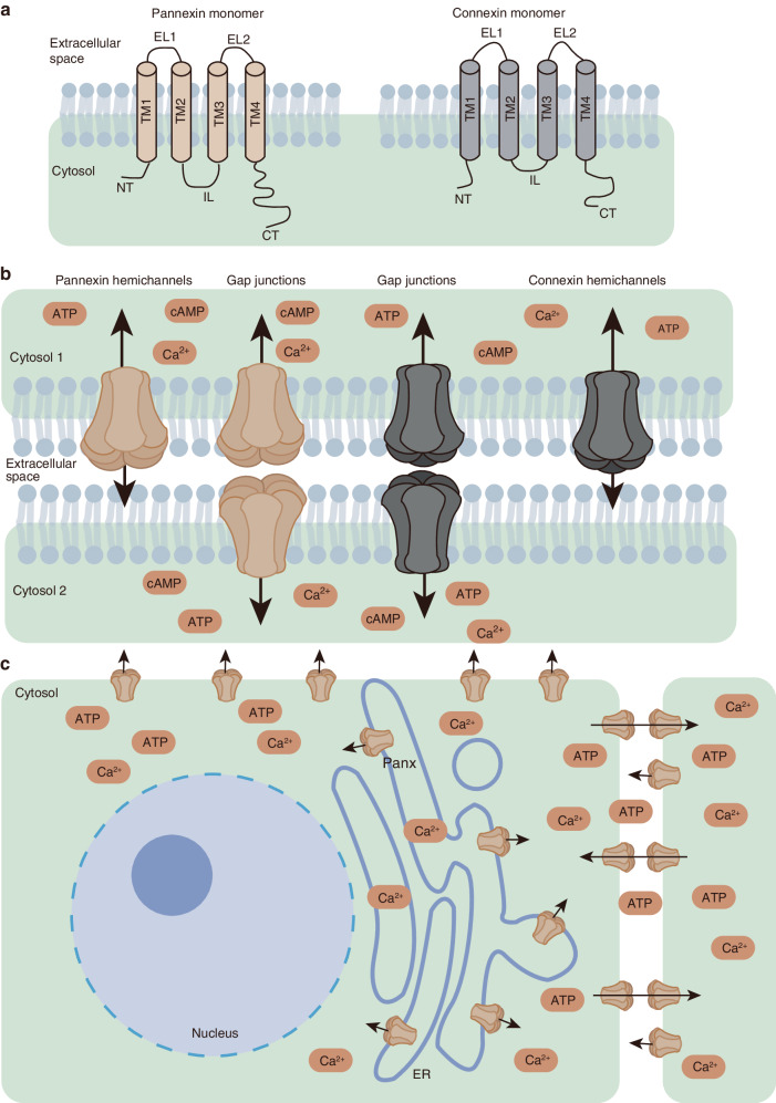
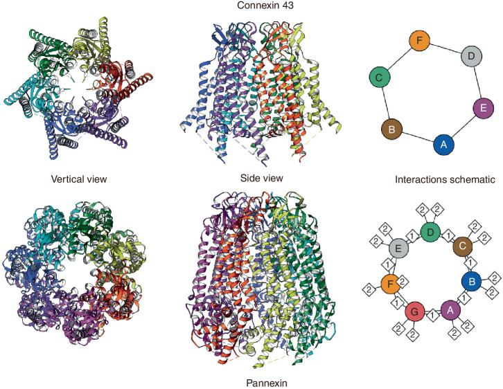
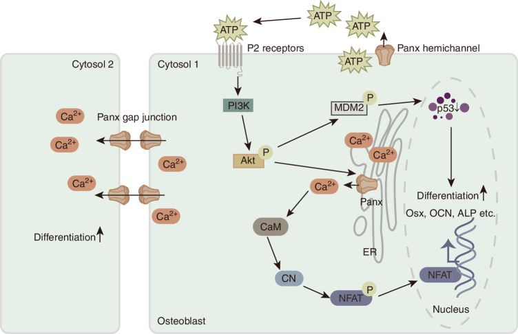
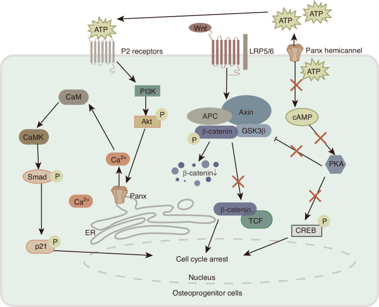
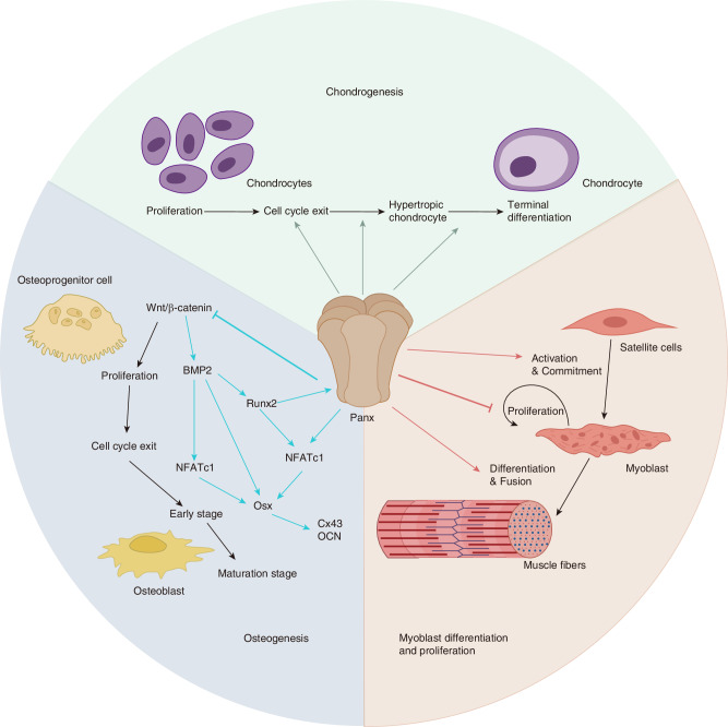
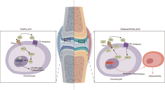
During cell differentiation, growth, and development, cells can respond to extracellular stimuli through communication channels. Pannexin (Panx) family and connexin (Cx) family are two important types of channel-forming proteins. Panx family contains three members (Panx1-3) and is expressed widely in bone, cartilage and muscle. Although there is no sequence homology between Panx family and Cx family, they exhibit similar configurations and functions. Similar to Cxs, the key roles of Panxs in the maintenance of physiological functions of the musculoskeletal system and disease progression were gradually revealed later. Here, we seek to elucidate the structure of Panxs and their roles in regulating processes such as osteogenesis, chondrogenesis, and muscle growth. We also focus on the comparison between Cx and Panx. As a new key target, Panxs expression imbalance and dysfunction in muscle and the therapeutic potentials of Panxs in joint diseases are also discussed.
© 2024. The Author(s).
Conflict of interest statement
The authors declare no competing interests.
Figures
Similar articles
-
Pannexins, distant relatives of the connexin family with specific cellular functions?Bioessays. 2009 Sep;31(9):953-74. doi: 10.1002/bies.200800236. Bioessays. 2009. PMID: 19644918 Review.
-
Differentiating connexin hemichannels and pannexin channels in cellular ATP release.FEBS Lett. 2014 Apr 17;588(8):1379-88. doi: 10.1016/j.febslet.2014.02.004. Epub 2014 Feb 15. FEBS Lett. 2014. PMID: 24548565 Free PMC article. Review.
-
Connexins and Pannexins in Vascular Function and Disease.Int J Mol Sci. 2018 Jun 5;19(6):1663. doi: 10.3390/ijms19061663. Int J Mol Sci. 2018. PMID: 29874791 Free PMC article. Review.
-
Pore positioning: current concepts in Pannexin channel trafficking.Channels (Austin). 2014;8(2):110-7. doi: 10.4161/chan.27287. Epub 2013 Dec 3. Channels (Austin). 2014. PMID: 24300303 Free PMC article. Review.
-
Mechanisms of Pannexin 1 (PANX1) Channel Mechanosensitivity and Its Pathological Roles.Int J Mol Sci. 2022 Jan 28;23(3):1523. doi: 10.3390/ijms23031523. Int J Mol Sci. 2022. PMID: 35163442 Free PMC article. Review.
Cited by
-
The emerging role of Panx1 as a potential therapeutic target for chronic pain.Mil Med Res. 2024 Jul 5;11(1):44. doi: 10.1186/s40779-024-00549-0. Mil Med Res. 2024. PMID: 38970139 Free PMC article. No abstract available.
-
Loosening the Lid on Shoulder Osteoarthritis: How the Transcriptome and Metabolic Syndrome Correlate with End-Stage Disease.Int J Mol Sci. 2025 Mar 28;26(7):3145. doi: 10.3390/ijms26073145. Int J Mol Sci. 2025. PMID: 40243895 Free PMC article.
-
Cuproptosis and its potential role in musculoskeletal disease.Front Cell Dev Biol. 2025 Apr 11;13:1570131. doi: 10.3389/fcell.2025.1570131. eCollection 2025. Front Cell Dev Biol. 2025. PMID: 40292330 Free PMC article. Review.
References
-
- Allen, M. R. & Burr, D. B. in Basic and Applied Bone Biology (eds David B. Burr & Matthew R. Allen) 75–90 (Academic Press, 2014).
-
- Stains JP, Civitelli R. Gap junctions in skeletal development and function. Biochim. Biophys. Acta. 2005;1719:69–81. - PubMed
-
- Bellido, T., Plotkin, L. I. & Bruzzaniti, A. in Basic and Applied Bone Biology (eds David B. Burr & Matthew R. Allen) 27–45 (Academic Press, 2014).
Publication types
MeSH terms
Substances
Grants and funding
LinkOut - more resources
Full Text Sources
Research Materials
