. 2024 May 5;14(1):10302.
doi: 10.1038/s41598-024-60949-z.

Coastal and deep-sea biodegradation of polyhydroxyalkanoate microbeads

Affiliations

Coastal and deep-sea biodegradation of polyhydroxyalkanoate microbeads

Natsumi Hyodo et al. Sci Rep. .

Abstract

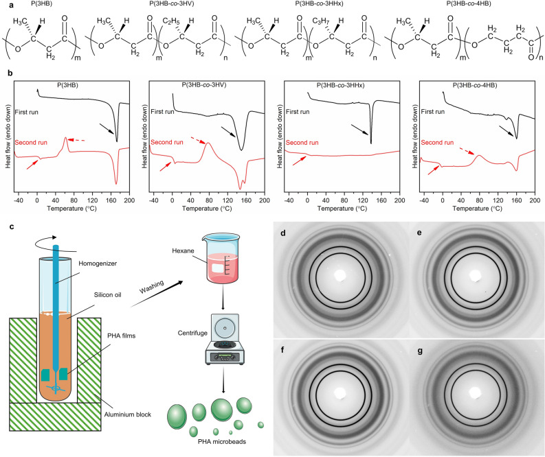
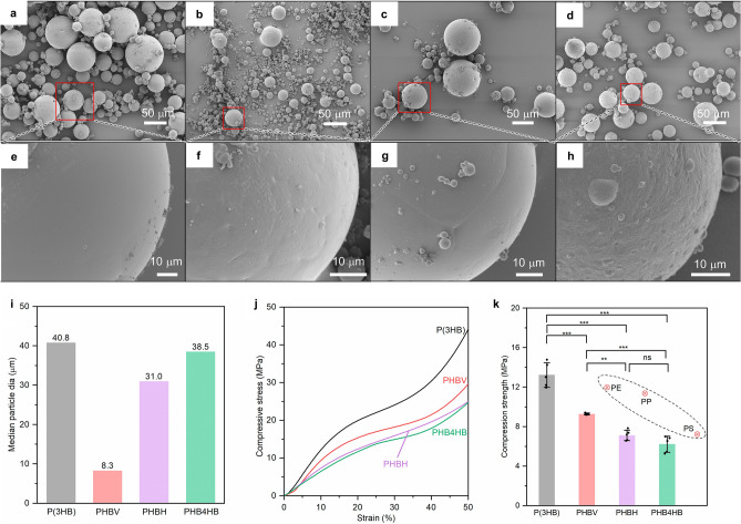
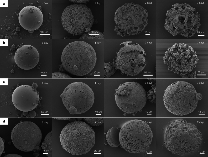
Microbeads find widespread usage in personal care items and cosmetics, serving as exfoliants or scrubbing agents. Their micro-scale size poses challenges in effective drainage capture and given their origin from non-biodegradable oil-based plastics, this contributes substantially to marine pollution. In this study, microbeads were prepared by a simple yet scalable melt homogenization method using four types of polyhydroxyalkanoates (PHA), namely poly[(R)-3-hydroxybutyrate] (P(3HB)), poly[(R)-3-hydroxybutyrate-co-(R)-3-hydroxyvalerate] (P(3HB-co-3HV)), poly[(R)-3-hydroxybutyrate-co-(R)-3-hydroxyhexanoate] (P(3HB-co-3HHx)) and poly[(R)-3-hydroxybutyrate-co-(R)-4-hydroxyvalerate] (P(3HB-co-4HB)). Microbeads with different surface smoothness, compressive strength (6.2-13.3 MPa) and diameter (from 1 ~ 150 μm) could be produced. The microbeads were subjected to a comprehensive degradation analysis using three techniques: enzymatic, Biochemical Oxygen Demand (BOD) evaluations, and in situ degradation tests in the deep-sea off Misaki Port in the northern Pacific Ocean (depth of 757 m). Qualitatively, results from enzymatic and in situ degradation demonstrated significant degradation within one week and five months, respectively. Quantitatively, BOD findings indicated that all PHA microbeads degraded similarly to cellulose (~ 85% biodegradability in 25 days). In conclusion, PHA microbeads from this study exhibit promising potential as alternatives to conventional non-biodegradable microbeads.


Conflict of interest statement

The authors declare no competing interests.

Figures

Figure 1
Figure 1
Processing, thermal and crystal properties of PHA microbeads. (a) Chemical structure of P(3HB), PHBV, PHBH and PHB4HB. (b) First and second run DSC curve of PHA neat samples. The arrows in the first run indicate the Tm. The solid and dotted arrows in the second run indicate Tg and Tc respectively. (c) Fabrication scheme of PHA microbeads (dg) 2D WAXD images of P(3HB), PHBV, PHBH and PHB4HB respectively. Refer Table 1 for the summary of thermal properties and crystallinity.
Figure 2
Figure 2
Morphological characteristics and compressive strength of PHA microbeads. (ah) Shows the SEM images of P(3HB) (a,e), PHBV (b,f), PHBH (c,g), PHB4HB beads (d,h). The images shown are representative of n = 3 samples. The morphology remained consistent irrespective of fabrication lots. (i) Median particle size of the prepared PHA microbeads. (j) Stress–strain curve of the PHA microbeads obtained during the compression test. The graph represents the stress–strain profile of the sample closest to the average value. (k) Compressive strength of P(3HB), PHBV, PHBH and PHB4HB microbeads at 10% strain. The value shown here is the average of n = 5 distinct samples picked randomly from different preparation lots. The error bar represents the standard deviation. The results were compared using a one-way ANOVA with post-hoc Tukey test. **p < 0.05, ***p < 0.01, ns not significant. The red marker represents the compressive strength of conventional microbeads.
Figure 3
Figure 3
Morphological evolution of PHA microbeads during the course of enzymatic degradation. Each row depicts the morphology of microbeads from different PHA. (a) Morphological characteristics of P(3HB) microbeads before and after 1-, 3- and 7-days of enzymatic degradation (bd) indicate the surface morphology of PHBV, PHBH, and PHB4HB, respectively. The images are representative of triplicate samples conducted simultaneously. The buffer and aqueous solution of the enzymes were replaced constantly to ensure the activity of the enzymes remain unchanged.
Figure 4
Figure 4
Marine degradation of PHA microbeads carried out under real-life conditions. (a) Map of Japan indicating the locations of in situ degradation test site (Bathyal seafloor off Misaki port) and seawater collection spot for BOD tests (near the Tokyo Bay). The map was created using GeoMapApp (geomapp.org) / CC BY (Ryan et al.). (b) Schematic representation of the environmental degradation tests of PHA microbeads. The submersion and recovery were carried out using the human operated Shinkai6500 submersible aboard its support vessel RV Yokosuka. (cj) SEM images of microbeads after 5 months of submersion in the deep-sea floor. (c,d P(3HB), e,f PHBV, g,h PHBH, i,j PHB4HB microbeads). The images are representative of n = 3 samples. (k,l) Surface morphology of PHB4HB after fixing microorganisms using formaldehyde solution. (m) BOD-biodegradability curve of PHA microbeads and cellulose standard using seawater from Tokyo bay. The curves are representative of n = 3 samples. Refer Table 2 for more details on the environmental degradation sites.

References

    1. Iwata T. Biodegradable and bio-based polymers: Future prospects of eco-friendly plastics. Angew. Chem. 2015;54:3210–3215. doi: 10.1002/anie.201410770. - DOI - PubMed
    1. Barnes DKA, Galgani F, Thompson RC, Barlaz M. Accumulation and fragmentation of plastic debris in global environments. Philos. Trans. R. Soc. B. 2009;364:1985–1998. doi: 10.1098/rstb.2008.0205. - DOI - PMC - PubMed
    1. Arthur, C., Baker, J. & Bamford, H. Proceedings of the International Research Workshop on the Occurrence, Effects, and Fate of Microplastic Marine Debris (2009).
    1. Cole M, Lindeque P, Halsband C, Galloway TS. Microplastics as contaminants in the marine environment: A review. Mar. Pollut. Bull. 2011;62:2588–2597. doi: 10.1016/j.marpolbul.2011.09.025. - DOI - PubMed
    1. Wagner M, et al. Microplastics in freshwater ecosystems: What we know and what we need to know. Environ. Sci. Eur. 2014;26:1–9. doi: 10.1186/s12302-014-0012-7. - DOI - PMC - PubMed

Publication types

MeSH terms

Substances