. 2024 May 5;14(1):10304.
doi: 10.1038/s41598-024-60460-5.

Contrasting neurofunctional correlates of face- and visuospatial-processing in children and adolescents with Williams syndrome: convergent results from four fMRI paradigms

Affiliations

Contrasting neurofunctional correlates of face- and visuospatial-processing in children and adolescents with Williams syndrome: convergent results from four fMRI paradigms

Madeline H Garvey et al. Sci Rep. .

Abstract

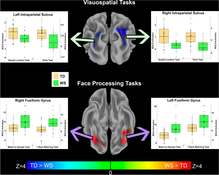
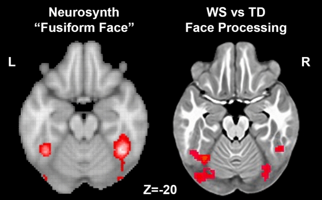
Understanding neurogenetic mechanisms underlying neuropsychiatric disorders such as schizophrenia and autism is complicated by their inherent clinical and genetic heterogeneity. Williams syndrome (WS), a rare neurodevelopmental condition in which both the genetic alteration (hemideletion of ~ twenty-six 7q11.23 genes) and the cognitive/behavioral profile are well-defined, offers an invaluable opportunity to delineate gene-brain-behavior relationships. People with WS are characterized by increased social drive, including particular interest in faces, together with hallmark difficulty in visuospatial processing. Prior work, primarily in adults with WS, has searched for neural correlates of these characteristics, with reports of altered fusiform gyrus function while viewing socioemotional stimuli such as faces, along with hypoactivation of the intraparietal sulcus during visuospatial processing. Here, we investigated neural function in children and adolescents with WS by using four separate fMRI paradigms, two that probe each of these two cognitive/behavioral domains. During the two visuospatial tasks, but not during the two face processing tasks, we found bilateral intraparietal sulcus hypoactivation in WS. In contrast, during both face processing tasks, but not during the visuospatial tasks, we found fusiform hyperactivation. These data not only demonstrate that previous findings in adults with WS are also present in childhood and adolescence, but also provide a clear example that genetic mechanisms can bias neural circuit function, thereby affecting behavioral traits.


Conflict of interest statement

The authors declare no competing interests.

Figures

Figure 1
Figure 1
fMRI paradigms used to investigate visuospatial and face processing and activation patterns in typically developing participants. Top and bottom sections show fMRI tasks used to study, respectively, visuospatial processing within the dorsal stream and face processing within the ventral stream, along with activation patterns for each task in our cohort of typically developing children thresholded at p < 0.001, FDR corrected; data are shown on inflated brain renderings with posterior views for the dorsal stream and inferior views for the ventral stream. Top left box depicts the Spatial Location Task, in which individuals judged the height of consecutively presented stimuli or responded to a sensorimotor control condition consisting of black boxes that were always at the same height. Top right box depicts the stimuli for the Tetris Task in which individuals moved a puzzle piece to fit into the lower image; “hard” trials included a complicated “landscape”, while “down” trials only required simple straight-down movements. Bottom left box depicts the Match-to-Sample Face Task, in which three faces or scrambled images were concurrently presented and participants indicated which face on the bottom matched the image at the top. Bottom right box depicts the One-Back Face-Matching Task in which a series of faces or scrambled images was presented and participants indicated whether each image matched the image shown immediately prior. Note that for the two visuospatial tasks the activation patterns demonstrate robust activation of the dorsal stream, particularly the bilateral intraparietal sulci, and for the two face processing tasks the activation patterns demonstrate robust activation of the ventral stream, including the bilateral fusiform gyri. The image on the left depicting dorsal and ventral streams was adapted from: https://commons.wikimedia.org/wiki/File:Ventral-dorsal_streams.svg.
Figure 2
Figure 2
Functional activation differences between children with WS compared to typically developing children in response to visuospatial tasks and face processing tasks. Top row Middle image displays a posterior view of the inflated brain surface with overlaid between-group meta-Z statistics of the two visuospatial tasks combined, showing bilateral hypoactivation of the intraparietal sulcus (IPS) in participants with WS (TD > WS, p < 0.001 FWE corrected). Green arrows point to plots of the average extracted BOLD from the identified significant IPS clusters for individuals with WS and TD controls for both visuospatial processing tasks, separately. Plots show that TD participants had greater activation than children with WS in the Spatial Location Task and the Tetris Task in both the right and left IPS regions. Bottom row Middle image displays an inferior view of the inflated brain surface with the overlaid color representing the between-group meta-Z statistics for both face processing tasks combined, showing bilateral hyperactivation of the fusiform gyri in children with WS (WS > TD, p < 0.001 FWE corrected). Purple arrows point to plots of the average extracted BOLD from the identified significant fusiform clusters for individuals with WS and TD controls for the two face processing tasks separately. Plots show that children with WS had greater activation than TD children in both the Match-to-Sample Face Task and the One-Back Face Matching Task in both the right and left fusiform regions.
Figure 3
Figure 3
Comparison of Neurosynth meta-analysis of “fusiform face” term with regions showing increased BOLD activation during face processing in children with WS in the present study. Left an axial brain slice at z = − 20 MNI coordinate with red colors representing the regions showing significant association (Z-statistic > 10) with the term “fusiform face” in 143 published studies. Right an axial brain slice, also at z = − 20, with red colors representing regions in the present study showing hyperactivation in WS during the combined meta-Z analysis of the two face processing tasks studied. Note the high degree of overlap between the left and right fusiform areas of activation. Neurosynth image was accessed from http://www.neurosynth.org.
Figure 4
Figure 4
Standardized BOLD activation differences between WS and TD groups derived from each significant cluster in the between-groups combined meta-Z analyses. Clusters were derived from combined analyses of visuospatial or face processing tasks (see Tables 2 and 3) and extracted from each task individually. Green indicates tasks that targeted visuospatial processing, and purple indicates tasks that targeted face processing. X-axis shows standardized mean activation difference between WS and TD groups, with negative numbers representing greater activation in TD children and positive numbers representing greater activation in children with WS. Lines for each cluster represent the 95% confidence interval of the standardized mean difference between groups. Note that the difference in BOLD activation consistently indicates greater activation in TD children than children with WS during visuospatial tasks and in children with WS than in TD children during face processing tasks.

Similar articles

Cited by

  • Face perception: a window into the social mind.
    Dalmaso M, Gobbini MI, Ricciardelli P, Ritchie KL, Pecchinenda A. Dalmaso M, et al. Sci Rep. 2025 Sep 3;15(1):32362. doi: 10.1038/s41598-025-17861-x. Sci Rep. 2025. PMID: 40903482 Free PMC article.

References

    1. Pearce E, Stringer C, Dunbar RI. New insights into differences in brain organization between Neanderthals and anatomically modern humans. Proc. Biol. Sci. 2013;280:20130168. doi: 10.1098/rspb.2013.0168. - DOI - PMC - PubMed
    1. Dilks DD, Hoffman JE, Landau B. Vision for perception and vision for action: Normal and unusual development. Dev. Sci. 2008;11:474–486. doi: 10.1111/j.1467-7687.2008.00693.x. - DOI - PubMed
    1. Kamps FS, et al. Dissociable cognitive systems for recognizing places and navigating through them: Developmental and neuropsychological evidence. J. Neurosci. 2023;43:6320–6329. doi: 10.1523/JNEUROSCI.0153-23.2023. - DOI - PMC - PubMed
    1. Dawson G, Webb SJ, McPartland J. Understanding the nature of face processing impairment in autism: insights from behavioral and electrophysiological studies. Dev. Neuropsychol. 2005;27:403–424. doi: 10.1207/s15326942dn2703_6. - DOI - PubMed
    1. Bertone A, Mottron L, Jelenic P, Faubert J. Enhanced and diminished visuo-spatial information processing in autism depends on stimulus complexity. Brain. 2005;128:2430–2441. doi: 10.1093/brain/awh561. - DOI - PubMed

Publication types