. 2024 Jan-Dec;16(1):2347722.
doi: 10.1080/19490976.2024.2347722. Epub 2024 May 5.

Microbiota-derived I3A protects the intestine against radiation injury by activating AhR/IL-10/Wnt signaling and enhancing the abundance of probiotics

Affiliations

Microbiota-derived I3A protects the intestine against radiation injury by activating AhR/IL-10/Wnt signaling and enhancing the abundance of probiotics

Li-Wei Xie et al. Gut Microbes. 2024 Jan-Dec.

Abstract

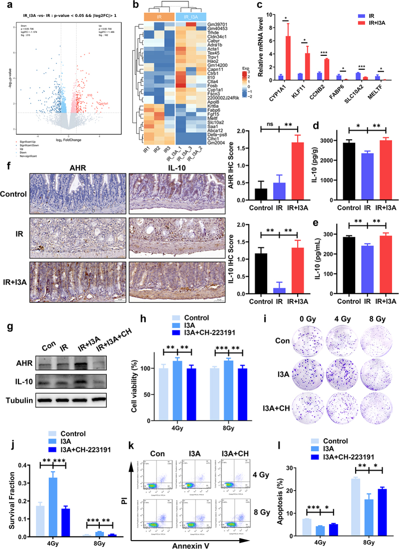
The intestine is prone to radiation damage in patients undergoing radiotherapy for pelvic tumors. However, there are currently no effective drugs available for the prevention or treatment of radiation-induced enteropathy (RIE). In this study, we aimed at investigating the impact of indole-3-carboxaldehyde (I3A) derived from the intestinal microbiota on RIE. Intestinal organoids were isolated and cultivated for screening radioprotective tryptophan metabolites. A RIE model was established using 13 Gy whole-abdominal irradiation in male C57BL/6J mice. After oral administration of I3A, its radioprotective ability was assessed through the observation of survival rates, clinical scores, and pathological analysis. Intestinal stem cell survival and changes in the intestinal barrier were observed through immunofluorescence and immunohistochemistry. Subsequently, the radioprotective mechanisms of I3A was investigated through 16S rRNA and transcriptome sequencing, respectively. Finally, human colon cancer cells and organoids were cultured to assess the influence of I3A on tumor radiotherapy. I3A exhibited the most potent radioprotective effect on intestinal organoids. Oral administration of I3A treatment significantly increased the survival rate in irradiated mice, improved clinical and histological scores, mitigated mucosal damage, enhanced the proliferation and differentiation of Lgr5+ intestinal stem cells, and maintained intestinal barrier integrity. Furthermore, I3A enhanced the abundance of probiotics, and activated the AhR/IL-10/Wnt signaling pathway to promote intestinal epithelial proliferation. As a crucial tryptophan metabolite, I3A promotes intestinal epithelial cell proliferation through the AhR/IL-10/Wnt signaling pathway and upregulates the abundance of probiotics to treat RIE. Microbiota-derived I3A demonstrates potential clinical application value for the treatment of RIE.

Keywords: Radiotherapy; gastrointestinal tract toxicity; gut microbiota; indole-3-carboxaldehyde; intestinal stem cells; tryptophan metabolites.


Conflict of interest statement

No potential conflict of interest was reported by the author(s).

Figures

Figure 1.
Figure 1.
I3A protects intestinal crypt organoids and HIEC-6 against radiation-induced injury. (a) Representative phase contrast images of intestinal organoids cultured with or without tryptophan metabolites at 0, 3, 5 or 7 d post-IR (scale bar = 200 μm). (b) The budding/total organoids (%) at 7 d post-IR. At least 50 intestinal organoids were counted in each group. (c) The number of damaged organoids per well at 7 d post-IR. At least 50 intestinal organoids were counted in each group. (d) Surface area per organoid at 7 d post-IR. At least 50 intestinal organoids were counted in each group. (e) Cytotoxicity of I3A in HIEC-6 cells. HIEC-6 cells were treated with the indicated concentrations of I3A for 24 h. Cell viability was determined using the CCK-8 assay. (f-g) Effects of I3A on cell viability post IR in HIEC-6 cells. HIEC-6 cells were pretreated with the indicated concentrations of I3A 1 h before 4 or 8 Gy IR, and cell viability was measured after 24 h. (h) Clonogenic survival assay of HIEC-6 cells pretreated with 100 μM I3A or vehicle 1 h following 0, 2, 4, 6, or 8 Gy radiation. (i) Effects of I3A on cell apoptosis post IR in HIEC-6 cells. Cell apoptosis was measured using Annexin V/PI staining in HIEC-6 cells treated with 100 μM I3A or vehicle 1 h before 4 or 8 Gy irradiation. Data are presented as the mean ± S.D. of three independent experiments. *p < .05; **p < .01; ***p < .001.
Figure 2.
Figure 2.
Oral gavage of I3A ameliorates TAI-associated intestinal injury. (a) Schematic diagram of I3A treatment. I3A (200 mg/kg) was administered by oral gavage into SPF C57BL/6J mice in three doses: 1 d before radiation, 1 h before radiation, and 1 d after radiation. Then the mice were received 13 Gy TAI. (b) Kaplan‒Meier analysis of C57BL/6J mice treated with I3A or vehicle following 13 Gy TAI (n = 20/group). p < .001 by log-rank test between IR and IR+I3A groups. (c) Clinical scores were determined as described in the materials and methods. (d-e) Colon length of mice in each group. The colon tissues were obtained at 3 d post-TAI. (f) The morphology of the small intestine was shown by H&E staining (scale bar = 100 μm). The small intestine tissues were obtained at 3 d post-TAI. (g) Histopathological scoring was performed as described in the materials and methods. (h) Villus height and (i) mucosal thickness of small intestines in each group. (j) Representative TUNEL-stained intestinal sections of various groups (scale bar = 50 μm). (k) Number of TUNEL+ cells per crypt at 6 h post-TAI. Data are presented as the mean ± S.D., *p < .05; **p < .01; ***p < .001; n = 6/group.
Figure 3.
Figure 3.
I3A promotes the proliferation and differentiation of crypt cells and maintains intestinal barrier integrity after TAI. (a) Representative images of Ki67 and Lgr5 IHC (in dark brown) and lysozyme IF (in red) staining of the vehicle- and I3A-treated mice at 3 d after TAI (scale bar = 20 μm). (b) Histogram showing the number of Lgr5+ ISCs per crypt. Lgr5+ ISCs were quantified in at least 30 crypts per mouse. (c-d) Histograms showing the number of Ki67+ cells per crypt and surviving crypts per circumference determined from panel a. At least 30 well-oriented crypts per mouse were counted. (e) The number of lysozyme+ cells in each crypt was detected. (f) Representative PAS-stained intestinal sections of various groups (scale bar = 50 μm). Histological staining by PAS was performed to show goblet cells per villus. (g) Number of PAS+ cells per villus at 3 d post-TAI. (h) Relative FITC-dextran level in the serum 3 d post-TAI. (i) Representative IF images of the small intestinal sections showing the expression of Claudin-3, E-cadherin, Occludin and ZO-1 at 3 d post-TAI (scale bar = 50 μm). (j-m) Relative fluorescence intensities of Claudin 3, E-cadherin, Occludin, and ZO-1, as determined from panel i (n = 6/group). Data are presented as the mean ± S.D., *p < .05; **p < .01; ***p < .001; n = 6/group.
Figure 4.
Figure 4.
I3A treatment affects the bacterial composition of the gut microbiota. (a) The concentration of I3A in the fecal pellets of SPF and GF C57BL/6J mice was measured at 0 or 3 d after 13 Gy TAI. (b) Wilcoxon plot of the microbial alpha diversity in each group at 3 d after 13 Gy TAI. (c) Principal coordinate analysis (PCoA) of gut microbiomes in each group at 3 d after 13 Gy TAI. (d) Linear discriminant analysis (LDA) effect size (LEfSe) of key genera that contribute to differences in the structure of mucosal microbiota between the control, IR + vehicle and IR + I3A groups. Only taxa meeting an LDA threshold > 3 were shown. (e) Hierarchical clustering heatmap of gut microbiota in the feces of all 3 groups. Red represents increased expression, while blue represents decreased expression. Relative abundances of Lactobacillus (f), Bifidobacterium (g), Alloprevotella (h), Lachnospiraceae_NK4A136 (i), Desulfovibrio (j) and Escherichia-shigella (k) in each group. Data are presented as the mean ± S.D., *p < .05; **p < .01; ***p < .001; n = 6/group.
Figure 5.
Figure 5.
The beneficial effect of I3A involves the AhR/IL-10 axis. (a) Volcano plot showing the DEGs between vehicle-treated vs. I3A-treated samples at 6 h post-TAI (n = 3/group). Genes with a fold change ≥ 2 were marked with red dots, and those with a fold change ≤ 0.5 were marked with blue dots. (b) Gene cluster map of the DEGs. (c) Quantitative RT-PCR validation of the DEGs. *p < .05; **p < .01; n = 3/group. (d-e) The concentration of IL-10 in intestinal tissues and serum. (f) Representative IHC images showing AhR and IL-10 expression in the small intestines of vehicle- and I3A-treated mice (scale bar = 50 μm); IHC scores were determined as described in materials and methods. (g) Verification of inhibition of AhR expression by its inhibitor CH-223191. Proteins of HIEC-6 cells pretreated with 100 μM I3A or with 10 μM CH-223191 were extracted, and Western blotting was performed to detect the protein levels of AhR and IL-10. Tubulin was used as a loading control. CH: CH-223191. (h) CCK-8 assay of HIEC-6 cells pretreated with vehicle, 100 μM I3A or 100 μM I3A +10 μM CH-223191 following 4 or 8 Gy radiation. (i-j) Colony formation of HIEC-6 cells treated with vehicle, 100 μM I3A or 100 μM I3A +10 μM CH-223191 subjected to 0, 2, 4, 6, and 8 Gy X-ray radiation. (k-l) Cell apoptosis was measured using Annexin V/PI staining in HIEC-6 cells pretreated with 100 μM I3A, and 10 μM CH-223191 following 4 or 8 Gy IR. Data are presented as the mean ± S.D. of three independent experiments. *p < .05; **p < .01; ***p < .001.
Figure 6.
Figure 6.
I3A activates the Wnt3/β-catenin signaling pathway. (a) Representative IF and IHC images showing Wnt3 and β-catenin expression in the small intestines of vehicle- and I3A-treated mice at 3 d post-TAI (scale bar = 20 μm). (b) Western blot results of IL-10, Wnt3 and β-catenin in HIEC-6 cells pretreated with I3A (100 μM) or CH-223191 (10 μM) with/without IL-10 (5 ng/ml) or anti-IL-10 (0.1 μg/ml) following 4 Gy IR. (c) CCK-8 assay of HIEC-6 cells pretreated with 100 μM I3A, 100 μM I3A +0.1 μg/ml anti-IL-10, 100 μM I3A +10 μM CH-223191 or 100 μM I3A +10 μM CH-223191 + 5 ng/ml IL-10 following 4 or 8 Gy radiation. (d-e) Colony formation results of HIEC-6 cells pretreated with 100 μM I3A, 100 μM I3A +0.1 μg/ml anti-IL-10, 100 μM I3A +10 μM CH-223191 or 100 μM I3A +10 μM CH-223191 + 5 ng/ml IL-10 for 1 h and then subjected to 0, 2, 4, 6, and 8 Gy X-ray radiation. (f) Representative phase contrast images of intestinal organoids in different groups at 1 or 5 d post-IR (scale bar = 200 μm). Mouse intestinal crypt organoids treated with vehicle, 100 μM I3A and/or 10 μM CH-223191, 0.1 μg/ml anti-IL-10 and 5 ng/ml IL-10 for 1 h and then subjected to 0 or 6 Gy X-ray radiation. (g) The budding organoids percentage of total organoids per well at 5 d post-IR. Data are presented as the mean ± S.D. of three independent experiments. **p < .05; **p < .01; ***p < .001.
Figure 7.
Figure 7.
I3A does not protect malignant tissues against radiation. (a, b) Effects of I3A on cell viability of HCT116 and SW620 cells post IR. HCT116 and SW620 cells were pretreated with 100 μM I3A 1 h before 4 or 8 Gy IR, and cell viability was measured after 24 h using the CCK-8 assay. (c, d) Clonogenic survival assay of HCT116 and SW620 cells pretreated with 100 μM I3A or vehicle 1 h following 0, 2, 4, 6, or 8 Gy radiation. (e) After 21 days, the mice were sacrificed, and the images of the tumors in each group were captured (n = 8 per group). (f) Tumor volume was recorded over time throughout the experiment. (g) Tumor weight in individual mice was detected. (h) Representative phase contrast images of human colorectal cancer organoids cultured with or without I3A at 0, 2, 4 or 6 d post-IR (scale bar = 200 μm). (i) Surface area per organoid at 6 d post-IR. At least 50 colorectal cancer organoids were counted in each group. (j) The budding organoids percentage of total organoids per well on day 6. At least 50 colorectal cancer organoids were counted in each group. Data are presented as the mean ± S.D. of three independent experiments. *p < .05; ***p < .001.
Figure 8.
Figure 8.
Gut microbiota – derived I3A protects the intestine against radiation injury. Exposure to ionizing radiation results in an imbalance of the intestinal flora, impairment of the integrity of the intestinal barrier, and induction of inflammation. However, the administration of I3A has been found to enhance the relative abundance of probiotics while reducing the abundance of pathogenic bacteria. Consequently, this intervention partially restored the diversity of the gut microbiota and its dysbiosis and facilitated the maintenance of a tightly interconnected intestinal barrier. Simultaneously, I3A stimulated intestinal epithelial cells to secrete IL-10 via AhR activation and then IL-10 activated Wnt3/β-catenin signaling to accelerate intestinal epithelial proliferation regeneration and thus maintain the epithelial barrier. This figure was drawn by Figdraw (www.figdraw.com).

References

    1. Hauer-Jensen M, Denham JW, Andreyev HJ.. Radiation enteropathy—pathogenesis, treatment and prevention. Nat Rev Gastroenterol Hepatol. 2014;11(8):470–23. doi:10.1038/nrgastro.2014.46. - DOI - PMC - PubMed
    1. Li H, Kucharavy HC, Hajj C, Zhao L, Hua G, Glass R, Paty PB, Fuks Z, Kolesnick R, Hubbard K. et al. Radiation-induced gastrointestinal (GI) syndrome as a function of age. Cell Death Discov. 2023;9(1):31. doi:10.1038/s41420-023-01298-0. - DOI - PMC - PubMed
    1. Wang K, Tepper JE. Radiation therapy-associated toxicity: etiology, management, and prevention. CA Cancer J Clin. 2021;71(5):437–454. doi:10.3322/caac.21689. - DOI - PubMed
    1. Garg M, Jones RM, Vaughan RB, Testro AG. Intestinal transplantation: current status and future directions. J Gastroenterol Hepatol. 2011;26(8):1221–1228. doi:10.1111/j.1440-1746.2011.06783.x. - DOI - PubMed
    1. Chelakkot C, Ghim J, Ryu SH. Mechanisms regulating intestinal barrier integrity and its pathological implications. Experimental & Molecular Medicine. 2018;50(8):1–9. doi:10.1038/s12276-018-0126-x. - DOI - PMC - PubMed

Publication types

MeSH terms

Substances