Sclerodermiform Cell Epithelioma of the Palpebromalar Region
- PMID: 38706470
- PMCID: PMC11068131
- DOI: 10.1097/GOX.0000000000005796
Sclerodermiform Cell Epithelioma of the Palpebromalar Region
Abstract
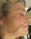
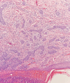
This report describes a recurrent sclerodermiform basal cell epithelioma of the malar region next to the inferior eyelid in a 57-year-old woman. Three interventions were necessary to obtain a clear margin of resection. The area of resection was closed with a local cutaneous flap. We report a rare basal cell carcinoma subtype underestimated in its aggressiveness with often inadequate medical and surgical management. This tumor, generally localized in the face, often requires aggressive surgery, and aesthetic results can be poor. The patients require close long-term follow-up even when margins are clear. General practitioners, dermatologists, and surgeons should be aware of sclerodermiform basal cell carcinoma, which is a malignant, aggressive, and recurrent tumor.
Copyright © 2024 The Authors. Published by Wolters Kluwer Health, Inc. on behalf of The American Society of Plastic Surgeons.
Conflict of interest statement
The authors have no financial interest to declare in relation to the content of this article.
Figures


References
-
- Brandt MG, Moore CC. Nonmelanoma skin cancer. Facial Plast Surg Clin North Am. 2019;27:1–13. - PubMed
-
- Agence Nationale d’Accreditation et Evaluation en Sante (ANAES). Good clinical practices. Diagnostic and therapeutic management of basal cell carcinoma in adults (extracts). Rev Stomatol Chir Maxillofac. 2005;106:83–88. - PubMed
-
- Loddé JP, Grangier Y, Le Roux P, et al. . Sclerodermiform basal cell carcinoma. Apropos of a study of 83 cases [in French]. Ann Chir Plast Esthet. 1998;43:373–382. - PubMed
-
- Conforti C, Pizzichetta MA, Vichi S, et al. . Sclerodermiform basal cell carcinomas vs. other histotypes: analysis of specific demographic, clinical and dermatoscopic features. J Eur Acad Dermatol Venereol. 2021;35:79–87. - PubMed
LinkOut - more resources
Full Text Sources
