. 2024 Apr 19:15:1375467.
doi: 10.3389/fgene.2024.1375467. eCollection 2024.

Human induced pluripotent stem cells (hiPSCs) derived cells reflect tissue specificity found in patients with Leigh syndrome French Canadian variant (LSFC)

Affiliations

Human induced pluripotent stem cells (hiPSCs) derived cells reflect tissue specificity found in patients with Leigh syndrome French Canadian variant (LSFC)

Roselle Gélinas et al. Front Genet. .

Abstract

Leigh syndrome French Canadian type (LSFC) is a recessive neurodegenerative disease characterized by tissue-specific deficiency in cytochrome c oxidase (COX), the fourth complex in the oxidative phosphorylation system. LSFC is caused by mutations in the leucine rich pentatricopeptide repeat containing gene (LRPPRC). Most LSFC patients in Quebec are homozygous for an A354V substitution that causes a decrease in the expression of the LRPPRC protein. While LRPPRC is ubiquitously expressed and is involved in multiple cellular functions, tissue-specific expression of LRPPRC and COX activity is correlated with clinical features. In this proof-of-principle study, we developed human induced pluripotent stem cell (hiPSC)-based models from fibroblasts taken from a patient with LSFC, homozygous for the LRPPRC*354V allele, and from a control, homozygous for the LRPPRC*A354 allele. Specifically, for both of these fibroblast lines we generated hiPSC, hiPSC-derived cardiomyocytes (hiPSC-CMs) and hepatocyte-like cell (hiPSC-HLCs) lines, as well as the three germ layers. We observed that LRPPRC protein expression is reduced in all cell lines/layers derived from LSFC patient compared to control cells, with a reduction ranging from ∼70% in hiPSC-CMs to undetectable levels in hiPSC-HLC, reflecting tissue heterogeneity observed in patient tissues. We next performed exploratory analyses of these cell lines and observed that COX protein expression was reduced in all cell lines derived from LSFC patient compared to control cells. We also observed that mutant LRPPRC was associated with altered expression of key markers of endoplasmic reticulum stress response in hiPSC-HLCs but not in other cell types that were tested. While this demonstrates feasibility of the approach to experimentally study genotype-based differences that have tissue-specific impacts, this study will need to be extended to a larger number of patients and controls to not only validate the current observations but also to delve more deeply in the pathogenic mechanisms of LSFC.

Keywords: Leigh syndrome French Canadian type; cardiomyocyte cells; disease modeling; hepatocyte-like cells; induced pluripotent stem cells; three germ layers.


Conflict of interest statement

The authors declare that the research was conducted in the absence of any commercial or financial relationships that could be construed as a potential conflict of interest.

Figures

FIGURE 1
FIGURE 1
Impact of LRPPRC*354V on LRPPRC and COXIV protein expression in LSFC hiPSCs. (A) Genotyping of LSFC and CTRL fibroblasts and hiPSCs for homozygous LRPPRC*354V mutation. (B, C) LRPPRC and COXIV average protein expression normalized to β-actin (LC) in CTRL and LSFC fibroblasts (two to three tech. rep. x one cell line) and hiPSCs (one to two tech. rep. x two clones). (D) ATP levels in CTRL and LSFC hiPSCs normalized to Hoechst 33342 viability stain (2 clones).
FIGURE 2
FIGURE 2
Characterization of cells from the three germ layers derived from LSFC hiPSCs. (A) Successful differentiation of LSFC and CTRL hiPSCs into the three germ layers demonstrated by IF staining of OTX2, SOX17 and Brachyury (red) as marker of ectoderm (ecto), definitive endoderm (endo) and mesoderm (meso), respectively. Nuclei (blue) were stained using DAPI (scale bar = 100 μm). (B) Gain in the expression of OTX2, SOX17 and Brachyury in the three germ layers derived from CTRL and LSFC hiPSCs demonstrated by WB (2 clones). (C) Protein levels of OTX2, SOX17 and Brachyury as well as LRPPRC in ectoderm, definitive endoderm and mesoderm derived from CTRL and LSFC hiPSCs. Bars are the average expression normalized to β-actin of two clones, except for ectoderm where the mean is representative of one clone from two independent differentiations.
FIGURE 3
FIGURE 3
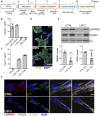
Characterization of hiPSC-HLCs from an LSFC patient. (A) Schematic representation of differentiation stages to obtain hiPSC-HLCs. (B) Pluripotency marker, (POU5F1) and (C) hepatic markers (AFP and ALB) relative mRNA expression normalized to HPRT in hiPSC-HLCs. Bars are the average expression of two clones from two independent differentiations. (D) Immunofluorescence staining of LDL-R (green) at the cell periphery of hiPSC-HLCs and nuclei (blue) using DAPI (scale = 10 µm). (E) LRPPRC and COXIV average protein levels in CTRL and LSFC hiPSC-HLCs of two clones from two independent differentiations. (F) IF staining of LRPPRC (red), TOM20 (green) as a marker of the mitochondria, LDL-R (grey) and nuclei (blue) in CTRL and LSFC hiPSC-HLCs (scale bar = 10 μm).
FIGURE 4
FIGURE 4
Characterization of hiPSC-CMs from an LSFC patient. (A) Schematic representation of differentiation stages to obtain beating hiPSC-CMs. (B) POU5F1 and (C) TNNT2 mRNA relative expression normalized to HPRT in hiPSC-CMs. (D) IF staining of cTnT (green) and nuclei (blue) using DAPI (scale bar = 100 µm) in CTRL and LSFC hiPSC-CMs. (E) LRPPRC and COXIV protein levels in CTRL and LSFC hiPSC-CMs (2 tech. rep. x one to two clones). (F) IF staining of LRPPRC (red) and TOM20 (green) in hiPSC-CMs. Phalloidin (grey) was used to stain striated F-actin and DAPI (blue) to stain nuclei (scale bar = 10 μm).

References

    1. Anderson N. S., Haynes C. M. (2020). Folding the mitochondrial UPR into the integrated stress response. Trends Cell. Biol. 30, 428–439. 10.1016/j.tcb.2020.03.001 - DOI - PMC - PubMed
    1. Burelle Y., Bemeur C., Rivard M. E., Thompson Legault J., Boucher G., Consortium L., et al. (2015). Mitochondrial vulnerability and increased susceptibility to nutrient-induced cytotoxicity in fibroblasts from leigh syndrome French canadian patients. PLoS One 10, e0120767. 10.1371/journal.pone.0120767 - DOI - PMC - PubMed
    1. Chaudhari N., Talwar P., Parimisetty A., Lefebvre D'Hellencourt C., Ravanan P. (2014). A molecular web: endoplasmic reticulum stress, inflammation, and oxidative stress. Front. Cell. Neurosci. 8, 213. 10.3389/fncel.2014.00213 - DOI - PMC - PubMed
    1. Cui J., Wang L., Ren X., Zhang Y., Zhang H. (2019). LRPPRC: a multifunctional protein involved in energy metabolism and human disease. Front. Physiol. 10, 595. 10.3389/fphys.2019.00595 - DOI - PMC - PubMed
    1. Cuillerier A., Ruiz M., Daneault C., Forest A., Rossi J., Vasam G., et al. (2021). Adaptive optimization of the OXPHOS assembly line partially compensates lrpprc-dependent mitochondrial translation defects in mice. Commun. Biol. 4, 989. 10.1038/s42003-021-02492-5 - DOI - PMC - PubMed