. 2024 Jan-Feb:12831:1283107.
doi: 10.1117/12.3005893. Epub 2024 Mar 12.

A dual-camera hyperspectral laparoscopic imaging system

Affiliations

A dual-camera hyperspectral laparoscopic imaging system

Kelden Pruitt et al. Proc SPIE Int Soc Opt Eng. 2024 Jan-Feb.

Abstract

Minimally invasive surgery (MIS) has expanded broadly in the field of abdominal and pelvic surgery. However, there are still prevalent issues surrounding intracorporeal surgery, such as iatrogenic injury, anastomotic leakage, or the presence of positive tumor margins after resection. Current approaches to address these issues and advance laparoscopic imaging techniques often involve fluorescence imaging agents, such as indocyanine green (ICG), to improve visualization, but these have drawbacks. Hyperspectral imaging (HSI) is an emerging optical imaging modality that takes advantage of spectral characteristics of different tissues. Various applications include tissue classification and digital pathology. In this study, we developed a dual-camera system for high-speed hyperspectral imaging. This includes the development of a custom application interface and corresponding hardware setup. Characterization of the system was performed, including spectral accuracy and spatial resolution, showing little sacrifice in speed for the approximate doubling of the covered spectral range, with our system acquiring 29 spectral images from 460-850 nm. Reference color tiles with various reflectance profiles were imaged and a RMSE of 3.56 ± 1.36% was achieved. Sub-millimeter resolution was shown at 7 cm working distance for both hyperspectral cameras. Finally, we image ex vivo tissues, including porcine stomach, liver, intestine, and kidney with our system and use a high-resolution, radiometrically calibrated spectrometer for comparison and evaluation of spectral fidelity. The dual-camera hyperspectral laparoscopic imaging system can have immediate applications in various surgeries.

Keywords: Hyperspectral imaging; image-guided intervention; laparoscopy; minimally invasive surgery.


Figures
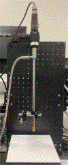
Figure 1.
Figure 1.
Dual-camera hyperspectral laparoscopic imaging setup.
Figure 2.
Figure 2.
Software application for concurrent hyperspectral imaging with the dual-camera system.
Figure 3.
Figure 3.
Demosaic methods visualized. The raw mosaic image (left) is processed into the demosaiced image (right).
Figure 4.
Figure 4.
Results of spectral accuracy of VIS camera (solid) against the tile standard reflectance (dashed). A) Red reflectance tile. B) Green reflectance tile. C) Blue reflectance tile. D) Yellow reflectance tile. E) Violet reflectance tile. F) Cyan reflectance tile. G) Orange reflectance tile. H) Purple reflectance tile.
Figure 5.
Figure 5.
Spectral footprint of liver, stomach, intestinal, and kidney tissue from laparoscopic system (solid) plotted alongside spectrometer readings (dotted). A) Pseudo-RGB endoscopic image of intestinal tissue. B) Pseudo-RGB endoscopic image of stomach tissue. C) Pseudo-RGB endoscopic image of liver tissue. D) Pseudo-RGB endoscopic image of kidney tissue.

References

    1. Alius C et al., “Indocyanine Green-Enhanced Colorectal Surgery—between Being Superfluous and Being a Game-Changer,” Diagnostics, vol. 10, no. 10, p. 742, 2020. - PMC - PubMed
    1. Grosek J and Tomazic A, “Key clinical applications for indocyanine green fluorescence imaging in minimally invasive colorectal surgery,” J Minim Access Surg, vol. 16, no. 4, pp. 308–314, Oct-Dec 2020, doi: 10.4103/jmas.JMAS_312_18. - DOI - PMC - PubMed
    1. Baltussen EJM et al., “Hyperspectral imaging for tissue classification, a way toward smart laparoscopic colorectal surgery,” J Biomed Opt, vol. 24, no. 1, pp. 1–9, Jan 2019, doi: 10.1117/1.JBO.24.1.016002. - DOI - PMC - PubMed
    1. Seidlitz S et al., “Robust deep learning-based semantic organ segmentation in hyperspectral images,” Med Image Anal, vol. 80, p. 102488, Aug 2022, doi: 10.1016/j.media.2022.102488. - DOI - PubMed
    1. Studier-Fischer A et al., “Spectral organ fingerprints for machine learning-based intraoperative tissue classification with hyperspectral imaging in a porcine model,” Sci Rep, vol. 12, no. 1, p. 11028, Jun 30 2022, doi: 10.1038/s41598-022-15040-w. - DOI - PMC - PubMed

LinkOut - more resources