. 2024 Apr 29:19:3861-3890.
doi: 10.2147/IJN.S445955. eCollection 2024.

Enzyme-Linked Lipid Nanocarriers for Coping Pseudomonal Pulmonary Infection. Would Nanocarriers Complement Biofilm Disruption or Pave Its Road?

Affiliations

Enzyme-Linked Lipid Nanocarriers for Coping Pseudomonal Pulmonary Infection. Would Nanocarriers Complement Biofilm Disruption or Pave Its Road?

Noha Nafee et al. Int J Nanomedicine. .

Abstract

Introduction: Cystic fibrosis (CF) is associated with pulmonary Pseudomonas aeruginosa infections persistent to antibiotics.

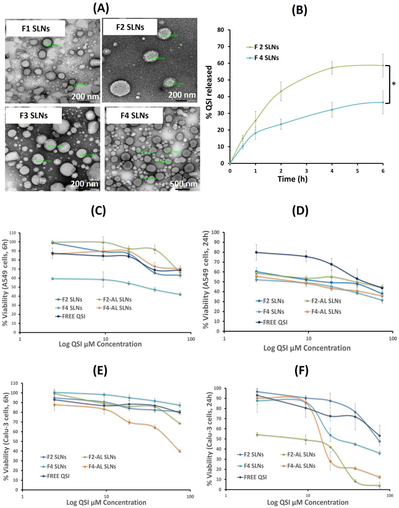
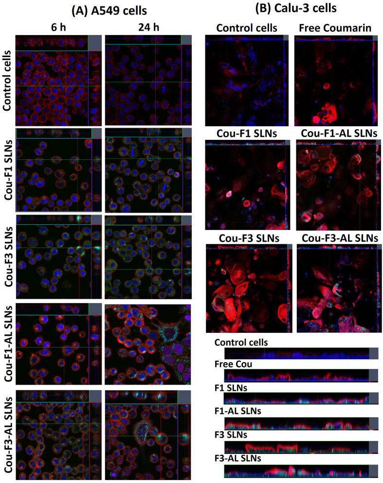
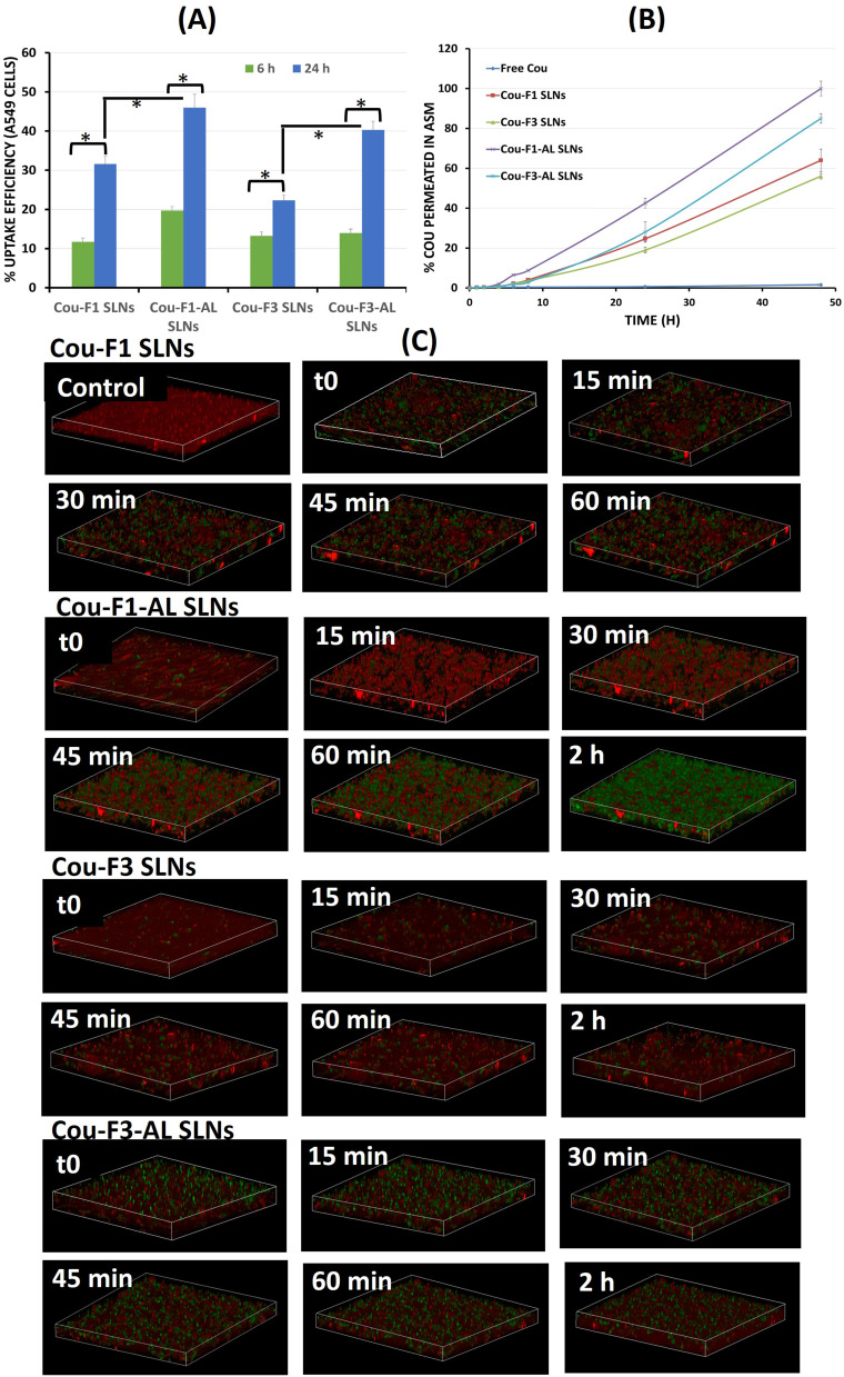
Methods: To eradicate pseudomonal biofilms, solid lipid nanoparticles (SLNs) loaded with quorum-sensing-inhibitor (QSI, disrupting bacterial crosstalk), coated with chitosan (CS, improving internalization) and immobilized with alginate lyase (AL, destroying alginate biofilms) were developed.

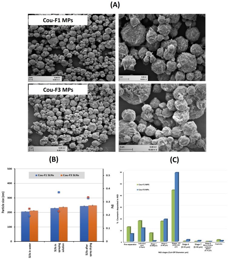
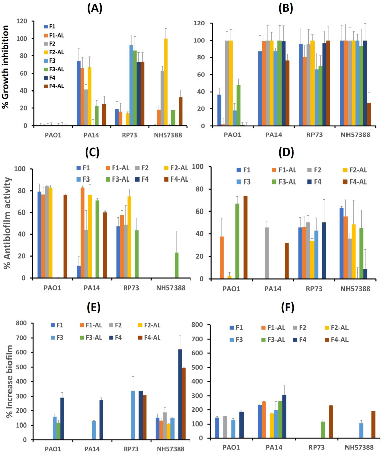
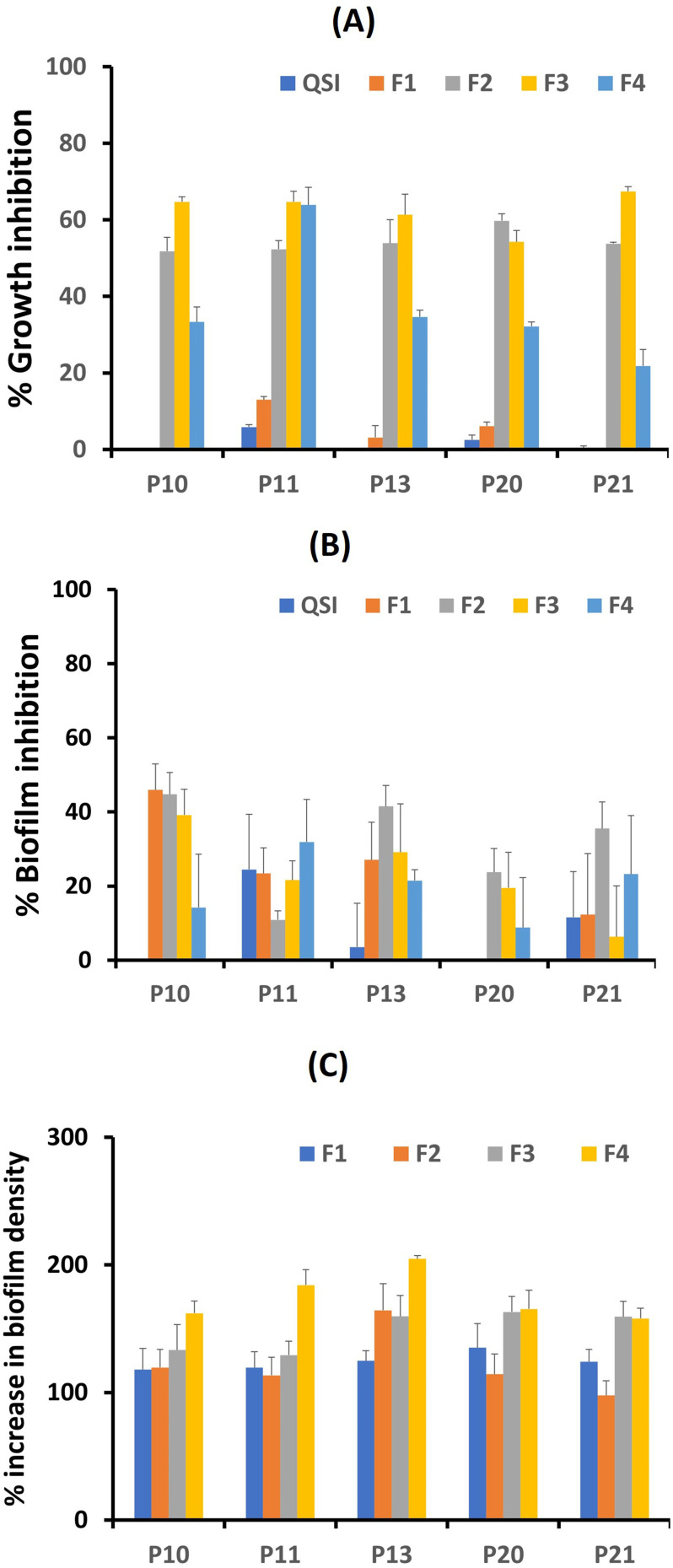
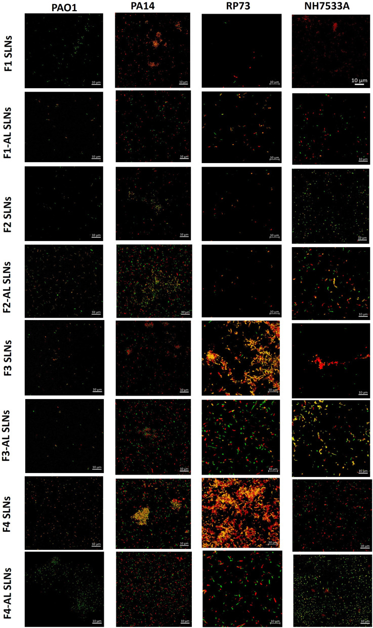
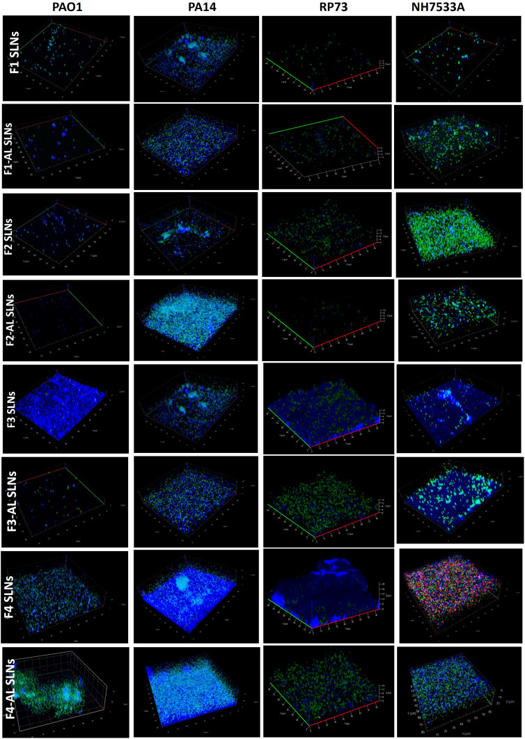
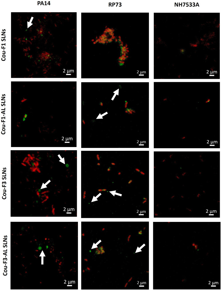
Results: SLNs (140-205 nm) showed prolonged release of QSI with no sign of acute toxicity to A549 and Calu-3 cells. The CS coating improved uptake, whereas immobilized-AL ensured >1.5-fold higher uptake and doubled SLN diffusion across the artificial biofilm sputum model. Respirable microparticles comprising SLNs in carbohydrate matrix elicited aerodynamic diameters MMAD (3.54, 2.48 µm) and fine-particle-fraction FPF (65, 48%) for anionic and cationic SLNs, respectively. The antimicrobial and/or antibiofilm activity of SLNs was explored in Pseudomonas aeruginosa reference mucoid/nonmucoid strains as well as clinical isolates. The full growth inhibition of planktonic bacteria was dependent on SLN type, concentration, growth medium, and strain. OD measurements and live/dead staining proved that anionic SLNs efficiently ceased biofilm formation and eradicated established biofilms, whereas cationic SLNs unexpectedly promoted biofilm progression. AL immobilization increased biofilm vulnerability; instead, CS coating increased biofilm formation confirmed by 3D-time lapse confocal imaging. Incubation of SLNs with mature biofilms of P. aeruginosa isolates increased biofilm density by an average of 1.5-fold. CLSM further confirmed the binding and uptake of the labeled SLNs in P. aeruginosa biofilms. Considerable uptake of CS-coated SLNs in non-mucoid strains could be observed presumably due to interaction of chitosan with LPS glycolipids in the outer cell membrane of P. aeruginosa.

Conclusion: The biofilm-destructive potential of QSI/SLNs/AL inhalation is promising for site-specific biofilm-targeted interventional CF therapy. Nevertheless, the intrinsic/extrinsic fundamentals of nanocarrier-biofilm interactions require further investigation.

Keywords: Pseudomonas aeruginosa; alginate lyase; chitosan; cystic fibrosis; quorum-sensing inhibitors; solid lipid nanoparticles.


Conflict of interest statement

Martin Empting reports a patent WO2020007938A1 pending to Helmholtz Centre for Infection Research (HZI), a patent EP20150104 pending to Helmholtz Centre for Infection Research (HZI), and a patent WO2021136805A1 pending to Helmholtz Centre for Infection Research (HZI). The authors report no other conflicts of interest in this work.

Figures

Figure 1
Figure 1
(A) TEM images F1 SLNs, F2 SLNs, F3 SLNs, and F4 SLNs; (B) In vitro release of QSI from F2 and F4 SLNs in SLF (*Statistically significant difference, two-way ANOVA with Tukey’s post-hoc test, p < 0.05); (C) Viability of A549 cells following 6 h incubation with QSI-loaded SLNs; (D) Viability of A549 cells following 24 h incubation with QSI-loaded SLNs; (E) Viability of Calu-3 cells following 6 h incubation with QSI-loaded SLNs; (F) Viability of Calu-3 cells following 24 h incubation with QSI-loaded SLNs. Data represent mean values (n = 3) ± SD.
Figure 2
Figure 2
Confocal images illustrating the uptake of coumarin-labeled SLNs in: (A) A549 cells following 6 and 24 h incubation, and (B) Calu-3 cells after 24 h incubation (bars at the bottom right represent z-stacks).
Figure 3
Figure 3
(A) Quantitative estimation of uptake efficiency of coumarin-labeled SLNs in A549 cells via Transwell technique following 6 and 24 h incubation (*Statistically significant difference, paired t-test, p <0.05); (B) Permeation efficiency of coumarin-labeled SLNs in artificial biofilm sputum medium; (C) 3D-time lapse confocal stacks showing diffusion of different coumarin-labeled SLNs in artificial biofilm sputum medium over 1–2 h.
Figure 4
Figure 4
(A) SEM micrographs of SLN-embedded microparticles (scale bar 2 µm); (B) Colloidal stability of SLNs in the spray drying solution and after spray drying; (C) Deposition pattern of cou-F1 MPs and cou-F3 MPs on stages of next generation impactor.
Figure 5
Figure 5
Antimicrobial activity of SLNs (equivalent to 18.75 µM QSI) on P. aeruginosa reference strains (A) P. aeruginosa planktonic reference strains in LB medium; (B) P. aeruginosa planktonic reference strains in ASM; (C) Antibiofilm activity in LB medium; (D) Antibiofilm activity in ASM; (E) Increase in biofilm formation in LB medium; (F) Increase in biofilm formation in ASM.
Figure 6
Figure 6
Antimicrobial activity of SLNs (equivalent to 18.75 µM QSI) on P. aeruginosa isolates (A) Growth inhibition of P. aeruginosa planktonic isolates; (B) Inhibition of biofilm formation of P. aeruginosa isolates; (C) Increase in biofilm formation of P. aeruginosa isolates.
Figure 7
Figure 7
CLSM micrographs of P. aeruginosa biofilms of PA01, PA14, RP37, and NH57388A strains following exposure to different SLNs (Live/Dead staining).
Figure 8
Figure 8
3D-time laps stacks (80 × 80×20 µm) of P. aeruginosa biofilms of PA01, PA14, RP37, and NH57388A strains showing live bacteria (stained green with Syto-9) and extracellular polysaccharides (stained blue with Calcofluor) following exposure to different SLNs.
Figure 9
Figure 9
Uptake of coumarin-labeled SLNs (green) in PA14, RP37, and NH57388A strains (dead bacteria stained red). Arrows represent SLN clusters around live non-stained bacteria. Scale bar 2 µm.

References

    1. Malhotra S, Hayes D, Wozniak Daniel J. Cystic fibrosis and pseudomonas aeruginosa: the host-microbe interface. Clin Microbiol Rev. 2019;32(3):e00138–00118. doi:10.1128/CMR.00138-18 - DOI - PMC - PubMed
    1. Flemming H-C, Wingender J. The biofilm matrix. Nat Rev Microbiol. 2010;8(9):623–633. doi:10.1038/nrmicro2415 - DOI - PubMed
    1. Hadinoto K, Cheow WS. Nano-antibiotics in chronic lung infection therapy against Pseudomonas aeruginosa. Colloids Surf B. 2014;116:772–785. doi:10.1016/j.colsurfb.2014.02.032 - DOI - PubMed
    1. Maunders E, Welch M. Matrix exopolysaccharides; the sticky side of biofilm formation. FEMS Microbiol Lett. 2017;364(13). doi:10.1093/femsle/fnx120 - DOI - PMC - PubMed
    1. Cotton LA, Graham RJ, Lee RJ. The role of alginate in P. aeruginosa PAO1 biofilm structural resistance to gentamicin and ciprofloxacin. J Exp Microbiol Immunol. 2009;13:58–62.

MeSH terms