. 2024 Aug;27(3):423-440.
doi: 10.1007/s10456-024-09917-9. Epub 2024 May 6.

IC100, a humanized therapeutic monoclonal anti-ASC antibody alleviates oxygen-induced retinopathy in mice

Affiliations

IC100, a humanized therapeutic monoclonal anti-ASC antibody alleviates oxygen-induced retinopathy in mice

Huijun Yuan et al. Angiogenesis. 2024 Aug.

Abstract

Background: Retinopathy of prematurity (ROP), which often presents with bronchopulmonary dysplasia (BPD), is among the most common morbidities affecting extremely premature infants and is a leading cause of severe vision impairment in children worldwide. Activations of the inflammasome cascade and microglia have been implicated in playing a role in the development of both ROP and BPD. Apoptosis-associated speck-like protein containing a caspase recruitment domain (ASC) is pivotal in inflammasome assembly. Utilizing mouse models of both oxygen-induced retinopathy (OIR) and BPD, this study was designed to test the hypothesis that hyperoxia induces ASC speck formation, which leads to microglial activation and retinopathy, and that inhibition of ASC speck formation by a humanized monoclonal antibody, IC100, directed against ASC, will ameliorate microglial activation and abnormal retinal vascular formation.

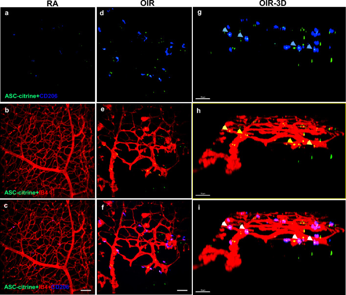
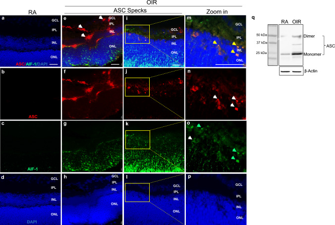
Methods: We first tested ASC speck formation in the retina of ASC-citrine reporter mice expressing ASC fusion protein with a C-terminal citrine (fluorescent GFP isoform) using a BPD model that causes both lung and eye injury by exposing newborn mice to room air (RA) or 85% O2 from postnatal day (P) 1 to P14. The retinas were dissected on P14 and retinal flat mounts were used to detect vascular endothelium with AF-594-conjugated isolectin B4 (IB4) and citrine-tagged ASC specks. To assess the effects of IC100 on an OIR model, newborn ASC citrine reporter mice and wildtype mice (C57BL/6 J) were exposed to RA from P1 to P6, then 75% O2 from P7 to P11, and then to RA from P12 to P18. At P12 mice were randomized to the following groups: RA with placebo PBS (RA-PBS), O2 with PBS (O2-PBS), O2 + IC100 intravitreal injection (O2-IC100-IVT), and O2 + IC100 intraperitoneal injection (O2-IC100-IP). Retinal vascularization was evaluated by flat mount staining with IB4. Microglial activation was detected by immunofluorescence staining for allograft inflammatory factor 1 (AIF-1) and CD206. Retinal structure was analyzed on H&E-stained sections, and function was analyzed by pattern electroretinography (PERG). RNA-sequencing (RNA-seq) of the retinas was performed to determine the transcriptional effects of IC100 treatment in OIR.

Results: ASC specks were significantly increased in the retinas by hyperoxia exposure and colocalized with the abnormal vasculature in both BPD and OIR models, and this was associated with increased microglial activation. Treatment with IC100-IVT or IC100-IP significantly reduced vaso-obliteration and intravitreal neovascularization. IC100-IVT treatment also reduced retinal microglial activation, restored retinal structure, and improved retinal function. RNA-seq showed that IC100 treatment corrected the induction of genes associated with angiogenesis, leukocyte migration, and VEGF signaling caused by O2. IC100 also corrected the suppression of genes associated with cell junction assembly, neuron projection, and neuron recognition caused by O2.

Conclusion: These data demonstrate the crucial role of ASC in the pathogenesis of OIR and the efficacy of a humanized therapeutic anti-ASC antibody in treating OIR mice. Thus, this anti-ASC antibody may potentially be considered in diseases associated with oxygen stresses and retinopathy, such as ROP.

Keywords: ASC; IC100; Microglia; Oxygen-induced retinopathy; Transcriptome.


Conflict of interest statement

JPdRV, WDD, and RWK are co-founders and managing members of InflamaCORE, LLC and have licensed patents on inflammasome proteins as biomarkers of injury and disease as well as on targeting inflammasome proteins for therapeutic purposes. JPdRV, WDD, and RWK are Scientific Advisory Board Members of ZyVersa Therapeutics. SW has a patent with a U.S. Patent Application No. 63/543,835 titled: Use of anti-ASC antibody in the treating of ocular disorders.

Figures

Fig. 1
Fig. 1
Immunofluorescent confocal microscopy images of BPD model ASC-citrine mouse whole mount retinas. ac Images of RA-exposed (RA) mouse whole mount retinas: a Green, ASC-citrine. b Normal vasculature, red (IB4 staining) c Merged image. df Images of 85% O2-exposed mouse whole mount retinas: d Green, ASC-citrine specks. e Disorganized proliferative vasculature, red (IB4 staining) f Merged image of d & e. gi Zoom-in images of df. g ASC-citrine specks (white arrow). h Proliferative vasculature, red (IB4 staining). i The merged image show ASC-citrine specks colocalized with abnormal vasculature (yellow, blue arrow). j Quantification of ASC-citrine speck images. ***P < 0.0001. n = 6 mice/group. scale bars: 50 µm
Fig. 2
Fig. 2
Immunofluorescent confocal microscopy images of OIR model ASC-citrine mouse retinal flat mount staining. ac Images of RA mouse whole mount retinas: a Merged image of ASC-citrine (green) and CD206 positive microglia (blue). b Merged image of normal vasculature (red, IB4 staining) and ASC-citrine. c Merged image of ASC-citrine, blood vessel and microglia. df Images of OIR mouse retinal flat mount staining: d Merged image of ASC-citrine specks (Green) and CD206 positive microglia (blue). e Merged image of neovascular tufts (red, IB4 staining) and ASC-citrine specks (green). f Merged image of OIR retinal neovascular tufts, ASC-citrine specks and CD206 positive microglia. gi 3D Images of images df scale bars: 70 µm
Fig. 3
Fig. 3
ASC speck formation in wildtype OIR model mouse retinas. Immunofluorescent microscopy images of mouse retinal cross-sections. ad Representative immunofluorescent microscopy images of RA mouse retinal sections. el Representative immunofluorescent microscopy images of OIR mouse retinal sections. mp Zoom-in images of il. ASC specks were detected in the intracellular in ganglion cell layer (GCL, e & i, white arrow), extracellular in inner plexiform layer (IPL, e & i, white arrow), and activated microglia cells in inner nuclear layer (INL, k & o, green arrow) in OIR mouse retinas. ASC specks, in the intracellular and/or extracellular matrix, (m, yellow arrow) colocalized with activated microglia (o, green arrow) in OIR mouse retinas. n = 5 mice/group. Scale bar: 25 µM. q Representative images of Western blot analysis of ASC from pooled 5 RA and 5 OIR mouse vitreous fluids
Fig. 4
Fig. 4
Anti-ASC antibody, IC100 reduced oxygen-induced pathological vaso-obliteration and intravitreal neovascularization. a Representative images of P18 mouse retinal flat mounts; control: RA + placebo PBS (RA-PBS), OIR: OIR treated with PBS (O2-PBS) by IVT injection, IC100 by IVT (O2-IC100-IVT), and IC100 by IP (O2-IC100-IP) injection. The avascular areas were circled in white. b The white boxes in a were enlarged eightfold, and the yellow arrows indicate vascular tufts. c Intravitreal neovascularization was highlighted by white dots generated by Zeiss microscope software based on vascular intensity. d Quantifications of avascular areas. The O2-PBS group had increased avascular areas compared to the RA-PBS group (****P < 0.0001). But treatment with IC100 by IVT and IP injection significantly reduced the avascular areas compared to the O2-PBS group (O2-IC100-IVT, ****P < 0.0001, O2-IC100-IP, *P < 0.05). e Measurements of neovascular tufts. The O2-PBS group had increased vascular tufts compared to the RA-PBS group (****P < 0.0001). But treatment with IC100 by IVT and IP injection significantly reduced the neovascular turfs compared to the O2-PBS group (****P < 0.0001). f Assessment of intravitreal neovascularization. The O2-PBS group had increased intravitreal neovascularization compared to the RA-PBS group (****P < 0.0001). But treatment with IC100 by IVT and IP injection significantly reduced the neovascular areas compared to the O2-PBS group (****P < 0.0001). RA-PBS, O2-PBS, and O2-IC100-IVT groups: n = 4 mice/group. O2-IC100-IP group: n = 10 mice/group. Scale bars: 50 μm
Fig. 5
Fig. 5
IC100 decreased microglial density and activation in OIR mice. a, d & g Images of retinal whole mount stained by AIF-1 (microglia, green). The microglia in RA-PBS retinas were small and organized (a). The microglia in O2-PBS retinas had large bodies and increased numbers compared to RA-PBS groups (d, white arrow). Treatment with IC100-IVT reduced the number of microglia in O2-exposed retinas that appear to have similar shapes as the RA-PBS group (g). b, e & h IB4 stained vasculature (red). The vasculature was normal in the RA-PBS group (b). The vasculature was abnormal with increased density and thickness in the O2-PBS group (e). Treatment with IC100-IVT improved vascular growth in O2-exposed retinas compared to the O2-PBS group (h). c, f & i Merged images. The retinas from O2-PBS group had abnormal microglia colocalized with the abnormal vasculature (f, blue arrows). j Quantification of microglia. The number of microglia in the O2-PBS retinas increased more than 70% compared to RA-PBS. While treatment with IC100-IVT drastically reduced the microglial counts by 75% compared to the O2-PBS group. **P < 0.01, ****P < 0.0001. n = 4–10 mice/group. Scale bars: 25 µm
Fig. 6
Fig. 6
Immunofluorescent confocal microscopy images of OIR model ASC-citrine mouse retinal flat mount staining. ad Images of RA-exposed mouse whole mount retinas: a ASC-citrine specks (green). b CD206 positive microglia (blue). c Normal vasculature (red, IB4 staining). d Merged image. e–h Images of OIR mouse retinal flat mount staining: e ASC-citrine specks (Green), f CD206 positive microglia (blue), g Avascular and neovascular tufts (red), and h Merged image. il Images of IC100 treated OIR mouse whole mount retinas: i Decreased ASC specks (green), j Suppression of CD206 positive microglia (blue), k Normal vasculature (red, IB4 staining), and l Merged image. m Graph of ASC speck quantification. n Graph of CD206 positive microglia quantification. n = 3 mice/group, 4 images/retina. ****P < 0.0001
Fig. 7
Fig. 7
IC100 treatment restored OIR model retinal structure formation. ac H&E-stained retinal cross-sections. The O2-PBS retinas (b) had disorganized GCL, and reduced thickness of the inner nuclear layer (INL), outer nuclear layer (ONL) and total thickness of retinal layer compared to the RA-PBS group (a). The O2-IC100-IVT group had retinal tissue layer thicknesses that were similar to the RA-PBS group (c). d Quantification of retinal INL thickness. e Quantification of retinal ONL. f Quantification of total retinal layer thickness. n = 9 mice. ****P < 0.0001. ns: no significance. Scale bar: 25 µm
Fig. 8
Fig. 8
IC100 improves OIR model mouse retinal function. a Average amplitude of all mice in each group. The amplitude was reduced to 12.09 µV in the O2-PBS retinas compared to 25.99 µV in the RA-PBS group, while treatment with IC100-IVT increased the amplitude to 21.77 µV compared to the O2-PBS group. b Average latency of all mice in each group. The latency had no significant difference among the three groups group. n = 6 mice/group. ***P < 0.001, ****P < 0.0001, ns: no significance
Fig. 9
Fig. 9
IC100 reduced expression of inflammatory cytokines, inflammasome-related molecules, and Vegf in OIR model mouse retinas. Real-time qRT-PCR of O2-PBS retinas showed gene expression of inflammasome cytokines, Il1b, Il6 and Tnf, respectively elevated 7.7, 4.3 and 4.7-fold compared to age-matched RA mouse retinas on P18. These genes were significantly decreased in O2-IC100-IVT mouse retinas (ac). Pycard/Asc, Gsdmd, and Vegf were increased 2.4 to threefold in O2-PBS retinas compared to RA mouse retinas but were significantly decreased in IC100-IVT treated OIR retinas (df). n = 5 mice/group. *P < 0.05, **P < 0.01, ***P < 0.001. Double immunofluorescence staining of anti-GSDMD or anti-IL-1β antibodies (red signal, red arrow) with an anti-AIF-1 antibody (green signal, green arrow) and DAPI staining (blue signal) (g). Upper panel: increased GSDMD was detected in AIF-1 positive cells (yellow signal, yellow arrow) in O2-PBS retinas, but this was decreased in O2-IC100 retinas. Lower panel: increased IL-1β was detected in AIF-1 positive cells (yellow signal, yellow arrow) in O2-PBS retinas, however, this was decreased in O2-IC100 retinas. Scale bar: 50 μm
Fig. 10
Fig. 10
Differential expression and clustering analyses show IC100 prevents hyperoxia modulation of gene pathways related to eye development, retinal angiogenesis, VEGF signaling, and leucocyte functions. a Principal component analysis (PCA) plot showing separation of RA-PBS, O2-PBS, and O2-IC100 by PC1 and PC2. b Heatmap of differentially expressed genes showing relative expression. c Expression patterns identified in the 9 clusters with the number of genes displayed next to the cluster numbers. d Gene pathways upregulated by hyperoxia but corrected by IC100 treatment to the near RA levels in cluster 4 include eye development, angiogenesis, leukocyte migration and cell–cell adhesion, endothelial cell differentiation, and VEGF signaling. e Gene pathways that were downregulated by hyperoxia but corrected by IC100 treatment to near RA levels in cluster 6 include cell junction assembly, synapse assembly, and neuron differentiation. n = 3–4 retinas/group

References

    1. Hartnett ME, Penn JS (2012) Mechanisms and management of retinopathy of prematurity. N Engl J Med 367(26):2515–2526 10.1056/NEJMra1208129 - DOI - PMC - PubMed
    1. Hartnett ME (2015) Pathophysiology and mechanisms of severe retinopathy of prematurity. Ophthalmology 122(1):200–210 10.1016/j.ophtha.2014.07.050 - DOI - PMC - PubMed
    1. Solebo AL, Teoh L, Rahi J (2017) Epidemiology of blindness in children. Arch Dis Child 102(9):853–857 10.1136/archdischild-2016-310532 - DOI - PubMed
    1. Bourne RR, Stevens GA, White RA, Smith JL, Flaxman SR, Price H et al (2013) Causes of vision loss worldwide, 1990–2010: a systematic analysis. Lancet Glob Health 1(6):e339–e349 10.1016/S2214-109X(13)70113-X - DOI - PubMed
    1. Chawanpaiboon S, Vogel JP, Moller AB, Lumbiganon P, Petzold M, Hogan D et al (2019) Global, regional, and national estimates of levels of preterm birth in 2014: a systematic review and modelling analysis. Lancet Glob Health 7(1):e37–e46 10.1016/S2214-109X(18)30451-0 - DOI - PMC - PubMed

MeSH terms

Substances

LinkOut - more resources