. 2024 Jun 27;128(25):4992-4998.
doi: 10.1021/acs.jpca.4c02509. Epub 2024 May 6.

The Ring-Closing Reaction of Cyclopentadiene Probed with Ultrafast X-ray Scattering

Affiliations

The Ring-Closing Reaction of Cyclopentadiene Probed with Ultrafast X-ray Scattering

Lisa Huang et al. J Phys Chem A. .

Abstract

The dynamics of cyclopentadiene (CP) following optical excitation at 243 nm was investigated by time-resolved pump-probe X-ray scattering using 16.2 keV X-rays at the Linac Coherent Light Source (LCLS). We present the first ultrafast structural evidence that the reaction leads directly to the formation of bicyclo[2.1.0]pentene (BP), a strained molecule with three- and four-membered rings. The bicyclic compound decays via a thermal backreaction to the vibrationally hot CP with a time constant of 21 ± 3 ps. A minor channel leads to ring-opened structures on a subpicosecond time scale.


Conflict of interest statement

The authors declare no competing financial interest.

Figures

Scheme 1
Scheme 1. Photochemical Reaction of Cyclopentadiene (CP) Proposed to Lead to Bicyclo[2.1.0]pentene (BP) and Tricyclo[2.1.0.0]pentane (TP)
Figure 1
Figure 1
Experimental setup for ultrafast X-ray scattering experiments. The optical UV pump pulse (5.10 eV) excites the CP molecules and induces a chemical reaction, the time evolution of which is probed by a hard X-ray pulse (16.2 keV). The scattering signals are measured as a function of the azimuthal angle (ϕ) and the momentum transfer vector (q) on the Jungfrau detector.
Figure 2
Figure 2
Time-dependent percent difference X-ray scattering signal of CP upon excitation at 243 nm as a function of momentum transfer vector and pump–probe delay. The isotropic component of the experimental pump–probe signal is represented as percent differences, with red regions signifying enhancements and blue areas indicating depletions. The time delay is separated into two segments, with the first stepping linearly from −0.5 to 2 ps and the second logarithmically from 2 to 25 ps. The scattering signal intensity, as shown by the color bar, varies on the order of 0.3%.
Figure 3
Figure 3
Proposed kinetic scheme for CP upon excitation by 243 nm light. CP* refers to the molecule rapidly evolving on the excited-state potential energy surfaces, CPhot is a highly vibrationally excited molecule in the ground electronic state, and BP is the bicyclic structure that results from cross-linking a chemical bond. RO represents any ring-open structure.
Figure 4
Figure 4
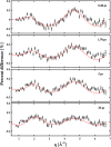
Experimental isotropic percent difference scattering signals at given time delays (0.40, 1.70, 5, and 25 ps) where the black dots are the experimental data and the red dashed lines are the fit results.
Figure 5
Figure 5
Comparison of the experimental pattern extracted from the fit for species BP with 1σ error bars (black circles) with the simulated theoretical percent difference pattern of vibrationally hot BP (blue line) and hot TP (orange dashed line).
Figure 6
Figure 6
Relative populations of the four components (electronically excited CP*, BP, CPhot, and RO) present in the reaction, as derived from the 2-D fits described above, presented on a logarithmic time scale.

References

    1. Emma P.; Akre R.; Arthur J.; Bionta R.; Bostedt C.; Bozek J.; Brachmann A.; Bucksbaum P.; Coffee R.; Decker F.-J.; et al. First lasing and operation of an ångstrom-wavelength free-electron laser. Nat. Photonics 2010, 4, 641–647. 10.1038/nphoton.2010.176. - DOI
    1. Yong H.; Xu X.; Ruddock J. M.; Stankus B.; Carrascosa A. M.; Zotev N.; Bellshaw D.; Du W.; Goff N.; Chang Y.; et al. Ultrafast X-ray scattering offers a structural view of excited-state charge transfer. Proc. Natl. Acad. Sci. U. S. A. 2021, 118, e2021714118.10.1073/pnas.2021714118. - DOI - PMC - PubMed
    1. Yong H.; Zotev N.; Stankus B.; Ruddock J. M.; Bellshaw D.; Boutet S.; Lane T. J.; Liang M.; Carbajo S.; Robinson J. S.; et al. Determining orientations of optical transition dipole moments using ultrafast X-ray scattering. J. Phys. Chem. Lett. 2018, 9, 6556–6562. 10.1021/acs.jpclett.8b02773. - DOI - PubMed
    1. Yong H.; Zotev N.; Ruddock J. M.; Stankus B.; Simmermacher M.; Carrascosa A. M.; Du W.; Goff N.; Chang Y.; Bellshaw D.; et al. Observation of the molecular response to light upon photoexcitation. Nat. Commun. 2020, 11, 2157.10.1038/s41467-020-15680-4. - DOI - PMC - PubMed
    1. Minitti M.; Budarz J.; Kirrander A.; Robinson J.; Ratner D.; Lane T.; Zhu D.; Glownia J.; Kozina M.; Lemke H.; et al. Imaging molecular motion: Femtosecond X-ray scattering of an electrocyclic chemical reaction. Phys. Rev. Lett. 2015, 114, 255501.10.1103/PhysRevLett.114.255501. - DOI - PubMed

LinkOut - more resources