Fuzhengjiedu San inhibits porcine reproductive and respiratory syndrome virus by activating the PI3K/AKT pathway
- PMID: 38709810
- PMCID: PMC11073700
- DOI: 10.1371/journal.pone.0283728
Fuzhengjiedu San inhibits porcine reproductive and respiratory syndrome virus by activating the PI3K/AKT pathway
Abstract
Background: Traditional Chinese medicine (TCM) has been garnering ever-increasing worldwide attention as the herbal extracts and formulas prove to have potency against disease. Fuzhengjiedu San (FZJDS), has been extensively used to treat viral diseases in pigs, but its bioactive components and therapeutic mechanisms remain unclear.
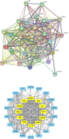
Methods: In this study, we conducted an integrative approach of network pharmacology and experimental study to elucidate the mechanisms underlying FZJDS's action in treating porcine reproductive and respiratory syndrome virus (PRRSV). We constructed PPI network and screened the core targets according to their degree of value. GO and KEGG enrichment analyses were also carried out to identify relevant pathways. Lastly, qRT-PCR, flow cytometry and western blotting were used to determine the effects of FZJDS on core gene expression in PRRSV-infected monkey kidney (MARC-145) cells to further expand the results of network pharmacological analysis.
Results: Network pharmacology data revealed that quercetin, kaempferol, and luteolin were the main active compounds of FZJDS. The phosphatidylinositol-3-kinase (PI3K)/Akt pathway was deemed the cellular target as it has been shown to participate most in PRRSV replication and other PRRSV-related functions. Analysis by qRT-PCR and western blotting demonstrated that FZJDS significantly reduced the expression of P65, JNK, TLR4, N protein, Bax and IĸBa in MARC-145 cells, and increased the expression of Bcl-2, consistent with network pharmacology results. This study provides that FZJDS has significant antiviral activity through its effects on the PI3K/AKT signaling pathway.
Conclusion: We conclude that FZJDS is a promising candidate herbal formulation for treating PRRSV and deserves further investigation.
Copyright: © 2024 Chang et al. This is an open access article distributed under the terms of the Creative Commons Attribution License, which permits unrestricted use, distribution, and reproduction in any medium, provided the original author and source are credited.
Conflict of interest statement
The authors have declared that no competing interests exist.
Figures


Similar articles
-
Antiviral mechanism of Fuzhengjiedu San against porcine reproductive and respiratory syndrome virus.Virology. 2025 Feb;603:110382. doi: 10.1016/j.virol.2024.110382. Epub 2025 Jan 6. Virology. 2025. PMID: 39798332
-
The Antimalaria Drug Artesunate Inhibits Porcine Reproductive and Respiratory Syndrome Virus Replication by Activating AMPK and Nrf2/HO-1 Signaling Pathways.J Virol. 2022 Feb 9;96(3):e0148721. doi: 10.1128/JVI.01487-21. Epub 2021 Nov 17. J Virol. 2022. PMID: 34787456 Free PMC article.
-
Control of the PI3K/Akt pathway by porcine reproductive and respiratory syndrome virus.Arch Virol. 2013 Jun;158(6):1227-34. doi: 10.1007/s00705-013-1620-z. Epub 2013 Feb 5. Arch Virol. 2013. PMID: 23381397 Free PMC article.
-
Huashi baidu granule alleviates inflammation and lung edema by suppressing the NLRP3/caspase-1/GSDMD-N pathway and promoting fluid clearance in a porcine reproductive and respiratory syndrome (PRRS) model.J Ethnopharmacol. 2025 Jan 31;340:119207. doi: 10.1016/j.jep.2024.119207. Epub 2024 Dec 7. J Ethnopharmacol. 2025. PMID: 39653102
-
Antiviral and Immune Enhancement Effect of Platycodon grandiflorus in Viral Diseases: A Potential Broad-Spectrum Antiviral Drug.Molecules. 2025 Feb 11;30(4):831. doi: 10.3390/molecules30040831. Molecules. 2025. PMID: 40005144 Free PMC article. Review.
Cited by
-
Antiviral effects and mechanism of Ma-Xing-Shi-Gan-San on porcine reproductive and respiratory syndrome virus.Front Microbiol. 2025 Apr 29;16:1539094. doi: 10.3389/fmicb.2025.1539094. eCollection 2025. Front Microbiol. 2025. PMID: 40365068 Free PMC article.
References
-
- Huang M, Wei Y, Dong J. Epimedin C modulates the balance between Th9 cells and Treg cells through negative regulation of noncanonical NF-κB pathway and MAPKs activation to inhibit airway inflammation in the ovalbumin-induced murine asthma model. Pulm Pharmacol Ther. 2020. Dec;65:102005. doi: 10.1016/j.pupt.2021.102005 - DOI - PubMed
Publication types
MeSH terms
Substances
LinkOut - more resources
Full Text Sources
Research Materials

