. 2024 Jan 8;18(1):wrae084.
doi: 10.1093/ismejo/wrae084.

Enantioselective transformation of phytoplankton-derived dihydroxypropanesulfonate by marine bacteria

Affiliations

Enantioselective transformation of phytoplankton-derived dihydroxypropanesulfonate by marine bacteria

Le Liu et al. ISME J. .

Abstract

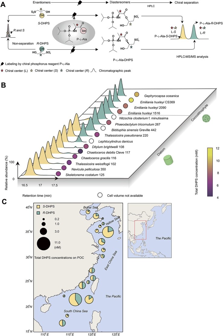
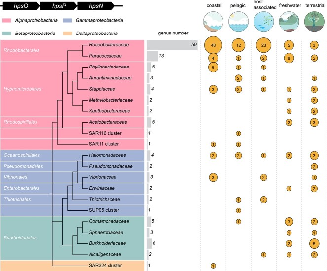
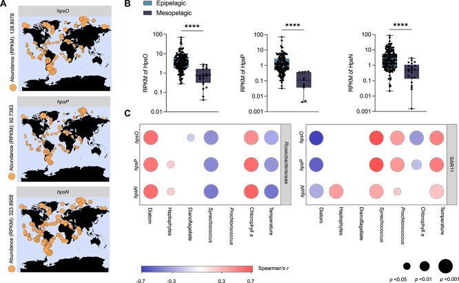
Chirality, a fundamental property of matter, is often overlooked in the studies of marine organic matter cycles. Dihydroxypropanesulfonate (DHPS), a globally abundant organosulfur compound, serves as an ecologically important currency for nutrient and energy transfer from phytoplankton to bacteria in the ocean. However, the chirality of DHPS in nature and its transformation remain unclear. Here, we developed a novel approach using chiral phosphorus-reagent labeling to separate DHPS enantiomers. Our findings demonstrated that at least one enantiomer of DHPS is present in marine diatoms and coccolithophores, and that both enantiomers are widespread in marine environments. A novel chiral-selective DHPS catabolic pathway was identified in marine Roseobacteraceae strains, where HpsO and HpsP dehydrogenases at the gateway to DHPS catabolism act specifically on R-DHPS and S-DHPS, respectively. R-DHPS is also a substrate for the dehydrogenase HpsN. All three dehydrogenases generate stable hydrogen bonds between the chirality-center hydroxyls of DHPS and highly conserved residues, and HpsP also form coordinate-covalent bonds between the chirality-center hydroxyls and Zn2+, which determines the mechanistic basis of strict stereoselectivity. We further illustrated the role of enzymatic promiscuity in the evolution of DHPS metabolism in Roseobacteraceae and SAR11. This study provides the first evidence of chirality's involvement in phytoplankton-bacteria metabolic currencies, opening a new avenue for understanding the ocean organosulfur cycle.

Keywords: Roseobacteraceae; bacteria; chirality; ocean; organosulfur cycle; phytoplankton.


Conflict of interest statement

The authors declare no competing financial interests.

Figures

Figure 1
Figure 1
Chiral separation and distribution of R-DHPS and S-DHPS in phytoplankton and seawater. (A) Schematic of the labelling of R- and S-DHPS by P-l-Ala and the separation via HPLC. Esterification between the C2-OH of R-, S-DHPS and carboxyl group of P-l-Ala converts the enantiomers to labeled diastereomers. The chromatographic peak of labeled S-DHPS (P-l-Ala-S-DHPS) has a relative shorter retention time compared to that of labeled R-DHPS (P-l-Ala-R-DHPS) on C18 column. (B) The distributions of R-DHPS and S-DHPS in diatoms and haptophytes. The chromatographic peaks represent the P-l-Ala-labeled R- and S-DHPS extracted from phytoplankton cells. The color of filled circles represent the concentration levels of total R- and S-DHPS within these phytoplankton cells. Empty circles indicate that the total R- and S-DHPS concentrations within these phytoplankton cells could not be calculated as cell volume was unknown. (C) The distributions of R-DHPS and S-DHPS in Bohai Sea, Yellow Sea, East China Sea, and South China Sea. The sizes of circles represent the total R- and S-DHPS concentrations in particulate organic carbon (POC). The mole ratios of R-DHPS to S-DHPS in the samples are represented as sectors in the circles.
Figure 2
Figure 2
The binding and oxidation of R- and S-DHPS by DHPS dehydrogenases. (A) SPR sensorgrams of RpHpsO binding R-DHPS with KD value of 0.33 mM. (B) Enzymatic activity assay monitoring NADH formation accompanying R-DHPS oxidation by RpHpsO (0.4 μM). (C) SPR sensorgrams of RpHpsN binding R-DHPS with KD value of 0.86 mM. (D) Enzymatic activity assay monitoring NADH formation accompanying R-DHPS oxidation by RpHpsN (0.4 μM). (E) SPR sensorgrams of RpHpsP binding S-DHPS with KD value of 0.74 mM. (F) Enzymatic activity assay monitoring NADH formation accompanying S-DHPS oxidation by RpHpsP (0.5 μM).
Figure 3
Figure 3
DHPS reaction products and suggested DHPS catabolic pathway. (A) Extracted ion chromatogram of R-DHPS in reaction buffer addition of RpHpsO to generate sulfolactaldehyde. The peak at 152.9852 m/z corresponds to the [M-H] ions of sulfolactaldehyde (C3H5O5S). (B) Extracted ion chromatogram of S-DHPS in reaction buffer addition of RpHpsP to generate sulfolactaldehyde. The peak at 152.9853 m/z corresponds to the [M-H] ions of sulfolactaldehyde (C3H5O5S). (C) Extracted ion chromatogram of R-DHPS in reaction buffer addition of RpHpsN to generate sulfolactate. The peak at 168.9812 m/z corresponds to the [M-H] ions of sulfolactate (C3H5O6S). (D) MS/MS fragmentation of sulfolactaldehyde generated in RpHpsO reaction with R-DHPS containing HSO3 (m/z 81) and [C3H3O2] (m/z 71) ions. (E) MS/MS fragmentation of sulfolactaldehyde generated in RpHpsP reaction with S-DHPS contains HSO3 (81) and [C3H3O2] (m/z 71) ions. (F) MS/MS fragmentation of sulfolactate generated in RpHpsN reaction with R-DHPS lead to loss of a water (−18) and to the formation HSO3 (m/z 81) and [C3H3O2] (m/z 71) ions. (G) R- and S-DHPS catabolic pathway. The enzymes and intermediates are shown, and the reactive groups are colored in red. HpsO, R-DHPS dehydrogenase; HpsP, S-DHPS dehydrogenase; HpsN, R-DHPS dehydrogenase; GabD-2, R- and S-sulfolactaldehyde dehydrogenase. SlcC, S-sulfolactate dehydrogenase; SlcD and ComC, R-sulfolactate dehydrogenase. SlcC and ComC convert S-sulfolactate and R-sulfolactate to sulfopyruvate, respectively [51, 73]. SuyAB, R-sulfolactate sulfolyase. Star symbols represent the updated steps in the previously assumed pathway [61]. Dashed lines indicate that the multiple-step reactions involve the conversion of sulfopyruvate to pyruvate, acetyl-CoA, and bisulfite. The representative bacteria of SAR11, Roseobacteraceae, and Burkholderiaceae contain this suggested DHPS catabolic pathway (Supplementary Data 2).
Figure 4
Figure 4
Structural insights of RpHpsO. (A) Overall structure of the RpHpsO tetramer in the asymmetric unit. The tetramer is composed of two dimers, each of which is formed by two identical monomers. (B) Cartoon diagram of the RpHpsO monomer with Rossmann fold. (C) the structural details of RpHpsO with NAD+ and R-DHPS after molecular dynamics simulation. Active site residues Ser145, Gln147, Tyr158, and Lys162 are shown as sticks. (D) the relative enzymatic activities of RpHpsO mutants toward R-DHPS. The activity of wild type (WT) RpHpsO toward R-DHPS was used as reference (100%). (E) The structure of RpHpsO docked with NAD+ and S-DHPS. (F) The structure of DsHpsP monomer in a ligand-free state and ligand-binding state after docking and molecular dynamics simulation. Loop1 (Gly249-Gly253) and loop2 (Asp46-Ala53) are color in red.(G) the structure of DsHpsP monomer with NAD+ and S-DHPS after molecular dynamics simulation. The monomer consists of two domains, the Rossman fold-like domain (Leu132-Ile270) colored in green and a catalytic domain (Met1-Pro131, Gly271-Pro327) colored in orange. The active sites are located in the cleft between these two domains. Active site residues (His59, Glu60, and Glu139) are shown as sticks. (H) The relative enzymatic activities of DsHpsP mutants toward S-DHPS. The activity of WT DsHpsP toward S-DHPS was used as reference (100%). (I) The structure of DsHpsP docked with NAD+ and R-DHPS. (J) The structure of the optimized RpHpsN monomer contains four domains (individually colored). Domain A (green), Val218-Leu374; domain B (purple), Ser18-Leu93, Val113-Arg217, Ser375-Tyr379; domain C (pink), Thr94-Pro112, Met380-Tyr393; domain D (orange), Lys394-Leu406. (K) Domain A contains five-stranded parallel β-sheets, sandwiched between five α-helices, forming a Rossmann-like fold. (L) Domain B folds into Rossmann-like fold at the core segment surrounded by two V-shaped α-helices. (M) The structural details of RpHpsN with NAD+ and R-DHPS after molecular dynamics simulation. Active site residues His320, Asp353, and His360 are shown as sticks. Metal atom (grey) represents the predicted site of Zn2+. (N) The structure of RpHpsN docked with NAD+ and S-DHPS. (O) the relative enzymatic activities of RpHpsN mutants toward R-DHPS. The activity of WT toward R-DHPS was used as reference (100%). Black dished lines represent hydrogen bonds. Green dashed line represents the distance between atoms. Orange dashed lines represent the coordinate bonds. ***, P < .001 (t-test). n.d., not detectable.
Figure 5
Figure 5
Distribution of homologs of R- and S-DHPS dehydrogenase genes. Maximum likelihood phylogenetic tree of bacteria containing candidate genes encoding hpsO, hpsP, and hpsN in an operon, and SAR11. The habitats of these genomes were classified as coastal, pelagic, host-associated, freshwater, and terrestrial environments. The size of the circles and figures within them indicate the number of genera isolated from the corresponding environments. Empty space indicates no genera were found in the respective environment.
Figure 6
Figure 6
Distribution of homologs of R- and S-DHPS dehydrogenase genes in Tara oceans. (A) Distribution of hpsO, hpsP, and hpsN in the epipelagic ocean (<200 m). The circle size indicates the relative transcripts abundance of genes. (B) The transcript abundances of hpsO, hpsP, and hpsN from Roseobacteraceae in the epipelagic ocean (<200 m) and mesopelagic ocean (200–1000 m). ****, P < .0001 (Mann Whitney test). (C) The correlation analysis among gene expression from Roseobacteraceae and SAR11 with environmental factors and phytoplankton. Color represents Spearman’s r. circle size represents statistical significance. Empty space indicates no significant correlation values (P ≥ .05).

Similar articles

Cited by

References

    1. Skolnick J, Zhou H, Gao M. On the possible origin of protein homochirality, structure, and biochemical function. Proc Natl Acad Sci USA 2019;116:26571–9. 10.1073/pnas.1908241116. - DOI - PMC - PubMed
    1. Xing P, Zhao Y. Controlling supramolecular chirality in multicomponent self-assembled systems. Acc Chem Res 2018;51:2324–34. 10.1021/acs.accounts.8b00312. - DOI - PubMed
    1. Bitchagno GTM, Nchiozem-Ngnitedem V-A, Melchert D, Fobofou SA. Demystifying racemic natural products in the homochiral world. Nat Rev Chem 2022;6:806–22. 10.1038/s41570-022-00431-4. - DOI - PMC - PubMed
    1. Zhang Y, van Dijk AD, Scaffidi Aet al. . Rice cytochrome P450 MAX1 homologs catalyze distinct steps in strigolactone biosynthesis. Nat Chem Biol 2014;10:1028–33. 10.1038/nchembio.1660. - DOI - PubMed
    1. Devínsky F. Chirality and the origin of life. Symmetry 2021;13:2277. 10.3390/sym13122277. - DOI

LinkOut - more resources