. 2024 Jun 10;52(10):6049-6065.
doi: 10.1093/nar/gkae330.

Structural snapshots of phenuivirus cap-snatching and transcription

Affiliations

Structural snapshots of phenuivirus cap-snatching and transcription

Harry M Williams et al. Nucleic Acids Res. .

Abstract

Severe fever with thrombocytopenia syndrome virus (SFTSV) is a human pathogen that is now endemic to several East Asian countries. The viral large (L) protein catalyzes viral transcription by stealing host mRNA caps via a process known as cap-snatching. Here, we establish an in vitro cap-snatching assay and present three high-quality electron cryo-microscopy (cryo-EM) structures of the SFTSV L protein in biologically relevant, transcription-specific states. In a priming-state structure, we show capped RNA bound to the L protein cap-binding domain (CBD). The L protein conformation in this priming structure is significantly different from published replication-state structures, in particular the N- and C-terminal domains. The capped-RNA is positioned in a way that it can feed directly into the RNA-dependent RNA polymerase (RdRp) ready for elongation. We also captured the L protein in an early-elongation state following primer-incorporation demonstrating that this priming conformation is retained at least in the very early stages of primer extension. This structural data is complemented by in vitro biochemical and cell-based assays. Together, these insights further our mechanistic understanding of how SFTSV and other bunyaviruses incorporate stolen host mRNA fragments into their viral transcripts thereby allowing the virus to hijack host cell translation machinery.


Figures

Graphical Abstract
Graphical Abstract
Figure 1.
Figure 1.
Establishing an in vitro cap-snatching reaction. (Left panel) The impact of primer capping in the presence of the wild-type SFTSV L protein was tested in vitro. The reactions were carried out with the L 5′ (1–20), L 3′-6A (1–26), and a 32 nt primer that was either diphosphorylated at the 5′ end or capped through the addition of an m7GTP cap under standard polymerase assay conditions (see Materials and Methods). (Central panel) The impact of primer length was then tested in vitro. The reactions were carried out with the wild-type L protein, which was incubated with the L 5′ (1–20), L 3′-6A (1–26) and a 24 nt primer that was capped through the addition of an m7GTP cap at the 5′ end as per our standard polymerase assay conditions (see Materials and Methods). (Right panel) To confirm the obtained 39 nt product was in fact a primer-incorporated product, the cap-snatching assay was repeated using a radiolabelled capped primer. The reactions were carried out with the wild-type L protein, which was incubated with the L 5′ (1–20), L 3′-6A (1–26) and a 24 nt primer that was capped through the addition of a radiolabelled m7GTP cap at the 5′ end (see Materials and Methods). For all reactions, samples were taken at regular time intervals between 1 and 60 min. Products were separated by denaturing gel electrophoresis and visualized by autoradiography.
Figure 2.
Figure 2.
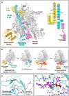
The SFTSV L protein transcription priming structure. (A) The SFTSV L protein (TRANSCRIPTION-PRIMING) is shown with key L protein domains coloured according to the following colour scheme: endonuclease (orange), cap-binding domain (cyan), and mid-link domain (light pink). RNAs are shown as sticks with surface overlaid (30% transparency) and coloured either yellow (5′ RNA), cyan (3′ RNA), or magenta (primer RNA). In addition, the RNAs present in each structure are shown schematically and labelled. (B) The conformational changes undergone by the L protein as it moves from transcription-priming to late-elongation are shown. L protein domains are coloured as in A. For clarity, RNA is not shown. (C) The position of the cap-binding domain (shown in cyan) blocking motif (composed of the Q1840 and R1843 sidechains) is shown sandwiched between the putative cap-binding residues (F1703 and Y1719) in the published LATE-ELONGATION structure (PDB:8asd, coloured dark blue) and in a retracted position in the TRANSCRIPTION-PRIMING structure published here (coloured light pink). (D) The cap-binding domain is shown with bound capped RNA. The cap-binding domain is coloured cyan, whereas the neighbouring mid-link domain is shown in light pink. The bound capped RNA is shown as sticks. Sidechains of key interacting amino acids, coloured according to the protein domain, are shown as sticks and labelled accordingly.
Figure 3.
Figure 3.
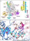
The transition from cap-snatching to early-elongation. (A) The SFTSV L (TRANSCRIPTION-EARLY-ELONGATION) is shown with key L protein domains coloured according to the following colour scheme: endonuclease (orange), cap-binding domain (cyan), and mid-link domain (light pink). RNAs are shown as sticks with surface overlaid (30% transparency) and coloured either yellow (5′ RNA), cyan (3′ RNA), or magenta (primer RNA). In addition, the RNAs present in each structure are shown schematically and labelled. (B) The active site in the TRANSCRIPTION-EARLY-ELONGATION structure is shown. Residues from the L protein are labelled and coloured according to the assigned domain (blue for fingers domain, and coral for the palm domain). RNAs are shown as sticks and coloured as in A.
Figure 4.
Figure 4.
Mutational analysis of capped RNA-interacting residues. RVFV mini-replicon data for L protein with mutations to capped RNA-interacting residues presenting luciferase reporter activity (in standardized relative light units relative to the wild-type L protein (WT)). Data were presented as mean values ± SD of three biological replicates (n = 3). All biological replicates are shown as black dots (top panel). Middle panels present Northern blotting results with signals for antigenomic viral RNA (AG, equal to cRNA), viral mRNA (mRNA) and 28 S ribosomal RNA (28 S) as a loading control, and the bottom panel shows Western blot detection of FLAG-tagged L proteins (L) to demonstrate general expressability of the mutants. Fast green staining of the membranes is included in the Supplementary data. The Western blots have not been used for quantification.
Figure 5.
Figure 5.
Structural insights into cap-independent primer elongation observed in vitro. The SFTSV L (TRANSCRIPTION-EARLY-ELONGATION (in vitro)) is shown with bound RNAs shown as sticks with surface overlaid (30% transparency) and coloured either yellow (5′ RNA), cyan (3′ RNA), magenta (primer RNA), and sea green (endonuclease-bound RNA). A zoomed-in view of the active site in the TRANSCRIPTION-EARLY-ELONGATION (in vitro) structure is also shown with residues from the SFTSV L protein labelled and coloured according to the assigned domain (blue for fingers domain, and coral for the palm domain).
Figure 6.
Figure 6.
Structure-based models of SFTSV and LACV transcription. L protein structures are displayed as surface with key domains coloured according to the following colour scheme: endonuclease (orange), cap-binding domain (cyan), mid-link domain (light pink), and C terminus (purple). In LACV, the C terminus is the zinc-binding domain (ZBD). The remaining protein is coloured grey. RNAs are represented by coloured solid/dashed lines and coloured either yellow (5′ RNA), cyan (3′ RNA), magenta (product/primer RNA). Key features are indicated. This process overview only includes states with known structural evidence for phenuiviruses and peribunyaviruses and therefore misses some steps such as primer-cleavage and termination.

References

    1. Casel M.A., Park S.J., Choi Y.K. Severe fever with thrombocytopenia syndrome virus: emerging novel phlebovirus and their control strategy. Exp. Mol. Med. 2021; 53:713–722. - PMC - PubMed
    1. Kim K.H., Yi J., Kim G., Choi S.J., Jun K.I., Kim N.H., Choe P.G., Kim N.J., Lee J.K., Oh M.D. Severe fever with thrombocytopenia syndrome, South Korea, 2012. Emerg. Infect. Dis. 2013; 19:1892–1894. - PMC - PubMed
    1. Takahashi T., Maeda K., Suzuki T., Ishido A., Shigeoka T., Tominaga T., Kamei T., Honda M., Ninomiya D., Sakai T. et al. . The first identification and retrospective study of Severe Fever with Thrombocytopenia Syndrome in Japan. J. Infect. Dis. 2014; 209:816–827. - PMC - PubMed
    1. Tran X.C., Yun Y., Van An L., Kim S.H., Thao N.T.P., Man P.K.C., Yoo J.R., Heo S.T., Cho N.H., Lee K.H. Endemic severe fever with thrombocytopenia syndrome, Vietnam. Emerg. Infect. Dis. 2019; 25:1029–1031. - PMC - PubMed
    1. Win A.M., Nguyen Y.T.H., Kim Y., Ha N.Y., Kang J.G., Kim H., San B., Kyaw O., Htike W.W., Choi D.O. et al. . Genotypic heterogeneity of Orientia tsutsugamushi in scrub typhus patients and thrombocytopenia syndrome co-infection, Myanmar. Emerg. Infect. Dis. 2020; 26:1878–1881. - PMC - PubMed

MeSH terms