. 2024 Jul:595:110096.
doi: 10.1016/j.virol.2024.110096. Epub 2024 Apr 30.

Commercial human 3D corneal epithelial equivalents for modeling epithelial infection in herpes keratitis

Affiliations

Commercial human 3D corneal epithelial equivalents for modeling epithelial infection in herpes keratitis

Tatiana Borodianskiy-Shteinberg et al. Virology. 2024 Jul.

Abstract

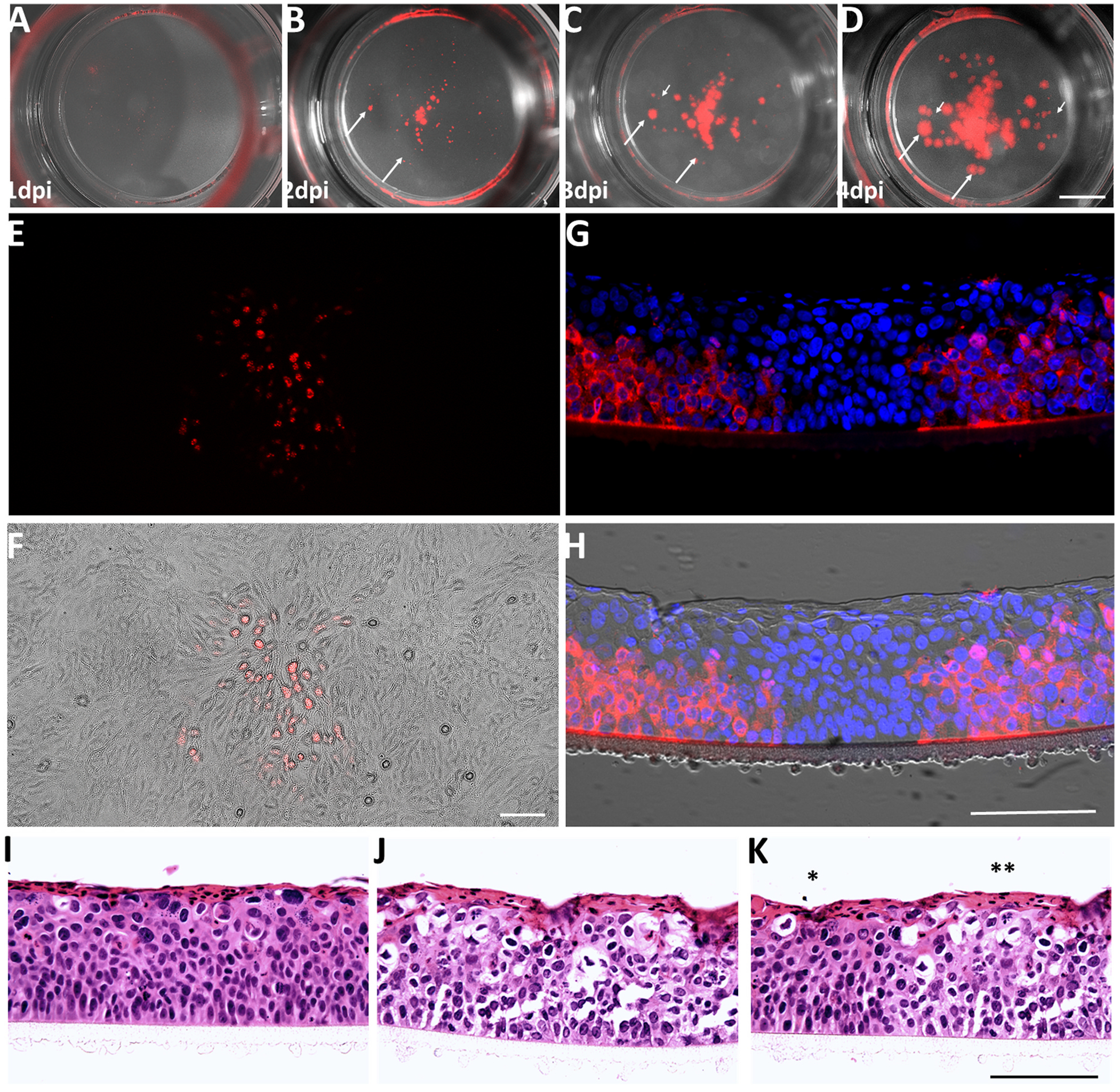
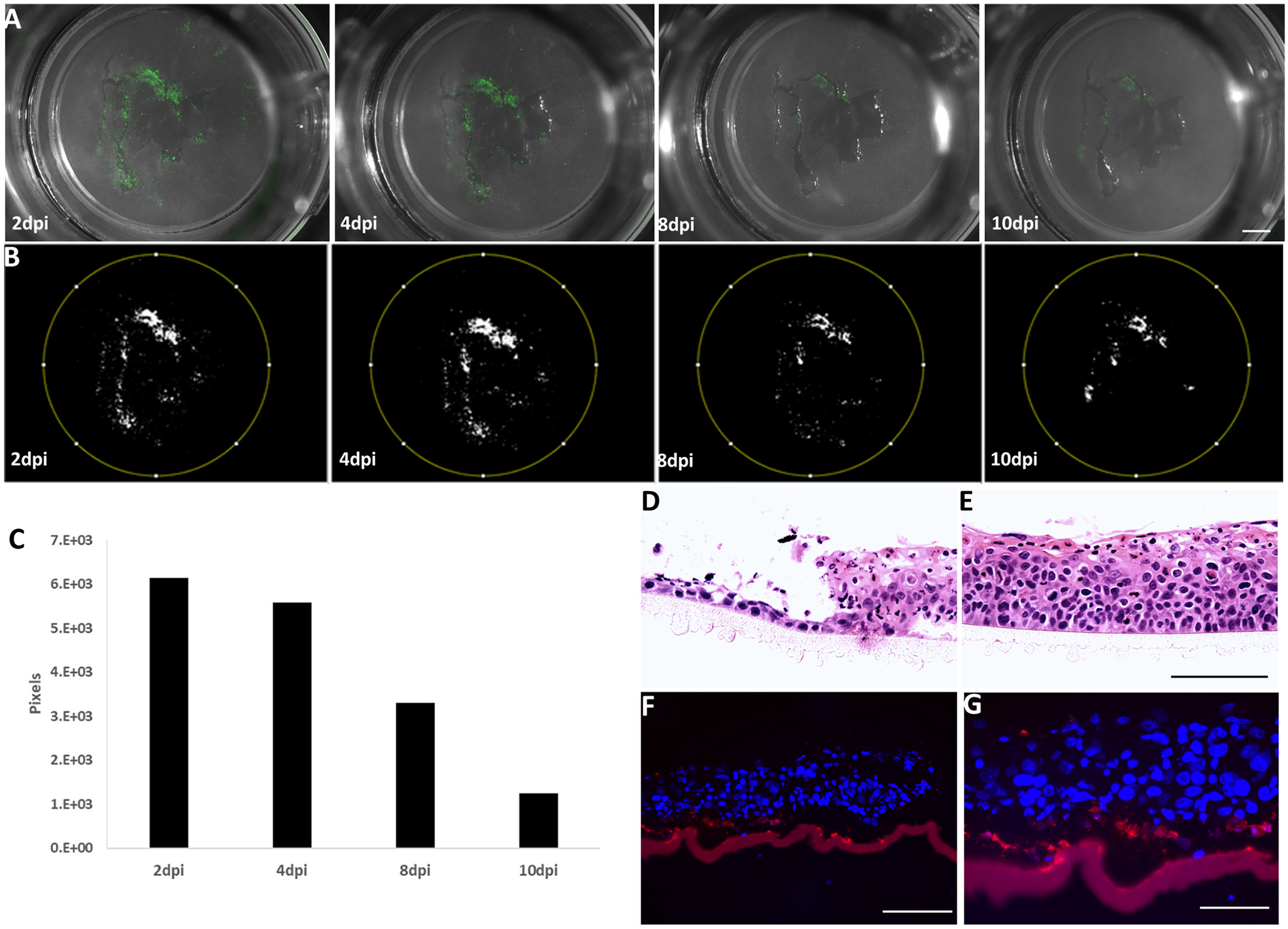
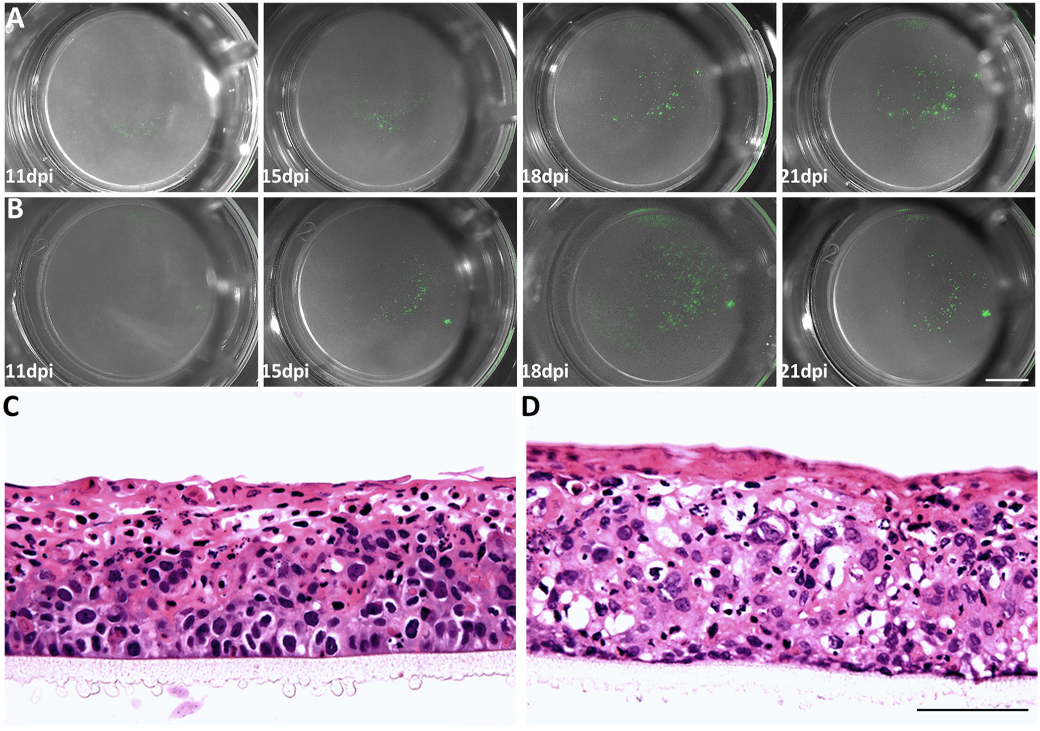
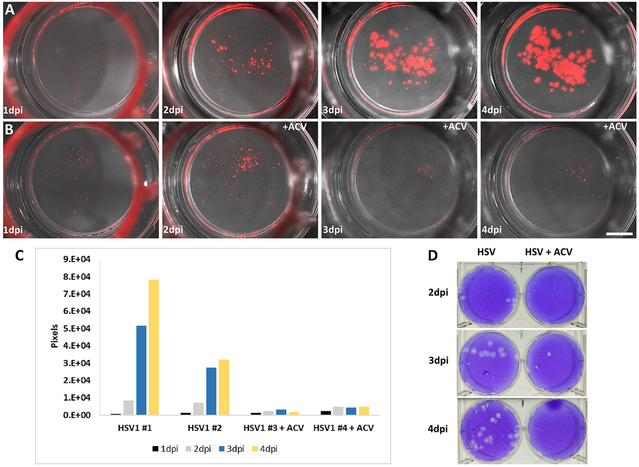
Herpes stromal keratitis is the leading cause of infectious blindness in the western world. Infection by HSV1 is most common, but VZV and hCMV also infect the cornea. Multiple models of HSV1 corneal infection exist, but none for VZV and hCMV because of their host specificity. Here, we used commercially available 3D human corneal epithelial equivalents (HCEE) to study infection by these herpesviruses. HCEE was infected by HSV-1 and hCMV without requiring scarification and resulted in spreading infections. Spread of HSV-1 infection was rapid, while that of hCMV was slow. In contrast, infections with VZV required damage to the HCEE and did not spread. Acyclovir dramatically reduced replication of HSV-1 in this model. We conclude that highly quality-controlled, readily available HCEE is a useful model to study human-restricted herpesvirus infection of the human corneal epithelium and for screening of antiviral drugs for treating HSK in an 3D model system.

Keywords: 3D culture; Drug testing; Herpes keratitis; Herpes zoster ophthalmicus; Herpesvirus.


Conflict of interest statement

Declaration of competing interest The authors declare that they have no known competing financial interests or personal relationships that could have appeared to influence the work reported in this paper.

Figures

Fig. 1.
Fig. 1.. HSV1 infects intact HCEE and releases infectious virus.
Panels A–D show micrographs of an HCEE treated with 104 PFU of HSV1 taken with a dissecting microscope on 1-4dpi. The infection is easily visualized by mCherry fluorescence (red). Images shown are merged white-light and fluorescence illumination photographs. Foci of infection apparent at 2dpi expand (arrows), and new foci appear on 3 and 4dpi (arrowheads). E-F shows HSV-1 infection of Vero cells by medium bathing infected HCEE for 24 h Scale bar = A-D 2 mm, 1 mm E&F = 100 μm (G&H) show the histology of HCEE infected with HSV1-mCherry Membrane inserts containing HSV1-infected HCEE 4dpi were cut out, fixed in paraformaldehyde and embedded in paraffin. (G) A photomicrograph of a section stained with anti-HSV1 ICP0 (red) and Hoechst nuclear stain (blue). H shows the same section with the merging of a bright-field image taken with Nomarski interference contrast optics showing the membrane and the cellular architecture of the section. Cells containing HSV1-ICP0 are present in the basal, but not superficial layers of the tissue despite the application of the infecting HSV1 to the superficial (“air”) surface. I–K hematoxylin and eosin-stained sections of HCEE 4dpi. I shows a section of a mock-infected HCEE with normal morphology and J&K of an infected HCEE with cytopathic changes including condensed nuclei and “holes” where pink-staining cytoplasm should be. In K, an asterisk indicates part of the section that was apparently not infected, and the two asterisks an infected area. Scale bars G-K = 100 μm.
Fig. 2.
Fig. 2.. VZV infects HCEE after scarification
Panels A-B show micrographs taken with a dissecting microscope of an HCEE treated with 3.2 × 10³ PFU of GFP-expressing VZV “debris” (see text) on 2,4,8 and 10 dpi. The infection tracks the damage to the epithelium. C shows the micrographs from A-B after thresholding for quantification. A graph of the number of fluorescent pixels counted on 2,4,8, and 10dpi shows that the GFP-expression as a proxy for infection is reduced over time, in contrast to the rapid spread of HSV1 infection of HCEE. Scale bar in A&B = 1 mm. Hematoxylin and eosin staining of sections from scarified and not-scarified HCEE 2dpi with VZV are shown in D and E respectively. Staining with a mix of anti-VZV protein antibodies (red) counterstained with Hoechst (blue) is shown in F&G. VZV proteins are present primarily in the basal layers, and the membrane upon which the epithelium was grown is artifactually autofluorescent and easily distinguished in the brightfield images. Scale bars D&E 100 μm, E&F 50 μm.
Fig. 3.
Fig. 3.. CMV infection of HCEE
Panels A-B show a series of micrographs taken in a dissecting microscope of two HCEE infected with CMV-GFP on 11–21 dpi (see text). CMV infects the epithelia without scarification and spreads at a much slower pace than the infection by HSV1. C&D Histology of mock infected HCEE 24 div and infected HCEE 24div/21dpi shows that although the tissue looks less “healthy” the normal stratification of the corneal remains. The infected tissue shows a distinct cytopathic effect. Scale bars: A&B – 2 mm, C&D - 50 μm.
Fig. 4.
Fig. 4.. HCEE can be used for rapid testing of anti-herpesvirus drugs.
2A&B are series of fluorescent images of HCEE from 1-4dpi. The images in A are from a control infected but untreated HCEE, and those in B from an HCEE that was bathed in 200 μM ACV from 2dpi. C is a graph representing the area of fluorescence in the epithelia over time showing the strong anti-viral effect of ACV treatment. D is an example of a plaque assay in wells of a 24 well plate made using the medium collected from infected (HSV column) and ACV-treated (HSV + ACV column) HCEE taken at 2,3, and 4dpi. The antiviral effect of the ACV is easily visualized and quantified both by image processing and plaque assays. Scale bar in A&B = 2 mm.

Similar articles

References

    1. Alépée N, Adriaens E, Abo T, Bagley D, Desprez B, Hibatallah J, Mewes K, Pfannenbecker U, Sala À, Van Rompay AR, Verstraelen S, McNamee P, 2019. Development of a defined approach for eye irritation or serious eye damage for neat liquids based on cosmetics Europe analysis of in vitro RhCE and BCOP test methods. Toxicol. Vitro 59, 100–114. 10.1016/j.tiv.2019.04.011. - DOI - PubMed
    1. Avivi C, Goldstein RS, 1999. Differential expression of Islet-1 in neural crest derived ganglia: islet-1+ dorsal root ganglion cells are post-mitotic and Islet-1+ sympathetic ganglion cells are still cycling. Dev. Brain Res 115, 89–92. - PubMed
    1. Azher TN, Yin X-T, Stuart PM, 2017. Understanding the role of chemokines and cytokines in experimental models of herpes simplex keratitis. J. Immunol. Res 2017, 7261980 10.1155/2017/7261980. - DOI - PMC - PubMed
    1. Bacon TH, Levin MJ, Leary JJ, Sarisky RT, Sutton D, 2003. Herpes simplex virus resistance to acyclovir and penciclovir after two decades of antiviral therapy. Clin. Microbiol. Rev 16, 114–128. 10.1128/CMR.16.1.114-128.2003. - DOI - PMC - PubMed
    1. Bleymehl K, Cinatl J, Schmidt-Chanasit J, 2011. Phenotypic and genetic characterization of varicella-zoster virus mutants resistant to acyclovir, brivudine and/or foscarnet. Med. Microbiol. Immunol 200, 193–202. 10.1007/s00430-011-0191-4. - DOI - PubMed

Publication types

MeSH terms

LinkOut - more resources