. 2024 Jul 22;33(15):1339-1355.
doi: 10.1093/hmg/ddae066.

Benfotiamine improves dystrophic pathology and exercise capacity in mdx mice by reducing inflammation and fibrosis

Affiliations

Benfotiamine improves dystrophic pathology and exercise capacity in mdx mice by reducing inflammation and fibrosis

Chantal A Coles et al. Hum Mol Genet. .

Abstract

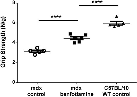
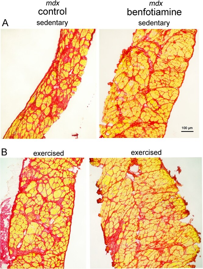
Duchenne Muscular Dystrophy (DMD) is a progressive and fatal neuromuscular disease. Cycles of myofibre degeneration and regeneration are hallmarks of the disease where immune cells infiltrate to repair damaged skeletal muscle. Benfotiamine is a lipid soluble precursor to thiamine, shown clinically to reduce inflammation in diabetic related complications. We assessed whether benfotiamine administration could reduce inflammation related dystrophic pathology. Benfotiamine (10 mg/kg/day) was fed to male mdx mice (n = 7) for 15 weeks from 4 weeks of age. Treated mice had an increased growth weight (5-7 weeks) and myofibre size at treatment completion. Markers of dystrophic pathology (area of damaged necrotic tissue, central nuclei) were reduced in benfotiamine mdx quadriceps. Grip strength was increased and improved exercise capacity was found in mdx treated with benfotiamine for 12 weeks, before being placed into individual cages and allowed access to an exercise wheel for 3 weeks. Global gene expression profiling (RNAseq) in the gastrocnemius revealed benfotiamine regulated signalling pathways relevant to dystrophic pathology (Inflammatory Response, Myogenesis) and fibrotic gene markers (Col1a1, Col1a2, Col4a5, Col5a2, Col6a2, Col6a2, Col6a3, Lum) towards wildtype levels. In addition, we observed a reduction in gene expression of inflammatory gene markers in the quadriceps (Emr1, Cd163, Cd4, Cd8, Ifng). Overall, these data suggest that benfotiamine reduces dystrophic pathology by acting on inflammatory and fibrotic gene markers and signalling pathways. Given benfotiamine's excellent safety profile and current clinical use, it could be used in combination with glucocorticoids to treat DMD patients.

Keywords: Duchenne; benfotiamine; inflammation; muscular dystrophy.


Figures

Figure 1
Figure 1
Timeline for mouse cohorts and growth data showing Benfotiamine increases mdx body weight and growth rate. (A) Schematic of timeline for three mouse cohorts used in study (B) sedentary from two independent experiments were weighed weekly for 15 weeks of benfotiamine treatment. Benfotiamine mdx mice weighed more than the mdx control from 5–7 weeks of age, then from 17–19 weeks of age. (C) The growth rate (grams per week) during the early mdx “growth” phase (4–8 weeks) was greater in benfotiamine mdx. (D) No difference in growth rate was found during the “adult” growth phase (9–15 weeks). The graphs show mean ± SEM. *indicates P < 0.05 (benfotiamine n = 13 compared to mdx control n = 11: Mice are from cohort 1 and cohort 3 pooled).
Figure 2
Figure 2
Benfotiamine increases myofibre size in quadriceps muscle. The quadriceps muscle from mdx control (A) and benfotiamine treated mdx (B) mice were stained with DAPI (blue) and laminin α2 antibody (red) to measure myofibre size (Feret’s minimum diameter). (C) Mean myofibre size in quadricep muscles was similar in untreated and benfotiamine treated mdx. Although no difference in mean myofiber size for minimum feret’s diameter, the coefficient of variation (% VC) was much higher in mdx (% VC 11.76%) compared with benfotiamine (% VC 4.35%) treated mice. (D) Frequency histogram showing there was a greater proportion of larger myofibres (70–90 μm diameter) and a reduced proportion of smaller myofibres (20–40 μm diameter) in benfotiamine mdx (D). *P < 0.05, **P < 0.001, n = 6. Scale bar, 200 μm. mdx control n = 6 mdx benfotiamine n = 7.
Figure 3
Figure 3
Benfotiamine reduces muscle damage and improves sarcolemma stability in mdx mice. Basal lamina (anti-laminin-α2 in red) and IgG (Alexa Fluor 488 in green) staining of transverse muscle sections were used to observe muscle integrity and damage respectively in mdx (A) and benfotiamine mdx (B) quadriceps. Nuclei are stained with DAPI (blue). Muscle architecture was visualised with hematoxylin and eosin in quadriceps of mdx (C) and benfotiamine mdx (D). (E) The percentage of damaged myofibres permeable to IgG in the quadriceps was not significantly reduced in benfotiamine mdx (P = 0.055). (F) Benfotiamine reduced the area of damage (including areas of necrosis and inflammatory cell infiltration compared to mdx control (P < 0.05). Muscle-specific creatine kinase in the serum, a marker of sarcolemmal damage, was not changed with benfotiamine treatment (G) (P = 0.073). (H) Benfotiamine reduced the proportion of fibres in mdx quadriceps muscle with central nucleation (P < 0.05). *P < 0.05, mdx control n = 6 mdx benfotiamine n = 7. Scale bar, 200 μM.
Figure 4
Figure 4
Benfotiamine increases grip strength in mdx mice. At 16 weeks of age, grip strength in benfotiamine treated mdx mice was improved towards wildtype (C57BL/10) levels. Grip strength was normalised to bodyweight. Graph shows mean ± SEM ****P < 0.0001 mdx control n = 5 mdx benfotiamine n = 6 Wildtype (WT) C557BL/10 n = 5.
Figure 5
Figure 5
Benfotiamine improves voluntary exercise. Voluntary exercise was assessed in control mdx (n = 5) and benfotiamine mdx (n = 5) mice. Mouse were housed individually with access to an exercise wheel for 3 weeks (age: 16–19 weeks). A number of parameters were recorded; mean daily distance, mean run time, mean bout distance, mean run rate. Benfotiamine treated mice ran further each day (P < 0.05) (A), ran for a longer period of time (P < 0.01) (B), covered a greater distance each time they ran (P < 0.05) (C) and ran at a faster rate (P < 0.05) (D) than control mdx. The graphs show mean ± SEM. *P < 0.05 **P < 0.01, mdx (n = 5) and benfotiamine mdx (n = 5).
Figure 6
Figure 6
Benfotiamine down-regulates gene expression of pro-inflammatory markers in the quadriceps of mdx mice. Reduced gene expression by RT-qPCR was found in treated mdx for the pan-macrophage marker Emr-1 (F480) (P < 0.05) (A), M2 macrophage marker (Cd163) (P = 0.05) (B), T lymphocyte markers Cd4 (P < 0.05) (C), and Cd8 (P < 0.01) (D). Ifng is a pro-inflammatory cytokine released from macrophages and naïve CD4 T cells (during Th2 differentiation), it’s expression was also downregulated with benfotiamine mdx (E) along with Postn (F) (P < 0.05), an extracellular matrix protein and marker of inflammation and fibrosis. Graphs show mean ± SEM. *Indicates P < 0.05 **P < 0.01. mdx (n = 6) and benfotiamine mdx (n = 7).
Figure 7
Figure 7
Heatmap of genes differentially expressed in mdx benfotiamine treated and mdx mice in the gastrocnemius muscle. Expression of differentially expressed (DE) genes (adj. P.Value < 0.05) was normalised across rows. The expression of DE genes in benfotiamine treated mdx shifts towards wildtype (WT) expression levels.
Figure 8
Figure 8
Gene set enrichment analysis (GSEA) shows benfotiamine reduces expression of inflammatory response genes in the gastrocnemius muscle of mdx. Barcode plots of the inflammatory response gene set in mdx vs WT show enrichment for upregulated genes (A). When mdx are treated with benfotiamine genes in the inflammatory response gene set are down regulated (B). (C) The top 20 genes ranked by t statistic, representing the most significantly upregulated inflammatory response genes mdx vs WT. (D) The top 20 genes ranked by t-statistic, representing the most significantly downregulated inflammatory response genes. *Genes present in the top 20 in both comparisons.
Figure 9
Figure 9
Gene set enrichment analysis shows benfotiamine impacts the myogenesis gene set in mdx gastrocnemius muscle. Barcode plots show the myogenesis gene set is upregulated in mdx vs WT (A) and down regulated in mdx treated with benfotiamine (B). The top 20 upregulated Hallmark myogenesis genes ranked by t statistic in mdx vs WT (C) and the top 20 downregulated myogenesis genes in benfotiamine treated mdx (D). *Genes present in the top 20 in both comparisons.
Figure 10
Figure 10
Benfotiamine treated mdx have reduced gene expression (RNAseq CPM) of extracellular matrix fibrosis markers and utrophin in the gastrocnemius muscle. Reduced gene expression of extracellular matrix proteins, in particular isoforms of collagen fibrils (Col1a1 (A), Col1a2 (B), Col4a5 (C), Col5a2 (D), Col6a2 (E), Col6a3(F)) and the small leucine-rich (SLRP) proteoglycan Lum (G) were found in treated mdx showing expression more towards wildtype levels (P < 0.05). Utrophin is overexpressed in mdx mice compensating for loss of dystrophin to stabilise muscle membranes from contraction induced injury, benfotamine treated mdx have reduced expression of Utrn (P < 0.05). Graphs show mean ± SEM. *Indicates P < 0.05 mdx (n = 4) and benfotiamine mdx (n = 4) CPM = counts per million.
Figure 11
Figure 11
Benfotiamine treated mdx show reduced fibrosis in the diaphragm. Picrosirius red stains collagen (red) in diaphragm of sedentary and exercised mdx mice, myofibres appear yellow. Increased accumulation of collagen (red) is observed in mdx mice (sedentary/exercised) compared to benfotiamine treated mdx. No difference is apparent between sedentary and exercised mice. Scale bar 100 μm.

References

    1. Mendell JR, Shilling C, Leslie ND. et al. Evidence-based path to newborn screening for Duchenne muscular dystrophy. Ann Neurol. 2012;71:304–313. - PubMed
    1. Hoffman EP, Brown RH Jr, Kunkel LM. Dystrophin: the protein product of the Duchenne muscular dystrophy locus. Cell 1987;51:919–928. - PubMed
    1. Campbell KP, Kahl SD. Association of dystrophin and an integral membrane glycoprotein. Nature 1989;338:259–262. - PubMed
    1. Ohlendieck K, Ervasti JM, Snook JB. et al. Dystrophin-glycoprotein complex is highly enriched in isolated skeletal muscle sarcolemma. J Cell Biol 1991;112:135–148. - PMC - PubMed
    1. Prins KW, Humston JL, Mehta A. et al. Dystrophin is a microtubule-associated protein. J Cell Biol 2009;186:363–369. - PMC - PubMed

Publication types

MeSH terms