. 2024 May 7;10(1):48.
doi: 10.1038/s41421-024-00679-8.

Mechanisms of ligand recognition and activation of melanin-concentrating hormone receptors

Affiliations

Mechanisms of ligand recognition and activation of melanin-concentrating hormone receptors

Qian He et al. Cell Discov. .

Abstract

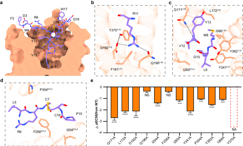
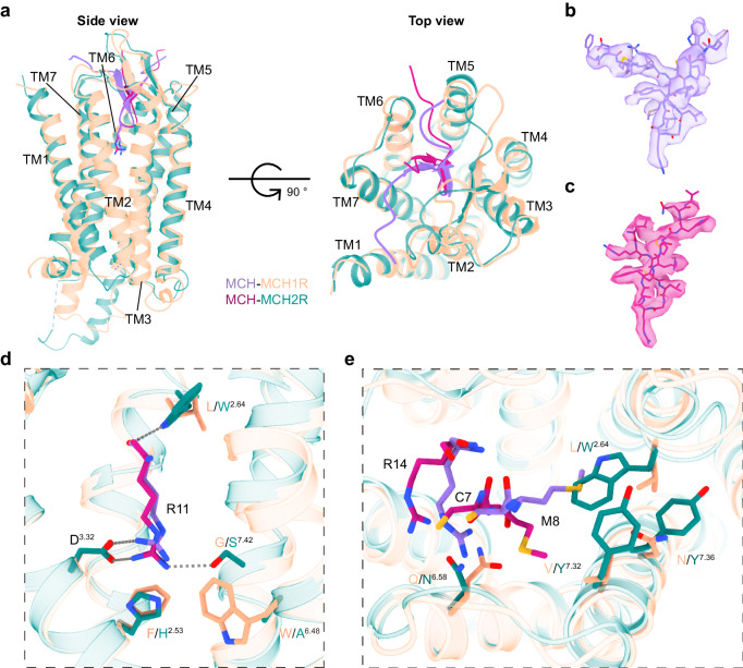
Melanin-concentrating hormone (MCH) is a cyclic neuropeptide that regulates food intake, energy balance, and other physiological functions by stimulating MCHR1 and MCHR2 receptors, both of which are class A G protein-coupled receptors. MCHR1 predominately couples to inhibitory G protein, Gi/o, and MCHR2 can only couple to Gq/11. Here we present cryo-electron microscopy structures of MCH-activated MCHR1 with Gi and MCH-activated MCHR2 with Gq at the global resolutions of 3.01 Å and 2.40 Å, respectively. These structures reveal that MCH adopts a consistent cysteine-mediated hairpin loop configuration when bound to both receptors. A central arginine from the LGRVY core motif between the two cysteines of MCH penetrates deeply into the transmembrane pocket, triggering receptor activation. Integrated with mutational and functional insights, our findings elucidate the molecular underpinnings of ligand recognition and MCH receptor activation and offer a structural foundation for targeted drug design.


Conflict of interest statement

The authors declare no competing interests.

Figures

Fig. 1
Fig. 1. Structural representations of the MCH‒MCHR1‒Gi and MCH‒MCHR2‒Gq complexes.
a Depiction of the MCH sequence and a schematic representation illustrating the G-protein coupling of MCHR1 and MCHR2. b, c Cryo-EM density map (b) and the corresponding molecular model (c) of the MCH‒MCHR1‒Gi complex. d, e Cryo-EM density map (d) and the corresponding molecular model (e) of the MCH‒MCHR2‒Gq complex.
Fig. 2
Fig. 2. Molecular basis of MCH recognition by MCHR1.
a Cross-section of the MCH-binding pocket in MCHR1. bd Detailed molecular interactions between MCH (depicted in medium purple) and MCHR1 (shown in light salmon). Hydrogen bonds are presented with gray dashed lines. e Functional implications of mutations within the MCHR1 binding pocket, represented as ΔpEC50 values. ΔpEC50 represents the difference in pEC50 values between the WT and the mutants of MCHR1. Data from three independent experiments, each of which was performed in triplicate, are presented as mean ± SEM. Statistical differences were determined by two-sided one-way ANOVA with Tukey’s test compared with WT. NA no activity; NS no significant difference; *P < 0.05; **P < 0.01; ***P < 0.001; ****P < 0.0001.
Fig. 3
Fig. 3. Molecular basis of MCH recognition by MCHR2.
a Cross-section of the MCH-binding pocket in MCHR2. bf Detailed molecular interactions between MCH (depicted in medium violet red) and MCHR2 (shown in teal). Hydrogen bonds are highlighted with gray dashed lines. g Functional data derived from alanine mutations of residues within the MCHR2 binding pocket, represented as ΔpEC50 values. ΔpEC50 represents the difference in pEC50 values between the WT and the mutants of MCHR2. Data are presented as mean ± SEM. from three independent experiments performed in triplicate and analyzed by two-sided one-way ANOVA with Tukey’s test compared with WT. NA no activity; *P < 0.05; **P < 0.01; ***P < 0.001; ****P < 0.0001.
Fig. 4
Fig. 4. Conformational differences of MCH recognition by MCHR1 and MCHR2.
a Structural superposition of MCH-bound MCHRs, presented from both side-view (the left) and top view (the right). b, c Mesh representations of MCH within the binding pockets of MCHR1 and MCHR2. d, e The differential interactions between MCH and MCHRs, focusing on R11 (d) and the C7, M8, and R14 residues of MCH (e).
Fig. 5
Fig. 5. Conformational changes in MCHR1 and MCHR2 upon activation.
a, b Structural superposition between cryo-EM structures of MCH-bound MCHR complexes and the AlphaFold2-predicted inactive models from the GPCR databank is shown from the side view and the cytoplasmic view. c Structural alignment highlighting the toggle switch mechanism. d Depiction of shifts in H/F2.53, D3.32, and Y7.43 at the base of the binding pocket. eg Illustration of the conserved motifs in MCH-activated MCHRs: DRY motif (e); NPxxY motif (f); PIF motif (g).
Fig. 6
Fig. 6. G-protein coupling of MCHR1 and MCHR2.
a Structural alignment between MCHR1‒Gi and MCHR2‒Gq complexes. b, c Comparative analysis of the Gα conformation in the α5 (b) and αN (c) domains. d Key residues in MCHR1 and MCHR2 that interact with the downstream Gαi or Gαq proteins. e Detailed interactions between the ICL2 of MCHR1 and MCHR2 with the αN domain of Gαi or Gαq. f IP-one assay data for wild-type and T22134.54R of MCHR1, with data presented as mean values ± SEM from three independent experiments performed in triplicate.
Fig. 7
Fig. 7. Distinct binding modes of cyclic peptides in receptors.
a Binding conformation of MCH in MCHR1. b Binding conformation of MCH in MCHR2. c Binding conformation of SST14 in SSTR2 (PDB: 7Y27). d Binding conformation of AVP in V2R (PDB: 7DW9).

References

    1. Kawauchi H, Kawazoe I, Tsubokawa M, Kishida M, Baker BI. Characterization of melanin-concentrating hormone in chum salmon pituitaries. Nature. 1983;305:321–323. doi: 10.1038/305321a0. - DOI - PubMed
    1. Skofitsch G, Jacobowitz DM, Zamir N. Immunohistochemical localization of a melanin concentrating hormone-like peptide in the rat brain. Brain Res. Bull. 1985;15:635–649. doi: 10.1016/0361-9230(85)90213-8. - DOI - PubMed
    1. Vaughan JM, Fischer WH, Hoeger C, Rivier J, Vale W. Characterization of melanin-concentrating hormone from rat hypothalamus. Endocrinology. 1989;125:1660–1665. doi: 10.1210/endo-125-3-1660. - DOI - PubMed
    1. Bittencourt JC, et al. The melanin-concentrating hormone system of the rat brain: an immuno- and hybridization histochemical characterization. J. Comp. Neurol. 1992;319:218–245. doi: 10.1002/cne.903190204. - DOI - PubMed
    1. Saito Y, Nothacker HP, Civelli O. Melanin-concentrating hormone receptor: an orphan receptor fits the key. Trends Endocrinol. Metab. 2000;11:299–303. doi: 10.1016/S1043-2760(00)00290-3. - DOI - PubMed