. 2024 May 6;14(1):10361.
doi: 10.1038/s41598-024-60646-x.

A Japanese herbal medicine (kampo), hochuekkito (TJ-41), has anti-inflammatory effects on the chronic obstructive pulmonary disease mouse model

Affiliations

A Japanese herbal medicine (kampo), hochuekkito (TJ-41), has anti-inflammatory effects on the chronic obstructive pulmonary disease mouse model

Masaaki Yuki et al. Sci Rep. .

Abstract

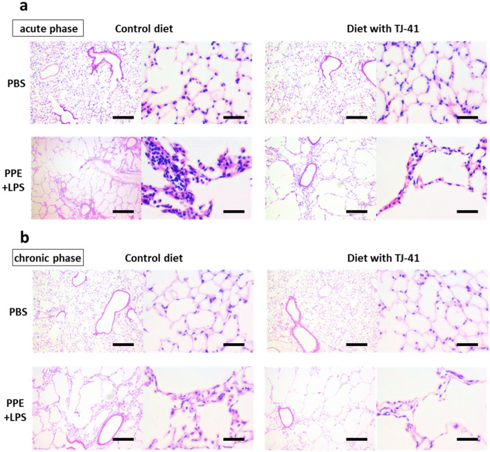
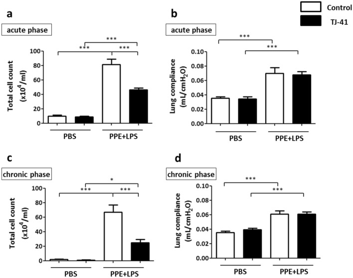
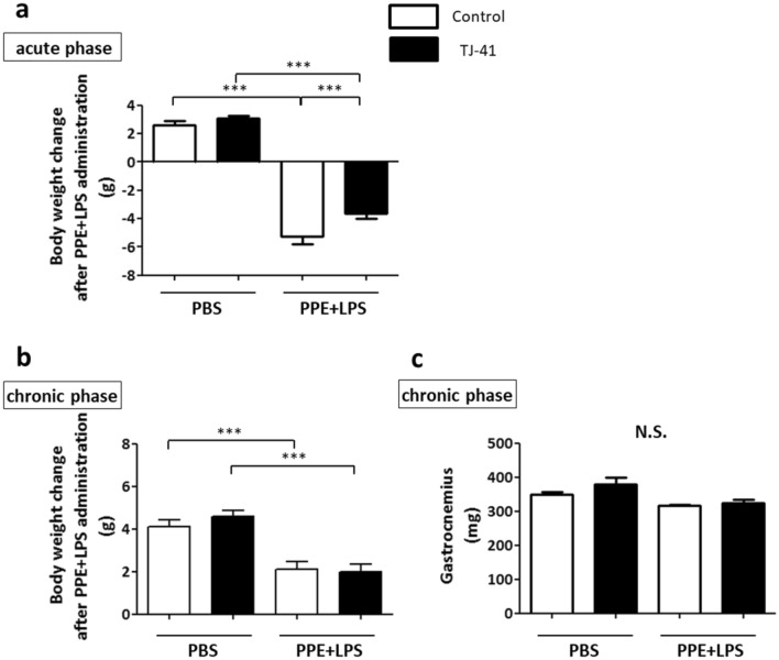
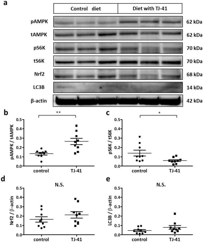
Chronic obstructive pulmonary disease (COPD) is a progressive disease that is characterized by chronic airway inflammation. A Japanese herbal medicine, hochuekkito (TJ-41), is prominently used for chronic inflammatory diseases in Japan. This study aimed to analyze the anti-inflammatory effect of TJ-41 in vivo and its underlying mechanisms. We created a COPD mouse model using intratracheal administration of porcine pancreatic elastase and lipopolysaccharide (LPS) and analyzed them with and without TJ-41 administration. A TJ-41-containing diet reduced inflammatory cell infiltration of the lungs in the acute and chronic phases and body weight loss in the acute phase. In vitro experiments revealed that TJ-41 treatment suppressed the LPS-induced inflammatory cytokines in BEAS-2B cells. Furthermore, TJ-41 administration activated the AMP-activated protein kinase (AMPK) pathway and inhibited the mechanistic target of the rapamycin (mTOR) pathway, both in cellular and mouse experiments. We concluded that TJ-41 administration reduced airway inflammation in the COPD mouse model, which might be regulated by the activated AMPK pathway, and inhibited the mTOR pathway.


Conflict of interest statement

AM, HI, and HT received research funding from Tsumura & Co. (Tokyo, Japan). TJ has received financial contributions from Tsumura & Co. because he has academic affiliations with the Department of Health Services Research, Graduate School of Medicine, and The University of Tokyo supported by Tsumura & Co. The authors declare no other competing interests in this work.

Figures

Figure 1
Figure 1
Hematoxylin–eosin staining of the lungs of COPD mice models with or without TJ-41 treatment in the acute phase (a) and the chronic phase (b). Magnification in the left image, × 40; magnification in the right image, × 200. The scale bar showed 250 µm in the left image and 50 µm in the right image.
Figure 2
Figure 2
BALF analysis in the acute (a) and chronic (c) phases revealed that TJ-41 administration mitigated the PPE- and LPS-induced increase in pulmonary inflammatory cells; N = 3–5 per group. (b and d) Lung compliance was increased by PPE and LPS administration in both phases. One-way analysis of variance and Tukey’s multiple comparison tests with *P < 0.05, **P < 0.01, and ***P < 0.001 were considered statistically significant; N = 7–9 in each group.
Figure 3
Figure 3
(a and b) The change in body weight at day 24 and day 49 in the acute (a) and chronic phases (b), respectively. (c) The weight of the gastrocnemius muscle at day 49; N = 7–9 in each group. One-way analysis of variance and Tukey’s multiple comparison tests with *P < 0.05 and ***P < 0.001 were considered statistically significant. N.S.: Not significant.
Figure 4
Figure 4
mRNA expressions of inflammatory cytokines, IL-6 (a), IL-8 (b), and TNF-α (c), in BEAS-2B cells after LPS treatment with or without TJ-41 administration. One-way analysis of variance and Tukey’s multiple comparison tests were performed in the three groups treated with LPS, and *P < 0.05 and ***P < 0.001 were considered statistically significant; N = 10 in each group. NC: negative control; N.S.: Not significant.
Figure 5
Figure 5
(ac) Western blotting analysis of BEAS-2B cells from 5 to 60 min after TJ-41 treatment. (df) Long-time analysis from 1 to 6 h of TJ-41-treated BEAS-2B cells. Unpaired t-tests were used for each antibody, and *P < 0.05, **P < 0.01, and ***P < 0.001 were considered statistically significant. N = 5 in each group.
Figure 6
Figure 6
(ae) Western blotting analysis using the lung tissues from wild-type mice that had been fed a TJ-41-containing diet. AMPK activity (b), mTOR activity (c), Nrf2 expression (d), and LC3B expression (e) were measured. Unpaired t-tests with **P < 0.01 were considered statistically significant for each antibody. N = 6 in each group. N.S.: not significant.
Figure 7
Figure 7
RNA sequencing analysis. (a) MA plot showing the RNA expression profile between control mice and COPD mice model. (b) The top 10 enriched biological process gene ontology terms are sorted by Enrichr combined score. (c) MA plot showing the RNA expression profile between the presence and absence of TJ-41 administration in the control mice. (d) The top 10 enriched biological process gene ontology terms. CPM: Counts per million; DEG: Differential expression gene; FC: Fold change; FDR: False discovery rate; GO: Gene ontology.
Figure 8
Figure 8
Study design for a COPD mice model. COPD mice model were administered PPE at 10 weeks of age (day 0), and they were treated with LPS three times every 3 days starting at 12 weeks of age (day 14). The 14-week-old mice (day 28) were considered an acute phase model of COPD exacerbation, whereas the 17-week-old mice (day 49) were considered chronic phase models of COPD.

References

    1. Celli B, et al. Definition and nomenclature of chronic obstructive pulmonary disease: time for its revision. Am. J. Respir. Crit. Care Med. 2022;206:1317–1325. doi: 10.1164/rccm.202204-0671PP. - DOI - PMC - PubMed
    1. Su B, et al. Inflammatory markers and the risk of chronic obstructive pulmonary disease: a systematic review and meta-analysis. PLoS ONE. 2016;11:e0150586. doi: 10.1371/journal.pone.0150586. - DOI - PMC - PubMed
    1. Wagner PD. Possible mechanisms underlying the development of cachexia in COPD. Eur. Respir. J. 2008;31:492–501. doi: 10.1183/09031936.00074807. - DOI - PubMed
    1. Gan WQ, Man SF, Senthilselvan A, Sin DD. Association between chronic obstructive pulmonary disease and systemic inflammation: a systematic review and a meta-analysis. Thorax. 2004;59:574–580. doi: 10.1136/thx.2003.019588. - DOI - PMC - PubMed
    1. Jones SE, et al. Sarcopenia in COPD: prevalence, clinical correlates and response to pulmonary rehabilitation. Thorax. 2015;70:213–218. doi: 10.1136/thoraxjnl-2014-206440. - DOI - PubMed

Publication types

MeSH terms