Comparative Study
. 2024 May 7;24(1):367.
doi: 10.1186/s12870-024-05033-4.

Elucidating the callus-to-shoot-forming mechanism in Capsicum annuum 'Dempsey' through comparative transcriptome analyses

Affiliations
Comparative Study

Elucidating the callus-to-shoot-forming mechanism in Capsicum annuum 'Dempsey' through comparative transcriptome analyses

Sang-Yun Han et al. BMC Plant Biol. .

Abstract

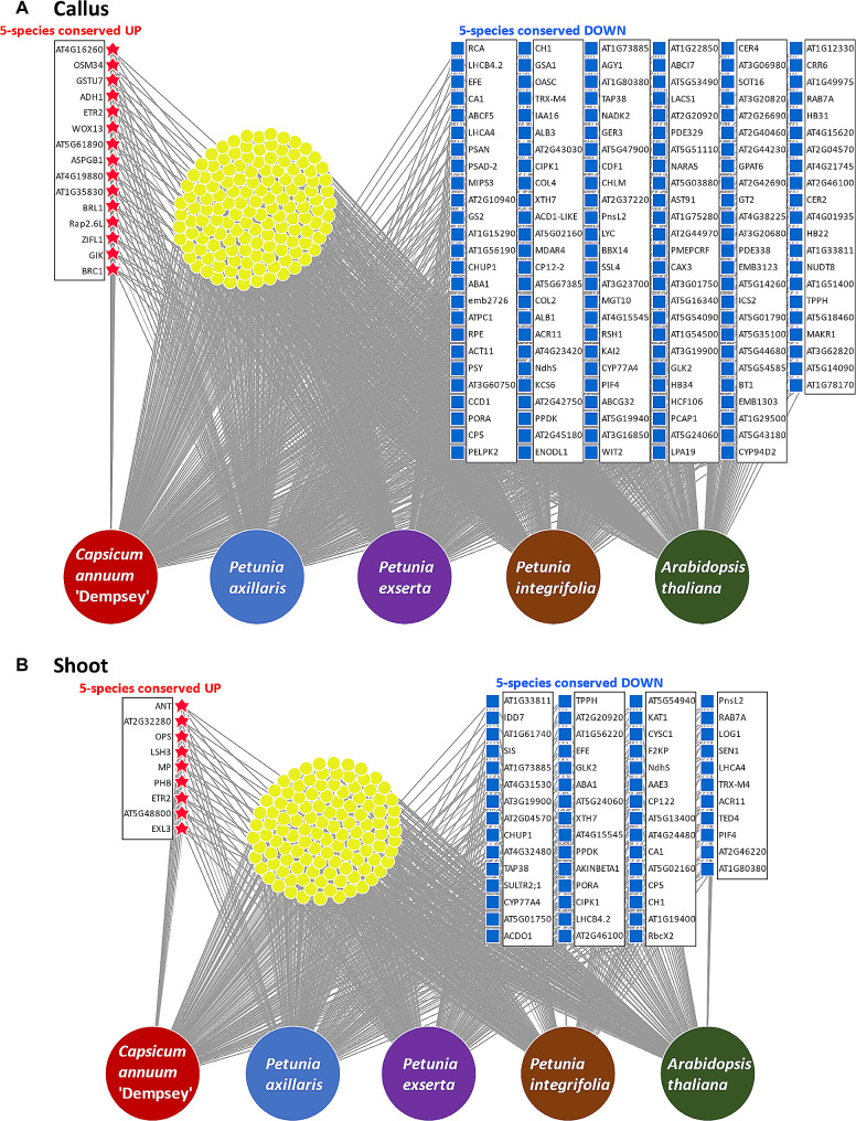
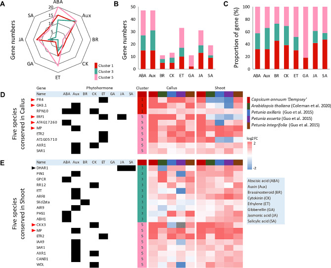
Background: The formation of shoots plays a pivotal role in plant organogenesis and productivity. Despite its significance, the underlying molecular mechanism of de novo regeneration has not been extensively elucidated in Capsicum annuum 'Dempsey', a bell pepper cultivar. To address this, we performed a comparative transcriptome analysis focusing on the differential expression in C. annuum 'Dempsey' shoot, callus, and leaf tissue. We further investigated phytohormone-related biological processes and their interacting genes in the C. annuum 'Dempsey' transcriptome based on comparative transcriptomic analysis across five species.

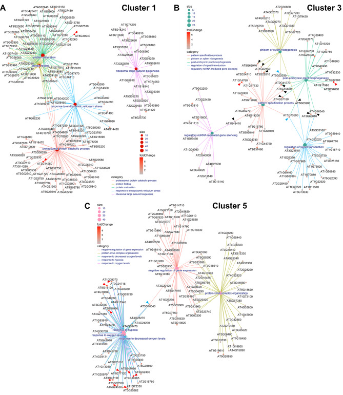
Results: We provided a comprehensive view of the gene networks regulating shoot formation on the callus, revealing a strong involvement of hypoxia responses and oxidative stress. Our comparative transcriptome analysis revealed a significant conservation in the increase of gene expression patterns related to auxin and defense mechanisms in both callus and shoot tissues. Consequently, hypoxia response and defense mechanism emerged as critical regulators in callus and shoot formation in C. annuum 'Dempsey'. Current transcriptome data also indicated a substantial decline in gene expression linked to photosynthesis within regenerative tissues, implying a deactivation of the regulatory system governing photosynthesis in C. annuum 'Dempsey'.

Conclusion: Coupled with defense mechanisms, we thus considered spatial redistribution of auxin to play a critical role in the shoot morphogenesis via primordia outgrowth. Our findings shed light on shoot formation mechanisms in C. annuum 'Dempsey' explants, important information for regeneration programs, and have broader implications for precise molecular breeding in recalcitrant crops.

Keywords: Capsicum annuum; Auxin redistribution; Bell pepper ‘Dempsey’; Defense mechanism; Hypoxia; Regeneration; Shoot formation; Transcriptome.


Conflict of interest statement

The authors declare no competing interests.

Figures

Fig. 1
Fig. 1
Analysis of DEGs from callus and shoot tissue in Capsicum annuum ‘Dempsey’. (A) Example images with the scale bar of samples from leaf (WT), callus, and shoot tissues used for RNA-seq analysis: ‘Dempsey’ leaf WT (left), leaf-derived callus tissue (middle), callus-derived emerging shoot tissue (right). (B) Volcano plots depicting the DEGs of callus versus WT (left) and shoot versus WT (right) comparisons. (C) Principal component analysis (PCA) plot of the TMM-normalized counts of the RNA-seq samples. (D) Correlation matrix plot (Corrplot) showing Pearson’s correlation efficient of RNA-seq samples. The filled fraction of the circle in each pie charts (upper) corresponds to the Pearson’s correlation coefficient (lower). Blue and red colors denote positive and negative correlations, respectively. (E) A heatmap of DEGs, which are grouped by K-means clustering into six clusters (colored bars) with numbers in brackets indicating the number of genes in each cluster. The X-axis represents the two biological replicates of RNA-seq samples taken from the three tissue types. The Y-axis represents individual gene expression levels, visualizing the variations in gene expression across tissue types and from the three tissue types. The Y-axis represents individual gene expression levels, visualizing the variations in gene expression across tissue types and samples. (F) Log2-transformed expression levels of genes in each K-means cluster. The X-axis represents the two biological replicates of RNA-seq samples taken from the three tissue types. The Y-axis represents the mean-centered log2 expression level of the genes. Each graph is marked by a line representing the mean log2 expression level in the color assigned to each cluster in panel E.
Fig. 2
Fig. 2
Visualization of GO terms (Y-axis) representing biological processes for K-means clusters in Capsicum annuum ‘Dempsey’. (A) Cluster 1; (B) Cluster 2; (C) Cluster 3; (D) Cluster 4; (E) Cluster 5; (F) Cluster 6. The dot color represents the adjusted p-value (p.adjust; −log10[FDR]). The dot size represents the number of DEGs representing each GO term (Count). The X-axis indicates the number of DEGs in each GO term relative to the total number of genes in each K-means cluster (GeneRatio)
Fig. 3
Fig. 3
Gene-concept network (Cnetplot) depicting gene-to-GO term relationships in Capsicum annuum ‘Dempsey’. (A) the callus-specific cluster (Cluster 1); (B) the shoot-specific cluster (Cluster 3); (C) the cluster representing both callus and shoot tissue DEGs (Cluster 5). The cnetplots visualize the top 5 significantly enriched GO terms and the genes related to those GO terms in each cluster (category). The size of dots at the center of each cluster represents the number of genes related to the associated GO term (size). The vertical color bar indicates the log2|fold change| in gene expression for each gene (foldChange). Red arrowheads indicate an extreme change in gene expression (log2|fold change| > 5). Blue arrowheads indicate key ternary or quaternion hub genes providing high connectivity among the morphogenesis or hypoxia-related GO terms. Black arrowheads indicate key binary hub genes providing high connectivity among the morphogenesis or hypoxia-related GO terms
Fig. 4
Fig. 4
DiVenn diagrams depicting the conserved gene regulation patterns of five species: Capsicum annuum ‘Dempsey’, Petunia axillaris, Petunia exserta, Petunia integrifolia, and Arabidopsis thaliana. (A) callus tissues of five species; (B) shoot tissues of five species. Red stars indicate the upregulated genes common among the five species (5-species conserved UP), while blue squares denote the common downregulated genes (5-species conserved DOWN)
Fig. 5
Fig. 5
Phytohormone-associated genes belonging to callus-specific Cluster 1 (red), shoot-specific Cluster 3 (aqua), and the cluster representing both callus and shoot tissue, Cluster 5 (pink), in C. annuum ‘Dempsey’. (A) a polar plot of phytohormone-related genes in each K-means cluster, with the eight phytohormones represented by each pole (see the light blue box for phytohormone abbreviations); (B) a stacked bar plot showing the gene numbers in each K-means cluster (colors) for each phytohormone (X-axis); (C) a proportional stacked bar plot of the genes in each K-means cluster (colors) for each phytohormone (X-axis); (D) heatmap of comparing the transcriptomes of the five species in callus tissue; (E) heatmap of comparing the transcriptomes of the five species in callus tissue. RNA-seq data were analyzed to identify phytohormone-related DEGs in each cluster with expression of C. annuum ‘Dempsey’ (red), A. thaliana (green), P. axillaris (blue), P. exserta (purple), and P. integrifolia (brown). The color scale bar of heat intensity indicates the log2-transformed fold change (log2|FC|) in expression (the grey box on the heatmap indicates no recorded expression). Red arrowheads indicate highly upregulated genes (log2|FC| > 2). The black arrowhead indicates the most upregulated gene (a log2|FC| of 1.5–2) for shoot formation (Cluster 3). The black boxes to the left of the heatmaps indicate the phytohormone(s) related to each gene
Fig. 6
Fig. 6
Schematic diagram of de novo shoot formation in C. annuum ‘Dempsey’ based on comparison of the transcriptomes of five species. The diagram illustrates how a hypoxic condition, caused by a low-oxygen-permeable lignin barrier, induces shoot development. This process allows for escaping oxygen and energy depletion, facilitating cell survival with ROS scavenging. For shoot morphogenesis at the escaping site, the loop of auxin-responsive regulators and the localization of auxin by the auxin efflux carrier accelerates auxin imbalance at the designated site for primordial growth and de novo shoot formation on the callus tissue. The antagonistic STM-CK and ANT-auxin pathways regulate the shoot apical meristem and primordia growth, respectively.At the same time, the inhibition of cell division by LSH3 establishes a boundary for the morphogenic site against the amorphous callus. Abbreviation: STM (SHOOT MERISTEMLESS), ANT (AINTEGUMENTA), LSH3 (LIGHT SENSITIVE HYPOCOTYLS 3), WUS (WUSCHEL), CKX3 (CYTOKININ OXIDASE 3), MP (MONOPTEROS), PIN1 (PIN-FORMED1), HD-ZIP III (class III homeodomain-leucine zipper), ROS (reactive oxygen species), CK (cytokinin)

Similar articles

References

    1. Duclercq J, Sangwan-Norreel B, Catterou M, Sangwan RS. De novo shoot organogenesis: from art to science. Trends Plant Sci. 2011;16(11):597–606. doi: 10.1016/j.tplants.2011.08.004. - DOI - PubMed
    1. Subban P, Kutsher Y, Evenor D, Belausov E, Zemach H, Faigenboim A, Bocobza S, Timko MP, Reuveni M. Shoot regeneration is not a single cell event. Plants. 2021;10(1):58. doi: 10.3390/plants10010058. - DOI - PMC - PubMed
    1. Shu H, Zhang Y, He C, Altaf MA, Hao Y, Liao D, Li L, Li C, Fu H, Cheng S, et al. Establishment of in vitro regeneration system and molecular analysis of early development of somatic callus in Capsicum chinense and Capsicum baccatum. Front Plant Sci. 2022;13:1025497. doi: 10.3389/fpls.2022.1025497. - DOI - PMC - PubMed
    1. Motte H, Vereecke D, Geelen D, Werbrouck S. The molecular path to in vitro shoot regeneration. Biotechnol Adv. 2014;32(1):107–21. doi: 10.1016/j.biotechadv.2013.12.002. - DOI - PubMed
    1. Ikeuchi M, Ogawa Y, Iwase A, Sugimoto K. Plant regeneration: cellular origins and molecular mechanisms. Development. 2016;143(9):1442–51. doi: 10.1242/dev.134668. - DOI - PubMed

Publication types

MeSH terms

Substances

LinkOut - more resources