. 2024 May 6;11(1):28.
doi: 10.1186/s40779-024-00529-4.

D-mannose alleviates intervertebral disc degeneration through glutamine metabolism

Affiliations

D-mannose alleviates intervertebral disc degeneration through glutamine metabolism

Zheng-Lin Dong et al. Mil Med Res. .

Abstract

Background: Intervertebral disc degeneration (IVDD) is a multifaceted condition characterized by heterogeneity, wherein the balance between catabolism and anabolism in the extracellular matrix of nucleus pulposus (NP) cells plays a central role. Presently, the available treatments primarily focus on relieving symptoms associated with IVDD without offering an effective cure targeting its underlying pathophysiological processes. D-mannose (referred to as mannose) has demonstrated anti-catabolic properties in various diseases. Nevertheless, its therapeutic potential in IVDD has yet to be explored.

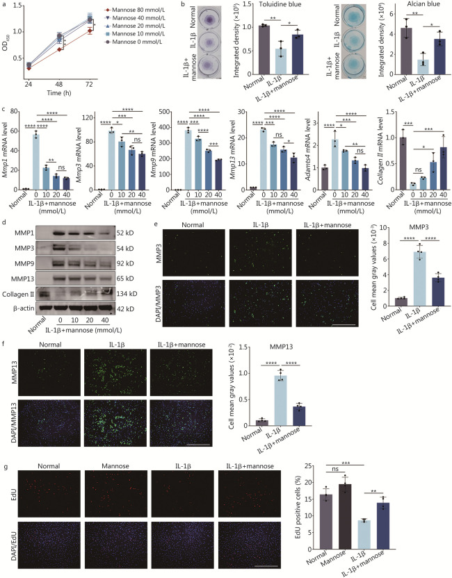
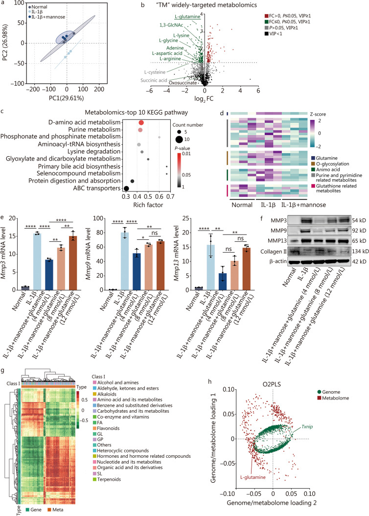
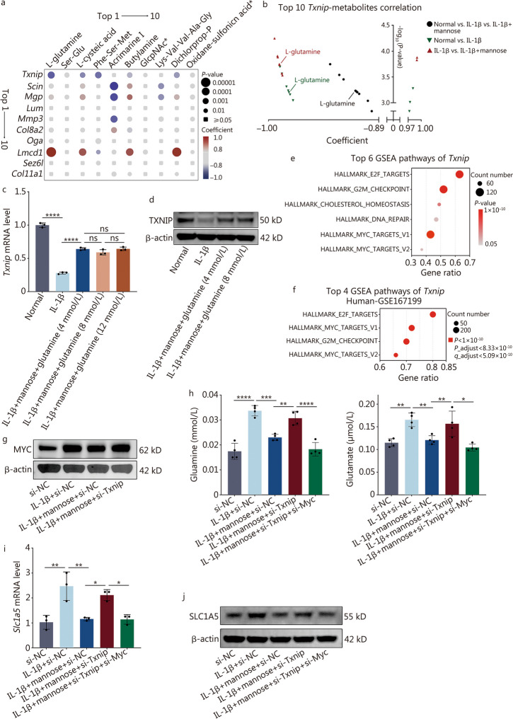
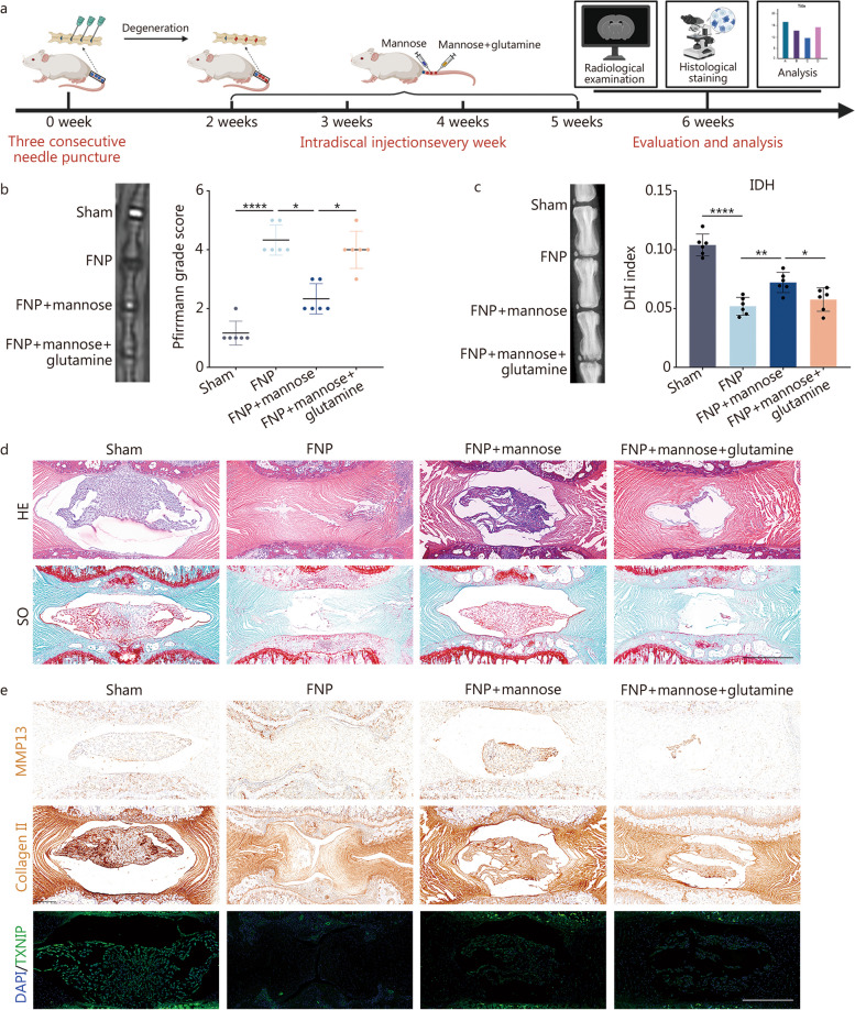
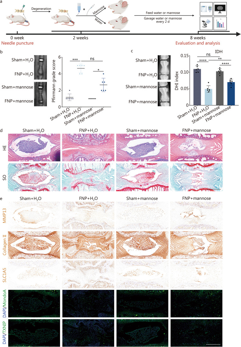
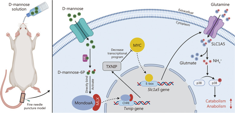
Methods: The study began with optimizing the mannose concentration for restoring NP cells. Transcriptomic analyses were employed to identify the mediators influenced by mannose, with the thioredoxin-interacting protein (Txnip) gene showing the most significant differences. Subsequently, small interfering RNA (siRNA) technology was used to demonstrate that Txnip is the key gene through which mannose exerts its effects. Techniques such as colocalization analysis, molecular docking, and overexpression assays further confirmed the direct regulatory relationship between mannose and TXNIP. To elucidate the mechanism of action of mannose, metabolomics techniques were employed to pinpoint glutamine as a core metabolite affected by mannose. Next, various methods, including integrated omics data and the Gene Expression Omnibus (GEO) database, were used to validate the one-way pathway through which TXNIP regulates glutamine. Finally, the therapeutic effect of mannose on IVDD was validated, elucidating the mechanistic role of TXNIP in glutamine metabolism in both intradiscal and orally treated rats.

Results: In both in vivo and in vitro experiments, it was discovered that mannose has potent efficacy in alleviating IVDD by inhibiting catabolism. From a mechanistic standpoint, it was shown that mannose exerts its anti-catabolic effects by directly targeting the transcription factor max-like protein X-interacting protein (MondoA), resulting in the upregulation of TXNIP. This upregulation, in turn, inhibits glutamine metabolism, ultimately accomplishing its anti-catabolic effects by suppressing the mitogen-activated protein kinase (MAPK) pathway. More importantly, in vivo experiments have further demonstrated that compared with intradiscal injections, oral administration of mannose at safe concentrations can achieve effective therapeutic outcomes.

Conclusions: In summary, through integrated multiomics analysis, including both in vivo and in vitro experiments, this study demonstrated that mannose primarily exerts its anti-catabolic effects on IVDD through the TXNIP-glutamine axis. These findings provide strong evidence supporting the potential of the use of mannose in clinical applications for alleviating IVDD. Compared to existing clinically invasive or pain-relieving therapies for IVDD, the oral administration of mannose has characteristics that are more advantageous for clinical IVDD treatment.

Keywords: D-mannose; Glutamine; Intervertebral disc degeneration; Thioredoxin-interacting protein (TXNIP).


Conflict of interest statement

The authors declare no potential conflicts of interest with respect to the research, authorship, or publication of this article.

Figures

Fig. 1
Fig. 1
Mannose suppresses catabolism in rat nucleus pulposus (NP) cells treated with IL-1β. a Effect of different concentrations of mannose on cell viability at various time points, as determined by a CCK-8 assay. The statistically significant differences between the 80 mmol/L and 0 mmol/L groups are shown. *P < 0.05. b Toluidine blue and alcian blue staining of NP cells treated with IL-1β and IL-1β + mannose. c RT-qPCR analysis of Mmp1, Mmp3, Mmp9, Mmp13, Adamts4 and collagen II in NP cells treated with different concentrations of mannose. d Western blotting of MMP1, MMP3, MMP9, MMP13 and collagen II in NP cells treated with different concentrations of mannose. e Immunofluorescence and quantitative analysis of MMP3 in NP cells treated with IL-1β and IL-1β + mannose (original magnification × 100; scale bar = 600 µm; n = 4). f Immunofluorescence and quantitative analysis of MMP13 in NP cells treated with IL-1β and IL-1β + mannose (original magnification × 100; scale bar = 600 µm; n = 4). g EdU staining of NP cells treated with mannose, IL-1β, and IL-1β + mannose (original magnification × 100; scale bar = 600 µm; n = 4). Unless otherwise specified, the IL-1β concentration was 10 ng/ml, the mannose concentration was 40 mmol/L, and n = 3. *P < 0.05, **P < 0.01, ***P < 0.001, ****P < 0.0001. ns non-significant, CCK-8 cell counting kit-8, IL-1β interleukin-1β, MMP1 matrix metalloproteinase 1, MMP3 matrix metalloproteinase 3, MMP9 matrix metalloproteinase 9, MMP13 matrix metalloproteinase 13, ADAMTS4 a disintegrin and metalloproteinase with thrombospondin motifs 4, EdU 5-ethynyl-2’-deoxyuridine, DAPI 4’,6-diamidino-2-phenylindole
Fig. 2
Fig. 2
Mannose inhibits catabolism by regulating TXNIP. a Principal component analysis (PCA) of normal group, IL-1β group and IL-1β + mannose group. b Transcriptome volcano plot of IL-1β group and IL-1β + mannose group. c Gene heatmap comparing IL-1β group and IL-1β + mannose group. d Transcriptome KEGG pathway ranking between IL-1β group and IL-1β + mannose group. RT-qPCR (e) and Western blotting (f) analysis of TXNIP in NP cells treated with IL-1β and IL-1β + mannose. RT-qPCR (g) and Western blotting (h) analysis of MMP3, MMP9, MMP13 and collagen II in NP cells treated with si-NC, IL-1β + si-NC, IL-1β + mannose + si-NC or IL-1β + mannose + si-Txnip. i UMAP plots of human single-cell RNA sequencing (scRNA-seq) data (GSE165722) showing differences in Txnip expression between Grade II–III and Grade IV–V intervertebral disc degeneration (IVDD) patients (n = 4). j The expression of Txnip in different cell types according to human scRNA-seq (GSE165722) between Grade II–III and Grade IV–V IVDD patients. Unless otherwise specified, 10 ng/ml IL-1β and 40 mmol/L mannose were used, n = 3. *P < 0.05, **P < 0.01, ***P < 0.001, ****P < 0.0001. ns non-significant, IL-1β interleukin-1β, MMP3 matrix metalloproteinase 3, MMP10 matrix metalloproteinase 10, MMP12 matrix metalloproteinase 12, MMP13 matrix metalloproteinase 13, SLC1A5 solute carrier family 1 member 5, GDH glutamate dehydrogenase 1, SLC7A5 solute carrier family 7 member 5, Col2a1 collagen type II alpha 1 chain, GLS glutaminase, Gls2 glutaminase 2, Col11a1 collagen type XI alpha 1 chain, TXNIP thioredoxin-interacting protein, KEGG Kyoto Encyclopedia of Genes and Genomes, PI3K-Akt phosphatidylinositol-3 kinase-protein kinase B, MAPK mitogen-activated protein kinase, JAK-STAT janus kinase-signal transducer and activator of transcription, WNT wingless/integrated, TNF tumor necrosis factor, AMPK AMP-activated protein kinase, Ras rat sarcoma protein, TGF-β transforming growth factor β, mTOR mammalian target of rapamycin, NF-κB nuclear factor kappa-light-chain-enhancer of activated B cells, MMPs matrix metalloproteinases, TCA tricarboxylic acid cycle
Fig. 3
Fig. 3
Mannose regulates TXNIP by directly targeting the MondoA transcription factor. a RT-qPCR analysis of Mmp13 and Txnip in NP cells treated with IL-1β, IL-1β + mannose or IL-1β + 2-DG (2 mmol/L). b Western blotting analysis of TXNIP in NP cells treated with IL-1β, IL-1β + mannose or IL-1β + 2-DG (2 mmol/L). c MondoA molecular docking experiments with glucose 6-phosphate and mannose 6-phosphate. d Confocal microscopy and quantitative analysis of the intracellular distribution of MondoA in NP cells treated with IL-1β and IL-1β + mannose (original magnification × 400, × 3600; scale bar = 100 µm, 33.33 µm; n = 4). RT-qPCR (e) and Western blotting (f) analysis of MMP13 and TXNIP in NP cells treated with oe-NC, IL-1β + oe-NC, IL-1β + mannose + oe-NC or IL-1β + mannose + oe-MPI. g Western blotting and quantitative analysis of nuclear-cytoplasmic MondoA protein in NP cells treated with oe-NC, IL-1β + oe-NC, IL-1β + mannose + oe-NC or IL-1β + mannose + oe-MPI. Unless otherwise specified, 10 ng/ml IL-1β and 40 mmol/L mannose were used, n = 3. *P < 0.05, **P < 0.01, ****P < 0.0001. ns non-significant, MMP13 matrix metalloproteinase 13, IL-1β interleukin-1β, 2-DG 2-deoxy-D-glucose, TXNIP thioredoxin-interacting protein, DAPI 4’,6-diamidino-2-phenylindole, MondoA max-like protein X-interacting protein, MPI mannose phosphate isomerase
Fig. 4
Fig. 4
Mannose suppresses catabolism by inhibiting glutamine in vitro. a Principal component analysis (PCA) of “TM” widely targeted metabolomics among normal group, IL-1β group and IL-1β + mannose group. b Volcano plot of the metabolomic data between IL-1β group and IL-1β + mannose group. c Comparison of the top 10 KEGG pathways associated with metabolism between IL-1β group and IL-1β + mannose group. d Metabolomic heatmap of IL-1β group and IL-1β + mannose group. e RT-qPCR analysis of Mmp3, Mmp9 and Mmp13 in NP cells treated with IL-1β and IL-1β + different concentrations of glutamine (4 mmol/L, 8 mmol/L, or 12 mmol/L). f Western blotting of MMP3, MMP9, MMP13 and collagen II in NP cells treated with IL-1β and IL-1β combined with different concentrations of glutamine (4 mmol/L, 8 mmol/L, or 12 mmol/L). g Correlation heatmap between genes and metabolites among the three groups. h O2PLS model analysis between transcriptomics and metabolomics. 10 ng/ml IL-1β and 40 mmol/L mannose were used, n = 3. *P < 0.05, **P < 0.01, ****P < 0.0001. ns non-significant, IL-1β interleukin-1β, VIP variable importance in projection, KEGG Kyoto Encyclopedia of Genes and Genomes, ABC adenosine triphosphate-binding cassette, O2PLS two-way orthogonal PLS, FA fatty acid, GL glucose, GP plycerol phosphate, SL sphingolipid, MMP3 matrix metalloproteinase 3, MMP9 matrix metalloproteinase 9, MMP13 matrix metalloproteinase 13
Fig. 5
Fig. 5
Mannose decreases the intracellular level of glutamine by upregulating TXNIP. a Correlation heatmap of the top 10 genes and top 10 metabolites among the three groups. GlcpNAc*: alpha-L-Fucp-(1- > 3)-[beta-D-Galp-(1- > 4)]-D-GlcpNAc; Oxidane-sulfonic acid*: [3-(5,7-dihydroxy-4-oxo-4H-chromen-2-yl)phenyl]oxidanesulfonic acid. b The top 10 metabolites associated with Txnip in different groups. RT-qPCR (c) and Western blotting (d) analysis of TXNIP in NP cells treated with IL-1β and IL-1β + different concentrations of glutamine. Top 6 GSEA pathways of Txnip in our transcriptomics dataset (e) and the top 4 GSEA pathways of Txnip in a human intervertebral disc degeneration tissue transcriptomics dataset (GSE167199, f). g Western blotting of MYC in NP cells treated with si-NC, IL-1β + si-NC, IL-1β + mannose + si-NC and IL-1β + mannose + si-Txnip. h Intracellular glutamine and glutamate levels in NP cells treated with si-NC, IL-1β + si-NC, IL-1β + mannose + si-NC, IL-1β + mannose + si-Txnip, or IL-1β + mannose + si-Txnip + si-Myc. RT-qPCR (i) and Western blotting (j) of SLC1A5 in NP cells treated with si-NC, IL-1β + si-NC, IL-1β + mannose + si-NC, IL-1β + mannose + si-Txnip, or IL-1β + mannose + si-Txnip + si-Myc. Unless otherwise specified, 10 ng/ml IL-1β and 40 mmol/L mannose were used, n = 3. *P < 0.05, **P < 0.01, ***P < 0.001, ****P < 0.0001. ns non-significant, Txnip thioredoxin-interacting protein, Scin scinderin, Mgp matrix Gla protein, Lum lumican, Mmp3 matrix metallopeptidase 3, Col8a2 collagen type VIII alpha 2 chain, Oga O-GlcNAcase, Lmcd1 LIM and cysteine-rich domains 1, Sez6l seizure-related 6 homolog-like, Col11a1 collagen type XI alpha 1 chain, IL-1β interleukin-1β, G2M gap 2 to mitosis phase, SLC1A5 solute carrier family 1 member 5
Fig. 6
Fig. 6
Glutamine induces catabolism through the NH4+-mediated MAPK pathway. a RT-qPCR analysis of Mmp13 in NP cells treated with IL-1β, IL-1β + mannose, or IL-1β + mannose + CB-839 (1 µmol/L). RT-qPCR (b) and Western blotting (c) of MMP13 in NP cells treated with IL-1β, IL-1β + mannose, or IL-1β + mannose + glutamate (200 μmol/L)/NH4Cl (2 mmol/L)/asparagine (1 mmol/L). d RT-qPCR analysis of Mmp13 in NP cells treated with IL-1β, IL-1β + mannose, or IL-1β + mannose + pyruvate (2 mmol/L)/DM-AKG (2 mmol/L) [dimethyl α-ketoglutarate (DM-AKG) is a cell-permeable derivative of α-ketoglutarate] [31]. e Intracellular ATP levels in NP cells from different groups. RT-qPCR (f) and Western blotting (g) of GDH in NP cells treated with glutamine, IL-1β, IL-1β + glutamine, IL-1β + mannose, or IL-1β + mannose + glutamine. h Western blotting of key MAPK pathway proteins in NP cells treated with si-NC, IL-1β + si-NC, IL-1β + mannose + si-NC, or IL-1β + mannose + si-Txnip/si-NC + glutamine/si-NC + NH4Cl. Unless otherwise specified, 10 ng/ml IL-1β, 40 mmol/L mannose, and 8 mmol/L glutamine were used, n = 3. *P < 0.05, **P < 0.01, ***P < 0.001, ****P < 0. 0001. ns non-significant, MMP13 matrix metalloproteinase 13, IL-1β interleukin-1β, TXNIP thioredoxin-interacting protein, DM-AKG dimethyl α-ketoglutarate, ATP adenosine triphosphate, GDH glutamate dehydrogenase 1, ERK extracellular signal-regulated kinases, JNK c-Jun N-terminal kinase
Fig. 7
Fig. 7
Moreover, glutamine reversed the attenuating effect of mannose on IVDD in vivo. a Rat intervertebral disc injection workflow diagram. b MR images and Pfirrmann disc degeneration grades of the sham group, FNP group, FNP + mannose group and FNP + mannose + glutamine group. c X-ray and IDH analyses of the sham group, FNP group, FNP + mannose group and FNP + mannose + glutamine group. d HE and SO staining of the sham group, FNP group, FNP + mannose group and FNP + mannose + glutamine group (original magnification × 7; scale bar = 800 µm). e Immunofluorescence staining of MMP13, collagen II and TXNIP in the sham group, FNP group, FNP + mannose group and FNP + mannose + glutamine group (original magnification × 7; scale bar = 800 µm). n = 6. *P < 0.05, **P < 0.01, ****P < 0.0001. FNP fine needle puncture, IDH intervertebral disc height, DHI disc height index, HE hematoxylin and eosin, SO safranin O, MMP13 matrix metalloproteinase 13, DAPI 4’,6-diamidino-2-phenylindole, TXNIP thioredoxin-interacting protein
Fig. 8
Fig. 8
IVDD can be reversed by the oral administration of mannose. a Rat oral feeding workflow diagram. b MR images and Pfirrmann disc degeneration grades of the sham + H2O group, FNP + H2O group, sham + mannose group, and FNP + mannose group. c X-ray and IDH analyses of the sham + H2O group, FNP + H2O group, sham + mannose group, and FNP + mannose group. d HE and SO staining of the sham + H2O group, FNP + H2O group, sham + mannose group, and FNP + mannose group (original magnification × 7; scale bar = 800 µm). e Immunofluorescence staining of MMP13, collagen II, SLC1A5, MondoA and TXNIP in the sham + H2O group, FNP + H2O group, sham + mannose group, and FNP + mannose group (original magnification × 7; scale bar = 800 µm). Unless otherwise specified, n = 6. *P < 0.05, **P < 0.01, ***P < 0.001, ****P < 0.0001. ns non-significant, FNP fine needle puncture, IDH intervertebral disc height, DHI disc height index, HE hematoxylin and eosin, SO safranin O, MMP13 matrix metalloproteinase 13, SLC1A5 solute carrier family 1 member 5, DAPI 4’,6-diamidino-2-phenylindole, MondoA max-like protein X-interacting protein, TXNIP thioredoxin-interacting protein
Fig. 9
Fig. 9
Schematic diagram of the alleviation of IVDD by mannose. HK hexokinase, MondoA max-like protein X-interacting protein, MLX max-like protein X, ChRE carbohydrate responsive element, TXNIP thioredoxin-interacting protein, E-box enhancer box, SLC1A5 solute carrier family 1 member 5

References

    1. Cieza A, Causey K, Kamenov K, Hanson SW, Chatterji S, Vos T. Global estimates of the need for rehabilitation based on the Global Burden of Disease study 2019: a systematic analysis for the Global Burden of Disease Study 2019. Lancet. 2021;396(10267):2006–2017. doi: 10.1016/S0140-6736(20)32340-0. - DOI - PMC - PubMed
    1. O’Keeffe M, George SZ, O’Sullivan PB, O’Sullivan K. Psychosocial factors in low back pain: letting go of our misconceptions can help management. Br J Sports Med. 2019;53(13):793–794. doi: 10.1136/bjsports-2018-099816. - DOI - PubMed
    1. Zhao Y, Qiu C, Wang W, Peng J, Cheng X, Shangguan Y, et al. Cortistatin protects against intervertebral disc degeneration through targeting mitochondrial ROS-dependent NLRP3 inflammasome activation. Theranostics. 2020;10(15):7015–7033. doi: 10.7150/thno.45359. - DOI - PMC - PubMed
    1. Wen ZQ, Lin J, Xie WQ, Shan YH, Zhen GH, Li YS. Insights into the underlying pathogenesis and therapeutic potential of endoplasmic reticulum stress in degenerative musculoskeletal diseases. Mil Med Res. 2023;10(1):54. - PMC - PubMed
    1. Vo NV, Hartman RA, Yurube T, Jacobs LJ, Sowa GA, Kang JD. Expression and regulation of metalloproteinases and their inhibitors in intervertebral disc aging and degeneration. Spine J. 2013;13(3):331–341. doi: 10.1016/j.spinee.2012.02.027. - DOI - PMC - PubMed

Publication types

LinkOut - more resources