Review
. 2024 May 1;137(9):jcs261776.
doi: 10.1242/jcs.261776. Epub 2024 May 7.

A short guide to the tight junction

Affiliations
Review

A short guide to the tight junction

Sandra Citi et al. J Cell Sci. .

Abstract

Tight junctions (TJs) are specialized regions of contact between cells of epithelial and endothelial tissues that form selective semipermeable paracellular barriers that establish and maintain body compartments with different fluid compositions. As such, the formation of TJs represents a critical step in metazoan evolution, allowing the formation of multicompartmental organisms and true, barrier-forming epithelia and endothelia. In the six decades that have passed since the first observations of TJs by transmission electron microscopy, much progress has been made in understanding the structure, function, molecular composition and regulation of TJs. The goal of this Perspective is to highlight the key concepts that have emerged through this research and the future challenges that lie ahead for the field.

Keywords: Actin; Barrier; Cingulin; Claudin; Epithelium; Myosin; Occludin; Permeability; Polarity; Tight junctions; ZO-1.


Conflict of interest statement

Competing interests The authors declare no competing or financial interests.

Figures
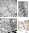
Fig. 1.
Fig. 1.
The ultrastructure of TJs. (A) Transmission electron micrograph of the AJC of mouse intestinal epithelial cells (within the jejunum), highlighting the position of the apical TJ and the neighboring AJ. (B) Detailed image of the TJ region highlighted in A, with sites of intimate plasma membrane apposition indicated by black arrows, and the electron-dense cytoplasmic plaque immediately beneath the plasma membrane indicated by white arrows. (C) FFEM image of the apical regions of epithelial intestinal (human jejunum) cells, showing the strands of a bTJ and a tTJ. (D) Cartoon depicting bTJ and tTJ structure, highlighting the positions of bTJ strands, joint tTJ strands and the central tube of a tTJ. Images in A and B provided by Kyoko Furuse, National Institute for Physiological Sciences, Okazaki, Japan. Image in C provided by Susanne M. Krug, Charité – Universitätsmedizin Berlin, Germany.
Fig. 2.
Fig. 2.
The barrier function of TJs, and claudin organization and structure. (A) Schematic of a sheet of polarized epithelial cells, highlighting the position of TJs and AJs. Arrows indicate three functionally defined paracellular pathways: (1) the ‘pore’ pathway formed by claudin-based channels (blue); (2) the ‘leak’ pathway formed by breaks within the bTJ and/or by opening of the tTJ central tube (green); and (3) the ‘unrestricted’ pathway caused by epithelial damage (red). The actin and microtubule (MT) cytoskeletons are outlined in a simplified organization within the cell in red and orange, respectively. (B) Schematic of a sheet of polarized epithelial cells (top), with an expanded section of the TJ region shown (bottom left) alongside top and side views of polymerized claudins within the TJs (bottom right). Note that individual claudin molecules interact in cis and in trans with other claudin molecules in this schematic drawing. Dashed boxes indicate claudin monomers. For more details, see Suzuki et al. (2015).
Fig. 3.
Fig. 3.
A model of the architecture of the AJC. Scheme showing a simplified model of the architectural organization of the AJC (bicellular junctions) and the associated actomyosin cytoskeleton. For simplicity, only a few major transmembrane proteins (occludin, claudins, JAM-A and cadherin), cytoplasmic scaffolds (ZO proteins), adaptors (cingulin) and cytoskeletal proteins (actin and myosin only, not microtubules) are shown. Occludin is representative of TAMPs, which include tricellulin at tTJs. JAM-A is representative of Ig-like CAMs, which include angulins at tTJs. Branched actin filaments associated with monomers or oligomers of myosin-2B are believed to tether the actomyosin belt to TJs, either by direct binding of actin filaments to ZO-1- and JAM-A-based complexes, and/or by cingulin-dependent tethering of myosin-2B to ZO-1. Proteins and protein complexes are not drawn to scale, and protein complexes are only shown at the section of membranes, whereas in vivo they are distributed continuously along the circumference of the AJC. This scheme was inspired by a similar scheme in Cartagena-Rivera et al. (2017), which is published under the terms of a CC-BY 4.0 license.

References

    1. Aijaz, S., D'Atri, F., Citi, S., Balda, M. S. and Matter, K. (2005). Binding of GEF-H1 to the tight junction-associated adaptor cingulin results in inhibition of Rho signaling and G1/S phase transition. Dev. Cell 8, 777-786. 10.1016/j.devcel.2005.03.003 - DOI - PubMed
    1. Arnold, T. R., Stephenson, R. E. and Miller, A. L. (2017). Rho GTPases and actomyosin: Partners in regulating epithelial cell-cell junction structure and function. Exp. Cell. Res. 358, 20-30. 10.1016/j.yexcr.2017.03.053 - DOI - PMC - PubMed
    1. Balda, M. S. and Matter, K. (2023). Tight junctions. Curr. Biol. 33, R1135-R1140. 10.1016/j.cub.2023.09.027 - DOI - PubMed
    1. Belardi, B., Hamkins-Indik, T., Harris, A. R., Kim, J., Xu, K. and Fletcher, D. A. (2020). A weak link with actin organizes tight junctions to control epithelial permeability. Dev. Cell 54, 792-804.e7. 10.1016/j.devcel.2020.07.022 - DOI - PMC - PubMed
    1. Bement, W. M., Goryachev, A. B., Miller, A. L. and von Dassow, G. (2024). Patterning of the cell cortex by Rho GTPases. Nat. Rev. Mol. Cell Biol. 25, 290-308. 10.1038/s41580-023-00682-z - DOI - PubMed

Publication types