. 2024 Jun 13;98(6):e0029524.
doi: 10.1128/jvi.00295-24. Epub 2024 May 7.

Characterization of virus‒host recombinant variants of the hepatitis E virus

Affiliations

Characterization of virus‒host recombinant variants of the hepatitis E virus

Olivia Paronetto et al. J Virol. .

Abstract

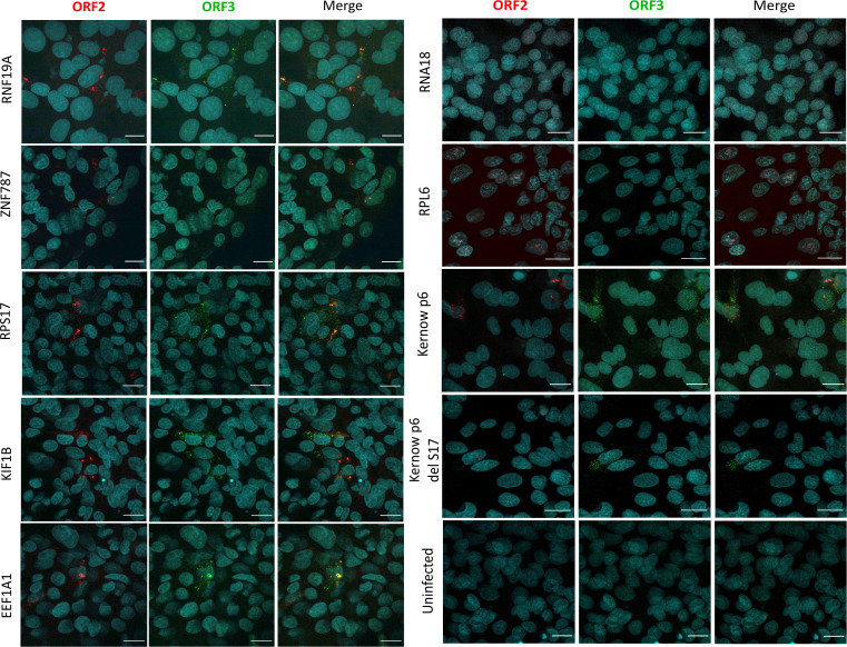
Hepatitis E virus is a single-strand, positive-sense RNA virus that can lead to chronic infection in immunocompromised patients. Virus-host recombinant variants (VHRVs) have been described in such patients. These variants integrate part of human genes into the polyproline-rich region that could introduce new post-translational modifications (PTMs), such as ubiquitination. The aim of this study was to characterize the replication capacity of different VHRVs, namely, RNF19A, ZNF787, KIF1B, EEF1A1, RNA18, RPS17, and RPL6. We used a plasmid encoding the Kernow strain, in which the fragment encoding the S17 insertion was deleted (Kernow p6 delS17) or replaced by fragments encoding the different insertions. The HEV RNA concentrations in the supernatants and the HepG2/C3A cell lysates were determined via RT-qPCR. The capsid protein ORF2 was immunostained. The effect of ribavirin was also assessed. The HEV RNA concentrations in the supernatants and the cell lysates were higher for the variants harboring the RNF19A, ZNF787, KIF1B, RPS17, and EEF1A1 insertions than for the Kernow p6 del S17, while it was not with RNA18 or RPL6 fragments. The number of ORF2 foci was higher for RNF19A, ZNF787, KIF1B, and RPS17 than for Kernow p6 del S17. VHRVs with replicative advantages were less sensitive to the antiviral effect of ribavirin. No difference in PTMs was found between VHRVs with a replicative advantage and those without. In conclusion, our study showed that insertions did not systematically confer a replicative advantage in vitro. Further studies are needed to determine the mechanisms underlying the differences in replicative capacity.

Importance: Hepatitis E virus (HEV) is a major cause of viral hepatitis. HEV can lead to chronic infection in immunocompromised patients. Ribavirin treatment is currently used to treat such chronic infections. Recently, seven virus-host recombinant viruses were characterized in immunocompromised patients. These viruses have incorporated a portion of a human gene fragment into their genome. We studied the consequences of these insertions on the replication capacity. We found that these inserted fragments could enhance virus replication for five of the seven recombinant variants. We also showed that the recombinant variants with replicative advantages were less sensitive to ribavirin in vitro. Finally, we found that the mechanisms leading to such a replicative advantage do not seem to rely on the post-translational modifications introduced by the human gene fragment that could have modified the function of the viral protein. The mechanisms involved in improving the replication of such recombinant viruses remain to be explored.

Keywords: hepatitis E virus; ribavirin; viral fitness; virus–host recombinant variants.


Conflict of interest statement

The authors declare no conflict of interest.

Figures

Fig 1
Fig 1
Schematic representation of the strategy used to obtain recombinant HEV strains. The nt sequences of the seven inserts were synthesized. The Kernow p6 plasmid was digested using the restriction sites StuI and NheI to insert different fragments of human genes.
Fig 2
Fig 2
Impact of the seven human fragments on the release of HEV RNA and ORF2 protein in the supernatant. HepG2/C3A cells were infected with 17 GEs/cell, with each of the recombinant variants. For 14 days, the culture supernatants were harvested on Days 0, 1 and every 2 days. (A) HEV RNA was extracted from supernatants, and the viral load was quantified via RT-PCR targeting ORF3. (B) The antigen HEV concentration was measured via a qualitative diagnostic approach. The antigen HEV concentration expressed in COI represents the signal/cutoff ratio generated by the analyzer. Samples were considered positive if the COI > 1. (A and B) The results are presented as the means ± SEMs from two independent experiments performed in triplicate. **P < 0.01 and ns, not significant. (C and D) Western blots of HEV ORF2 proteins in supernatants of HepG2/C3A cells 10 days (C) or 15 days (D) after infection with each of the VHRVs and Kernow p6 del S17.
Fig 3
Fig 3
Impact of the seven human fragments on the production of HEV RNA and ORF2 protein in the cell lysate. HepG2/C3A cells were infected with 17 GEs/cell by each of the recombinant HEV variants, and the cells were lysed on Days 0, 5, 10, and 15 post-infection. (A) HEV RNA was extracted, and the viral load was quantified via RT-PCR using primers targeting ORF3. (B) The antigen HEV concentration was measured via a qualitative diagnostic approach. The antigen HEV concentration expressed in the COI represents the signal/cutoff ratio generated by the analyzer. Samples were considered positive if the COI > 1. (A and B) The results are presented as the means ± SDs from two independent experiments performed in triplicate. (C and D) Western blots of HEV ORF2 proteins in HepG2/C3A cells 10 days (C) or 15 days (D) after infection with each of the VHRVs and Kernow p6 del S17. Tubulin served as the loading control.
Fig 4
Fig 4
Quantification of the FFU in infected cells. HepG2/C3A cells were fixed and permeabilized before staining with an anti-ORF2 antibody on Day 5 post-infection. (A) FFUs (red) were viewed with a Leica sp8 confocal microscope with a 63× objective. The scale bar represents 20 µm. (B) The number of HEV FFUs was calculated with the software ImageJ v1.52p from the images of five fields acquired on Apotome (Zeiss). These results were obtained from 12 images from two independent experiments. The results are presented as the means ± SDs from two independent experiments performed in triplicate. *P < 0.05, **P < 0.01, ***P < 0.001, and ns, not significant.
Fig 5
Fig 5
Characterization of the HEV particles released in supernatants. (A) Supernatants containing 7.5 × 10⁴ IU/mL of HEV RNA were loaded onto a 7.5%–40% iodixanol gradient, ultracentrifuged, and fractionated. The percentage of HEV RNA in each fraction (density) is shown. The density gradients are representative of the results of one experiment. (B) Supernatants containing 106 IU/mL were used to infect HepG2/C3A cells, corresponding to two GEs/cell. The viral load in the supernatants was quantified by RT-qPCR every 2 days. The results are presented as the means ± SDs from two independent experiments performed in triplicate.
Fig 6
Fig 6
Sensitivity of the different virus–host recombinant variants to ribavirin. HepG2/C3A cells were infected with 17 GEs/cell for each of the recombinant viruses, and after 6 h, 1, 10, 100, or 500 µM ribavirin was added to the culture medium. (A) Viability of the uninfected cells was assessed in the cell lysates on Day 10 based on luminescence measurements. The cell viability for each ribavirin concentration was normalized to that of the untreated wells. (B) The culture supernatants were harvested 10 days post-infection, and the HEV RNA was quantified via RT-qPCR. The ratio of viral production corresponds to the ratio of infected cells treated/untreated infected cells. (A and B) The results are presented as the means ± SDs from two independent experiments performed in triplicate.
Fig 7
Fig 7
Impact of the seven human fragments on the nuclear localization of the ORF2 protein. HepG2/C3A infected cells were fixed and permeabilized. In addition, the nuclei were also permeabilized before staining with an anti-ORF2 and an anti-ORF3 antibody on Day 5 post-infection. ORF2 expression (red) and ORF3 expression (green) were viewed with a Leica sp8 confocal microscope with a 63× objective. The scale bar represents 20 µm.

References

    1. Kamar N, Izopet J, Pavio N, Aggarwal R, Labrique A, Wedemeyer H, Dalton HR. 2017. Hepatitis E virus infection. Nat Rev Dis Primers 3:17086. doi:10.1038/nrdp.2017.86 - DOI - PubMed
    1. Lhomme S, Marion O, Abravanel F, Izopet J, Kamar N. 2020. Clinical manifestations, pathogenesis and treatment of hepatitis E virus infections. J Clin Med 9:331. doi:10.3390/jcm9020331 - DOI - PMC - PubMed
    1. Kamar N, Rostaing L, Legrand-Abravanel F, Izopet J. 2013. How should hepatitis E virus infection be defined in organ-transplant recipients? Am J Transplant 13:1935–1936. doi:10.1111/ajt.12253 - DOI - PubMed
    1. Kamar N, Selves J, Mansuy J-M, Ouezzani L, Péron J-M, Guitard J, Cointault O, Esposito L, Abravanel F, Danjoux M, Durand D, Vinel J-P, Izopet J, Rostaing L. 2008. Hepatitis E virus and chronic hepatitis in organ-transplant recipients. N Engl J Med 358:811–817. doi:10.1056/NEJMoa0706992 - DOI - PubMed
    1. Kamar N, Mansuy J-M, Cointault O, Selves J, Abravanel F, Danjoux M, Otal P, Esposito L, Durand D, Izopet J, Rostaing L. 2008. Hepatitis E virus-related cirrhosis in kidneyand kidney-pancreas-transplant recipients. Am J Transplant 8:1744–1748. doi:10.1111/j.1600-6143.2008.02286.x - DOI - PubMed

MeSH terms

LinkOut - more resources