. 2024 May;311(2):e230750.
doi: 10.1148/radiol.230750.

Evaluation of a Cascaded Deep Learning-based Algorithm for Prostate Lesion Detection at Biparametric MRI

Affiliations

Evaluation of a Cascaded Deep Learning-based Algorithm for Prostate Lesion Detection at Biparametric MRI

Yue Lin et al. Radiology. 2024 May.

Abstract

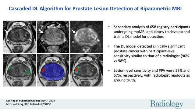
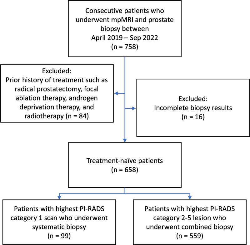
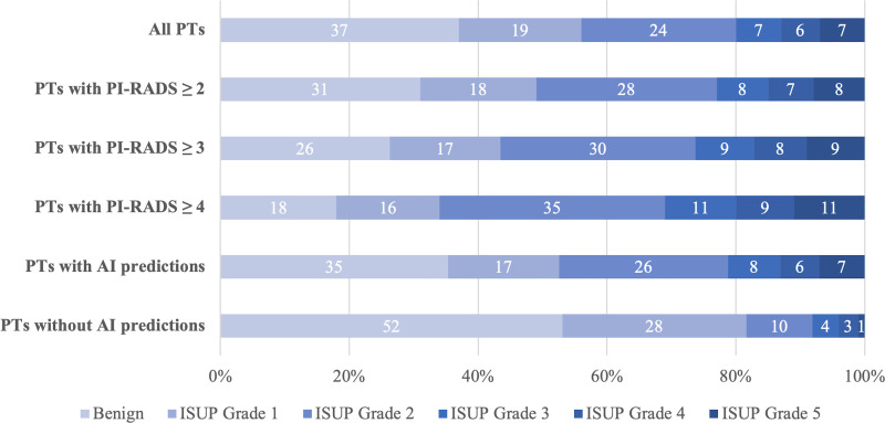
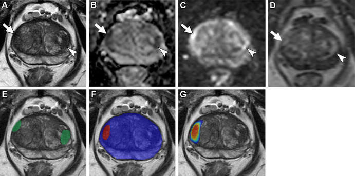
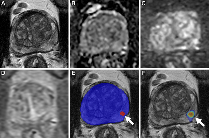
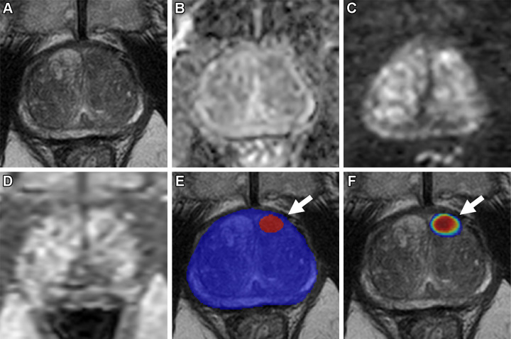
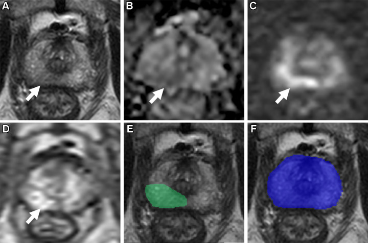
Background Multiparametric MRI (mpMRI) improves prostate cancer (PCa) detection compared with systematic biopsy, but its interpretation is prone to interreader variation, which results in performance inconsistency. Artificial intelligence (AI) models can assist in mpMRI interpretation, but large training data sets and extensive model testing are required. Purpose To evaluate a biparametric MRI AI algorithm for intraprostatic lesion detection and segmentation and to compare its performance with radiologist readings and biopsy results. Materials and Methods This secondary analysis of a prospective registry included consecutive patients with suspected or known PCa who underwent mpMRI, US-guided systematic biopsy, or combined systematic and MRI/US fusion-guided biopsy between April 2019 and September 2022. All lesions were prospectively evaluated using Prostate Imaging Reporting and Data System version 2.1. The lesion- and participant-level performance of a previously developed cascaded deep learning algorithm was compared with histopathologic outcomes and radiologist readings using sensitivity, positive predictive value (PPV), and Dice similarity coefficient (DSC). Results A total of 658 male participants (median age, 67 years [IQR, 61-71 years]) with 1029 MRI-visible lesions were included. At histopathologic analysis, 45% (294 of 658) of participants had lesions of International Society of Urological Pathology (ISUP) grade group (GG) 2 or higher. The algorithm identified 96% (282 of 294; 95% CI: 94%, 98%) of all participants with clinically significant PCa, whereas the radiologist identified 98% (287 of 294; 95% CI: 96%, 99%; P = .23). The algorithm identified 84% (103 of 122), 96% (152 of 159), 96% (47 of 49), 95% (38 of 40), and 98% (45 of 46) of participants with ISUP GG 1, 2, 3, 4, and 5 lesions, respectively. In the lesion-level analysis using radiologist ground truth, the detection sensitivity was 55% (569 of 1029; 95% CI: 52%, 58%), and the PPV was 57% (535 of 934; 95% CI: 54%, 61%). The mean number of false-positive lesions per participant was 0.61 (range, 0-3). The lesion segmentation DSC was 0.29. Conclusion The AI algorithm detected cancer-suspicious lesions on biparametric MRI scans with a performance comparable to that of an experienced radiologist. Moreover, the algorithm reliably predicted clinically significant lesions at histopathologic examination. ClinicalTrials.gov Identifier: NCT03354416 © RSNA, 2024 Supplemental material is available for this article.


Conflict of interest statement

Disclosures of conflicts of interest: Y.L. No relevant relationships. E.C.Y. Associate editor on the Radiology In Training Editorial Board. M.J.B. No relevant relationships. S.A.H. Institution (National Cancer Institute) has a cooperative research and development agreement with NVIDIA. J.T. This work was completed under a cooperative research and development agreement between the National Institutes of Health (NIH) and NVIDIA. T.E.P. No relevant relationships. K.M.M. No relevant relationships. L.H. No relevant relationships. C.G. No relevant relationships. D.Y. No relevant relationships. Z.X. No relevant relationships. N.S.L. This research supported by NIH Intramural Research Program; receives shared royalties from NIH for an unrelated prostate cancer detection system patent (US patent no. US11200667B2). A.T. No relevant relationships. M.J.M. No relevant relationships. D.X. No relevant relationships. Y.M.L. No relevant relationships. S.G. No relevant relationships. B.J.W. Institution (NIH) and NVIDIA have a cooperative research and development agreement; NIH and Philips have a cooperative research and development agreement that includes work in the space of prostate cancer and also artificial intelligence; principal investigator on cooperative research and development agreements between NIH and Philips, Celsion, Biocompatibles, Boston Scientific, Siemens, NVIDIA, XACT Robotics, and Promaxo; support for research from 3T Technologies (devices), Exact Imaging (data), AngioDynamics (equipment), AstraZeneca (pharmaceuticals, National Cancer Institute cooperative research and development agreement), ArciTrax (devices and equipment), Johnson & Johnson (equipment), Medtronic (equipment), Theromics (supplies), Profound Medical (equipment and supplies), QT Imaging (equipment and supplies), Boston Scientific (equipment), Varian (materials), Combat Medical (equipment), Clinical LaserThermia Systems (equipment), MediView (equipment), CIVCO Medical Solutions (equipment), Galvanize (equipment); royalties from a patent licensing agreement between Philips and NIH; licensing agreements between NIH and NVIDIA and Canon; NIH and CIVCO are coinventors of intellectual property; NIH and ArciTrax are coinventors of intellectual property; NIH and Philips are coinventors of intellectual property; NIH and Boston Scientific are coinventors of intellectual property; NIH and NVIDIA are coinventors of intellectual property; NIH may own intellectual property in the field; another nonlicensed family of patents available upon request (includes 50 patents); editor of Springer book, Interventional Urology, 2nd Edition, which may contain indirect mention of the topic of this work; NIH liaison to Academy for Radiology & Biomedical Imaging Research; and NIH liaison to Society of Interventional Oncology. P.L.C. Patents planned, issued, or pending with Philips Medical Systems and NVIDIA (author does not receive royalty payments). P.A.P. Royalties from a licensing agreement between Philips and institution (NIH); cooperative research and development agreement between NIH and Philips; NIH has intellectual property in the field including, among other patents and patent applications, System and Method for Prostate Cancer Detection and Distribution Mapping (US patent no. 8,447,384) and System and Method for Computer Aided Cancer Detection Using T2-weighted and High-Value Diffusion-weighted Magnetic Resonance Imaging (US patent no. 10,215,830); and licensing agreement with NIH and Philips (Invivo). B.T. Cooperative research and development agreement with NVIDIA and Philips and receives royalties from NIH.

Figures

None
Graphical abstract
Participant flow diagram. mpMRI = multiparametric MRI, PI-RADS =
Prostate Imaging Reporting and Data System.
Figure 1:
Participant flow diagram. mpMRI = multiparametric MRI, PI-RADS = Prostate Imaging Reporting and Data System.
Distribution of combined biopsy outcomes (percentage) based on highest
International Society of Urological Pathology (ISUP) grade group per
participant (PT). AI = artificial intelligence, PI-RADS = Prostate Imaging
Reporting and Data System.
Figure 2:
Distribution of combined biopsy outcomes (percentage) based on highest International Society of Urological Pathology (ISUP) grade group per participant (PT). AI = artificial intelligence, PI-RADS = Prostate Imaging Reporting and Data System.
Axial multiparametric MRI scans in a 72-year-old male participant with
a serum prostate-specific antigen level of 9.1 ng/mL: (A) T2-weighted image,
(B) apparent diffusion coefficient map, (C) high-b-value diffusion-weighted
image (b = 1500 sec/mm2), (D) dynamic contrast-enhanced image (frame 17 of
54 acquired at 5.6-second intervals), (E) T2-weighted image with
radiologist-segmented lesions (green contours) overlaid, (F) T2-weighted
image with artificial intelligence (AI) prediction map overlaid (red contour
is positive prediction; blue contour is AI prostate organ segmentation), and
(G) T2-weighted image with AI probability map overlaid (red indicates higher
probability). Two distinct lesions were detected by the radiologist and
represented the ground truth. Lesion 1 (1.6 cm; arrow in A–D) was in
the right midgland transition zone and was designated Prostate Imaging
Reporting and Data System (PI-RADS) category 4. Lesion 2 (1.5 cm; arrowhead
in A–D) was in the left midgland transition zone and was designated
PI-RADS category 3. Lesion 1 was correctly detected (true positive), while
lesion 2 was missed by the AI algorithm (false negative). Based on targeted
biopsy samples, lesion 1 was positive for Gleason score 7 (3 + 4) prostate
adenocarcinoma, and lesion 2 was benign.
Figure 3:
Axial multiparametric MRI scans in a 72-year-old male participant with a serum prostate-specific antigen level of 9.1 ng/mL: (A) T2-weighted image, (B) apparent diffusion coefficient map, (C) high-b-value diffusion-weighted image (b = 1500 sec/mm2), (D) dynamic contrast-enhanced image (frame 17 of 54 acquired at 5.6-second intervals), (E) T2-weighted image with radiologist-segmented lesions (green contours) overlaid, (F) T2-weighted image with artificial intelligence (AI) prediction map overlaid (red contour is positive prediction; blue contour is AI prostate organ segmentation), and (G) T2-weighted image with AI probability map overlaid (red indicates higher probability). Two distinct lesions were detected by the radiologist and represented the ground truth. Lesion 1 (1.6 cm; arrow in A–D) was in the right midgland transition zone and was designated Prostate Imaging Reporting and Data System (PI-RADS) category 4. Lesion 2 (1.5 cm; arrowhead in A–D) was in the left midgland transition zone and was designated PI-RADS category 3. Lesion 1 was correctly detected (true positive), while lesion 2 was missed by the AI algorithm (false negative). Based on targeted biopsy samples, lesion 1 was positive for Gleason score 7 (3 + 4) prostate adenocarcinoma, and lesion 2 was benign.
Axial multiparametric MRI scans in a 64-year-old male participant with
a serum prostate-specific antigen level of 8.1 ng/mL: (A) T2-weighted image,
(B) apparent diffusion coefficient map, (C) high-b-value diffusion-weighted
image (b = 1500 sec/mm2), (D) dynamic contrast-enhanced image (frame 45 of
54 acquired at 5.6-second intervals), (E) T2-weighted image with artificial
intelligence (AI) prediction map overlaid (red contour is positive
prediction; blue contour is AI prostate organ segmentation), and (F)
T2-weighted image with AI probability map overlaid (red indicates higher
probability). No distinct lesion was detected by the radiologist (Prostate
Imaging Reporting and Data System category 1). One lesion was called by the
AI algorithm in the left midgland peripheral zone (arrow in E and F),
representing a false positive based on the radiologist ground truth.
Systematic biopsy obtained from this site (left midgland lateral) was
positive for Gleason score 7 (3 + 4) prostate adenocarcinoma.
Figure 4:
Axial multiparametric MRI scans in a 64-year-old male participant with a serum prostate-specific antigen level of 8.1 ng/mL: (A) T2-weighted image, (B) apparent diffusion coefficient map, (C) high-b-value diffusion-weighted image (b = 1500 sec/mm2), (D) dynamic contrast-enhanced image (frame 45 of 54 acquired at 5.6-second intervals), (E) T2-weighted image with artificial intelligence (AI) prediction map overlaid (red contour is positive prediction; blue contour is AI prostate organ segmentation), and (F) T2-weighted image with AI probability map overlaid (red indicates higher probability). No distinct lesion was detected by the radiologist (Prostate Imaging Reporting and Data System category 1). One lesion was called by the AI algorithm in the left midgland peripheral zone (arrow in E and F), representing a false positive based on the radiologist ground truth. Systematic biopsy obtained from this site (left midgland lateral) was positive for Gleason score 7 (3 + 4) prostate adenocarcinoma.
Axial multiparametric MRI scans in a 69-year-old male participant with
a serum prostate-specific antigen level of 7.3 ng/mL: (A) T2-weighted image,
(B) apparent diffusion coefficient map, (C) high-b-value diffusion-weighted
image (b = 1500 sec/mm2), (D) dynamic contrast-enhanced image (frame 25 of
54 acquired at 5.6-second intervals), (E) T2-weighted image with artificial
intelligence (AI) prediction map overlaid (red contour is positive
prediction; blue contour is AI prostate organ segmentation), and (F)
T2-weighted image with AI probability map overlaid (red indicates higher
probability). One lesion was called by the AI algorithm in the left midgland
anterior transition zone (arrow in E and F), representing a false positive
based on the radiologist ground truth. A systematic biopsy sample obtained
from this site (left midgland medial) was benign.
Figure 5:
Axial multiparametric MRI scans in a 69-year-old male participant with a serum prostate-specific antigen level of 7.3 ng/mL: (A) T2-weighted image, (B) apparent diffusion coefficient map, (C) high-b-value diffusion-weighted image (b = 1500 sec/mm2), (D) dynamic contrast-enhanced image (frame 25 of 54 acquired at 5.6-second intervals), (E) T2-weighted image with artificial intelligence (AI) prediction map overlaid (red contour is positive prediction; blue contour is AI prostate organ segmentation), and (F) T2-weighted image with AI probability map overlaid (red indicates higher probability). One lesion was called by the AI algorithm in the left midgland anterior transition zone (arrow in E and F), representing a false positive based on the radiologist ground truth. A systematic biopsy sample obtained from this site (left midgland medial) was benign.
Axial multiparametric MRI scans in a 74-year-old male participant with
a serum prostate-specific antigen level of 12.9 ng/mL: (A) T2-weighted
image, (B) apparent diffusion coefficient map, (C) high-b-value
diffusion-weighted image (b = 1500 sec/mm2), (D) dynamic contrast-enhanced
image (frame 16 of 54 acquired at 5.6-second intervals), (E) T2-weighted
image with radiologist-segmented lesion (green contour) overlaid, and (F)
T2-weighted image with artificial intelligence (AI) prediction map overlaid
(no positive prediction; blue contour is AI prostate organ segmentation).
One lesion was detected by the radiologist and represented the ground truth.
The lesion (1.9 cm; arrow in A–D) was in the right apical midgland
peripheral zone and was designated Prostate Imaging Reporting and Data
System category 4. This lesion was missed by the AI algorithm, representing
a false negative. A targeted biopsy sample obtained from the lesion was
positive for Gleason score 7 (3 + 4) prostate adenocarcinoma.
Figure 6:
Axial multiparametric MRI scans in a 74-year-old male participant with a serum prostate-specific antigen level of 12.9 ng/mL: (A) T2-weighted image, (B) apparent diffusion coefficient map, (C) high-b-value diffusion-weighted image (b = 1500 sec/mm2), (D) dynamic contrast-enhanced image (frame 16 of 54 acquired at 5.6-second intervals), (E) T2-weighted image with radiologist-segmented lesion (green contour) overlaid, and (F) T2-weighted image with artificial intelligence (AI) prediction map overlaid (no positive prediction; blue contour is AI prostate organ segmentation). One lesion was detected by the radiologist and represented the ground truth. The lesion (1.9 cm; arrow in A–D) was in the right apical midgland peripheral zone and was designated Prostate Imaging Reporting and Data System category 4. This lesion was missed by the AI algorithm, representing a false negative. A targeted biopsy sample obtained from the lesion was positive for Gleason score 7 (3 + 4) prostate adenocarcinoma.

Similar articles

Cited by

References

    1. Kasivisvanathan V , Rannikko AS , Borghi M , et al. . MRI-targeted or standard biopsy for prostate-cancer diagnosis . N Engl J Med 2018. ; 378 ( 19 ): 1767 – 1777 . - PMC - PubMed
    1. Ahdoot M , Wilbur AR , Reese SE , et al. . MRI-targeted, systematic, and combined biopsy for prostate cancer diagnosis . N Engl J Med 2020. ; 382 ( 10 ): 917 – 928 . - PMC - PubMed
    1. Ahmed HU , El-Shater Bosaily A , Brown LC , et al. . Diagnostic accuracy of multi-parametric MRI and TRUS biopsy in prostate cancer (PROMIS): a paired validating confirmatory study . Lancet 2017. ; 389 ( 10071 ): 815 – 822 . - PubMed
    1. Turkbey B , Rosenkrantz AB , Haider MA , et al. . Prostate Imaging Reporting and Data System version 2.1: 2019 update of Prostate Imaging Reporting and Data System version 2 . Eur Urol 2019. ; 76 ( 3 ): 340 – 351 . - PubMed
    1. Greer MD , Shih JH , Lay N , et al. . Interreader variability of Prostate Imaging Reporting and Data System version 2 in detecting and assessing prostate cancer lesions at prostate MRI . AJR Am J Roentgenol 2019. ; 212 ( 6 ): 1197 – 1205 . - PMC - PubMed

Publication types

Associated data