. 2024 Aug;30(4):380-392.
doi: 10.1007/s13365-024-01207-w. Epub 2024 May 7.

Characterization of HIV variants from paired Cerebrospinal fluid and Plasma samples in primary microglia and CD4+ T-cells

Affiliations

Characterization of HIV variants from paired Cerebrospinal fluid and Plasma samples in primary microglia and CD4+ T-cells

Stephanie B H Gumbs et al. J Neurovirol. 2024 Aug.

Abstract

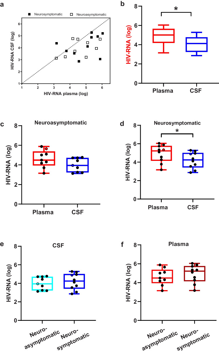
Despite antiretroviral therapy (ART), HIV persistence in the central nervous system (CNS) continues to cause a range of cognitive impairments in people living with HIV (PLWH). Upon disease progression, transmigrating CCR5-using T-cell tropic viruses are hypothesized to evolve into macrophage-tropic viruses in the CNS that can efficiently infect low CD4-expressing cells, such as microglia. We examined HIV-1 RNA concentration, co-receptor usage, and CSF compartmentalization in paired CSF and blood samples from 19 adults not on treatment. Full-length envelope CSF- and plasma-derived reporter viruses were generated from 3 subjects and phenotypically characterized in human primary CD4+ T-cells and primary microglia. Median HIV RNA levels were higher in plasma than in CSF (5.01 vs. 4.12 log10 cp/mL; p = 0.004), and coreceptor usage was mostly concordant for CCR5 across the paired samples (n = 17). Genetically compartmentalized CSF viral populations were detected in 2 subjects, one with and one without neurological symptoms. All viral clones could replicate in T-cells (R5 T cell-tropic). In addition, 3 CSF and 1 plasma patient-derived viral clones also had the capacity to replicate in microglia/macrophages and, therefore have an intermediate macrophage tropic phenotype. Overall, with this study, we demonstrate that in a subset of PLWH, plasma-derived viruses undergo genetic and phenotypic evolution within the CNS, indicating viral infection and replication in CNS cells. It remains to be studied whether the intermediate macrophage-tropic phenotype observed in primary microglia represents a midpoint in the evolution towards a macrophage-tropic phenotype that can efficiently replicate in microglial cells and propagate viral infection in the CNS.

Keywords: CSF; Compartmentalization; HIV; Microglia; Plasma.


Figures

Fig. 1
Fig. 1
Relationship of HIV-1 RNA levels (log 10cp/mL) measured in paired plasma and CSF samples in neuroasymptomatic and neurosymptomatic subjects. a Plots depicts the correlation between HIV RNA CSF and HIV RNA plasma for all subjects. Black dashed line represents line of identity. b, c, d Boxes depict median HIV RNA levels and IQR, measured in plasma (red) and CSF (blue) in all patients, neuroasymptomatic and neurosymptomatic subjects. e, f Boxes depict CSF (blue) and plasma (red) median HIV RNA levels and IQR between neuroasymptomatic and neurosymptomatic subjects. * = Statistically significant (p < 0.05)
Fig. 2
Fig. 2
CSF- and plasma-derived viruses can efficiently infect and replicate in CD4+ T-cells. a-c CD4+ T-cells were infected with 10 ng (p24 Gag) CSF- or plasma-derived virus generated from subjects 8, 19 and 27. Scattered dot plots depict luciferase activity measured in supernatant collected on day 14 post-infection, whereas horizontal black lines represent median luminescence. FPR values for each clone are included at the bottom of the figure. An FPR value below 3.5% is predicted to be X4-tropic virus. An FPR value above 3.5% is predicted to be R5-tropic virus. (d-f) CD4+ T-cells were untreated (no ART) or treated with 100 nM Maraviroc (MVC) prior to infection with 10 ng (p24 Gag) CSF- or plasma-derived virus generated from subjects 8, 19 and 27. Bar graphs depict the maximum infection measured on day 14 post-infection with (grey) and without (white) treatment with MVC. Black error bars depict standard error of the means. Lab strains and viruses derived from plasma (red) or CSF (blue) are separated by vertical dotted lines
Fig. 3
Fig. 3
CSF-derived viruses have a modest enhancement for infecting primary microglia. a-c Primary microglia were infected with 10 ng (p24 Gag) virus generated from subject 8, 19 and 27. Graphs represent luciferase activity measured in supernatant collected on Day 17 post-infection. Black line depicts the median luminescence measured. Lab strains and viruses derived from plasma (red) or CSF (blue) are separated by vertical dotted lines
Fig. 4
Fig. 4
Majority of the CSF and plasma viral population displayed the typical R5 T-cell tropic phenotype. Graphs represent luciferase activity measured in supernatant collected on Day 14 post-infection (D14 p.i.) in primary microglia and CD4+ T-cells. Laboratory strains are depicted in black, CSF-derived clones in blue and plasma-derived clones in red. Black arrows indicate viral clones with an intermediate M-tropic phenotype

References

    1. Arrildt KT, LaBranche CC, Joseph SB, Dukhovlinova EN, Graham WD, Ping LH, Schnell G, Sturdevant CB, Kincer LP, Mallewa M, Heyderman RS, Van Rie A, Cohen MS, Spudich S, Price RW, Montefiori DC, Swanstrom R (2015) Phenotypic correlates of HIV-1 macrophage tropism. J Virol 89:11294–11311. 10.1128/jvi.00946-15 - PMC - PubMed
    1. Bai F, Iannuzzi F, Merlini E, Borghi L, Tincati C, Trunfio M, Bini T, d’Arminio Monforte A, Marchetti G (2017) Clinical and viro-immunological correlates of HIV associated neurocognitive disorders (HAND) in a cohort of antiretroviral-naïve HIV-infected patients. AIDS 31:311–314. 10.1097/QAD.0000000000001346 - PubMed
    1. Bednar MM, Sturdevant CB, Tompkins LA, Arrildt KT, Dukhovlinova E, Kincer LP, Swanstrom R (2015) Compartmentalization, viral evolution, and viral latency of HIV in the CNS. Curr HIV/AIDS Rep 12:262–271. 10.1007/s11904-015-0265-9 - PMC - PubMed
    1. Boom R, Sol JA, Salimans MMM, Jansen CL, Wertheim-Van Dillen PME, Van Der Noordaa J (1990) Rapid and simple method for purification of nucleic acids. J Clin Microbiol 28:495–503 - PMC - PubMed
    1. Borrajo A, Svicher V, Salpini R, Pellegrino M, Aquaro S (2021) Crucial role of central nervous system as a viral anatomical compartment for hiv-1 infection. Microorganisms 9:2537. 10.3390/microorganisms9122537 - PMC - PubMed

Publication types

MeSH terms