Review
. 2024 Oct 1;103(10):955-964.
doi: 10.1097/PHM.0000000000002515. Epub 2024 Apr 6.

Quantitative Musculoskeletal Imaging of the Pediatric Shoulder

Affiliations
Review

Quantitative Musculoskeletal Imaging of the Pediatric Shoulder

Caleb M A Cordes et al. Am J Phys Med Rehabil. .

Abstract

Pediatric acquired and congenital conditions leading to shoulder pain and dysfunction are common. Objective, quantitative musculoskeletal imaging-based measures of shoulder health in children lag recent developments in adults. We review promising applications of quantitative imaging that tend to be available for common pediatric shoulder pathologies, especially brachial plexus birth palsy and recurrent shoulder instability, and imaging-related considerations of musculoskeletal growth and development of the shoulder. We highlight the status of quantitative imaging practices for the pediatric shoulder and highlight gaps where better care may be provided with advances in imaging technique and/or technology.


Conflict of interest statement

Financial disclosure statements have been obtained, and no conflicts of interest have been reported by the authors or by any individuals in control of the content of this article.

Figures

Figure 1:
Figure 1:
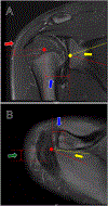
Secondary ossification centers of the developing shoulder joint complex. A: Anterolateral view of glenoid secondary ossification centers. The yellow arrow indicates the subcoracoid glenoid secondary ossification center. The blue arrows indicate the inferior glenoid secondary ossification centers. The black arrow indicates the acromion process. B: Superior axial view of ossification centers of the distal clavicle and acromion process. The green arrows indicate the secondary ossification centers of the acromion process. The red arrow indicates a secondary ossification center of the distal clavicle.
Figure 2:
Figure 2:
Imaging-based measures of glenohumeral conformation. A: Axial T2-weighted gradient echo image of the bilateral shoulders in 18-month-old patient with left-sided brachial plexus birth injury. Imaging performed due to downstream glenohumeral dysplasia. The yellow dotted lines indicate the glenoid line, a line demarking the anterior and posterior rims of the glenoid, on each side. Red dotted lines indicate the Friedman line, a line from the medial tip of the scapular spine through the midpoint of the glenoid cavity. The yellow alpha indicates the glenoid version angle for the unaffected side (right), and the red alpha indicates the glenoid version angle for the affected side (left). Glenoid version is equal to 90° - alpha. Note the greater version on the pathologic side. B: Noncontrast axial proton density fat suppressed image of the right shoulder at the level of the glenohumeral joint. The dotted red line indicates the Friedman line. The dotted yellow line passes through the center of the humeral head perpendicular to the Friedman line. The intersection of the dotted yellow line and dotted red line is the geometric axis of rotation of the humeral head. The solid yellow line is drawn from the geometric axis of rotation to the border of the biceps tendon closest to the humeral head. The yellow delta indicates the angular measurement between the dotted red and solid yellow lines as a measurement of internal/external rotation conformation of the glenohumeral joint. C: Transverse musculoskeletal ultrasound images of affected right and unaffected left side in 5-month-old patient with right-sided brachial plexus birth injury illustrating posterior subluxation of humeral head. Dotted red lines are drawn parallel to the plane of the scapula at the posterior margin. Yellow dotted lines are drawn tangential to the humeral head and posterior aspect of the glenoid. The red alpha and yellow alpha indicate the alpha angle for the right and left sides, respectively. The alpha angle is normal on the left, 24°, and abnormal on the right, 51°.
Figure 3:
Figure 3:
Three-dimensional reformatting of clinical images with axial views of the humeral head and superolateral scapula. The dotted red line represents the glenoid line, drawn between the anterior and posterior rims of the glenoid cavity. The solid green line represents the Friedman line. The dotted black line is drawn through the center of the humeral head perpendicular to the Friedman line. The red alpha indicates the angular measurement between Friedman’s line and the glenoid line. Glenoid version is equal to 90° - alpha angle. The yellow arrow indicates a point (a) on the anterior humeral head on the dotted black line passing through the center of the humeral head. The green arrow indicates a point (b) at the intersection of Friedman’s line and the dotted black line passing through the center of the humeral head. The blue arrow indicates a point (c) on the posterior humeral head on the dotted black line passing through the center of the humeral head. Percentage of the humeral head anterior to the scapular line (PHHA) is computed as (ab/ac)×100. A: Original axial image with measurements of glenoid version and PHHA. Glenoid version is equal to 21° and PHHA is equal to 11% as measured on the original clinical image. B: Anatomically-reformatted image, transformed to orient with the plane of the scapula. Glenoid version is equal to 32° and PHHA is equal to 15% as measured on the reformatted image.
Figure 4:
Figure 4:
Three-dimensional glenohumeral conformation. Components of translational and angular measurements of humeral conformation are indicated. Humeral head translation is defined as the distance from the humeral head center, defined as the center of a best fit ellipsoid, (red dot) to the glenoid center (yellow dot). Three-dimensional (3D) glenoid version (alpha) is defined as the angle between the average normal vector to the glenoid cavity (solid yellow line) and the scapular plane (solid red dotted line), defined by the glenoid center, trigonum spinae scapulae, and the angulus inferior. A: Coronal T2-weighted fat suppressed image of the right shoulder. The red arrow indicates the superoinferior component of translation of the humeral head. The blue arrow indicates the mediolateral component of translation of the humeral head. Alpha indicates measurement of 3D glenoid version in the coronal plane, measured between the solid yellow line average normal vector to the glenoid plane and the solid red line representing the scapular plane. B: Axial proton density fat suppressed image of the right shoulder at the level of the glenohumeral joint. The blue arrow indicates the mediolateral component of humeral head translation. The green arrow represents the anteroposterior component of humeral head translation. Alpha indicates 3D glenoid version in the axial plane, measured between the solid yellow line representing the average normal vector to the glenoid plane and the solid red line representing the scapular plane.

References

    1. Lin CL, Chen YW, Lin LF, Chen CP, Liou TH, Huang SW. Accuracy of the Critical Shoulder Angle for Predicting Rotator Cuff Tears in Patients With Nontraumatic Shoulder Pain. Orthop J Sports Med. 2020;8(5):2325967120918995. - PMC - PubMed
    1. Lee ECS, Roach NT, Clouthier AL, et al. Three-dimensional scapular morphology is associated with rotator cuff tears and alters the abduction moment arm of the supraspinatus. Clin Biomech (Bristol, Avon). 2020;78:105091. - PMC - PubMed
    1. Chang KV, Chen WS, Wang TG, Hung CY, Chien KL. Quantitative ultrasound facilitates the exploration of morphological association of the long head biceps tendon with supraspinatus tendon full thickness tear. PLoS One. 2014;9(11):e113803. - PMC - PubMed
    1. Arend CF, Arend AA, da Silva TR. Diagnostic value of tendon thickness and structure in the sonographic diagnosis of supraspinatus tendinopathy: room for a two-step approach. Eur J Radiol. 2014;83(6):975–979. - PubMed
    1. Bullock GS, Garrigues GE, Ledbetter L, Kennedy J. A Systematic Review of Proposed Rehabilitation Guidelines Following Anatomic and Reverse Shoulder Arthroplasty. J Orthop Sports Phys Ther. 2019;49(5):337–346. - PubMed

MeSH terms

LinkOut - more resources