. 2024 May 7;22(5):e3002634.
doi: 10.1371/journal.pbio.3002634. eCollection 2024 May.

Systematic characterization of all Toxoplasma gondii TBC domain-containing proteins identifies an essential regulator of Rab2 in the secretory pathway

Affiliations

Systematic characterization of all Toxoplasma gondii TBC domain-containing proteins identifies an essential regulator of Rab2 in the secretory pathway

Justin J Quan et al. PLoS Biol. .

Abstract

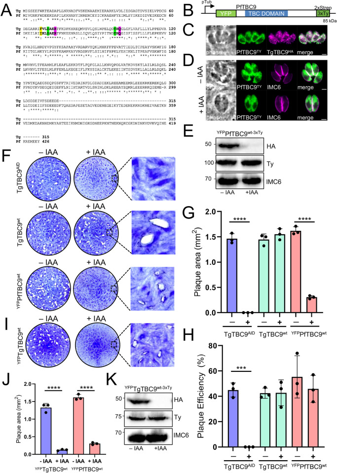
Toxoplasma gondii resides in its intracellular niche by employing a series of specialized secretory organelles that play roles in invasion, host cell manipulation, and parasite replication. Rab GTPases are major regulators of the parasite's secretory traffic that function as nucleotide-dependent molecular switches to control vesicle trafficking. While many of the Rab proteins have been characterized in T. gondii, precisely how these Rabs are regulated remains poorly understood. To better understand the parasite's secretory traffic, we investigated the entire family of Tre2-Bub2-Cdc16 (TBC) domain-containing proteins, which are known to be involved in vesicle fusion and secretory protein trafficking. We first determined the localization of all 18 TBC domain-containing proteins to discrete regions of the secretory pathway or other vesicles in the parasite. Second, we use an auxin-inducible degron approach to demonstrate that the protozoan-specific TgTBC9 protein, which localizes to the endoplasmic reticulum (ER), is essential for parasite survival. Knockdown of TgTBC9 results in parasite growth arrest and affects the organization of the ER and mitochondrial morphology. TgTBC9 knockdown also results in the formation of large lipid droplets (LDs) and multi-membranous structures surrounded by ER membranes, further indicating a disruption of ER functions. We show that the conserved dual-finger active site in the TBC domain of the protein is critical for its GTPase-activating protein (GAP) function and that the Plasmodium falciparum orthologue of TgTBC9 can rescue the lethal knockdown. We additionally show by immunoprecipitation and yeast 2 hybrid analyses that TgTBC9 preferentially binds Rab2, indicating that the TBC9-Rab2 pair controls ER morphology and vesicular trafficking in the parasite. Together, these studies identify the first essential TBC protein described in any protozoan and provide new insight into intracellular vesicle trafficking in T. gondii.


Conflict of interest statement

The authors have declared that no competing interests exist.

Figures

Fig 1
Fig 1. Toxoplasma gondii contains 18 TBC proteins.
Diagram showing the domains present in the Toxoplasma TBC proteins. The approximate position of the TBC and other domains as predicted by SMART, PFAM, and NCBI conserved protein domain search tools are shown [–30]. TBC, Tre2–Bub2–Cdc16 (TBC) domain-containing proteins; Sec7, Sec7-domain-containing; STK, Serine/Threonine protein kinase; TLDc, TBC, LysM, Domain Catalytic domain; CC, coil-coil domain; TM, transmembrane; SMART, Simple Modular Architecture Research Tool.
Fig 2
Fig 2. T. gondii TBC proteins localize to discrete regions of the secretory pathway and cytoplasmic vesicles.
IFAs of endogenously epitope tagged TgTBC1-18 parasites. (A) Diagram of TgTBC1-18 showing the epitope tag and selectable marker. (B) IFA of endogenously tagged TgTBC1, 2, 3, and 18 stained with antibodies against epitope tags and colocalized with GRASP55. The yellow and white arrowheads in the bottom 2 panels denote subtle differences between the TBC protein and GRASP55. Green = endogenously tagged TBC proteins, Magenta = GRASP55-mCherry. Quantification of all colocalizations were quantified by calculating the Pearson’s correlation coefficient (R). Mean values and respective standard deviation of 10–15 parasites are indicated next to the respective image (see also S1 Data). (C) IFA of TgTBC6, 9, and 14 stained with anti-HA and colocalized with SERCA. Green = rabbit anti-HA, Magenta = mouse anti-SERCA. (D) IFA of TgTBC 5, 12, 13, 16, and 17 shown partially colocalizing with SERCA. Green = rabbit anti-HA, Magenta = mouse anti-SERCA. (E) IFA of TgTBC10 shows cytoplasmic and nuclear staining with partial colocalization with SERCA. The white arrowheads show that TgTBC10 is not nuclear excluded. Green = rabbit anti-HA, Magenta = mouse anti-SERCA. (F) IFA of TgTBC11 and TgTBC15 colocalizing with endogenously tagged IMC293xV5 in daughter buds. Magenta = mouse anti-HA, Green = rabbit anti-V5. (G) IFA of TgTBC8 shows staining central portion of the maternal IMC as accessed by IMC6 staining. The white arrowheads indicate basal portions of the parasite where TgTBC8 is absent. Magenta = mouse anti-HA, Green = rabbit anti-IMC6. (H) IFA of TgTBC4 and TgTBC7 with no detectable smHA staining. Magenta = mouse anti-HA, Green = rabbit anti-IMC6. Scale bars for all images, 2 μm. (I) Diagram and PCR of endogenous TgTBC4 and TgTBC7 tagged parasites. Primers labeled as red arrows were used to test gDNA from parental and tagged strains. IFA, indirect immunofluorescence assay; IMC, inner membrane complex; smHA, spaghetti monster HA; TBC, Tre2-Bub2-Cdc16.
Fig 3
Fig 3. TgTBC9 is essential for parasite survival and organization of the ER and mitochondria.
(A) Maximum likelihood tree based on the TBC domains of TgTBC9 and its orthologs from Plasmodium falciparum (Pf), Trypanosoma brucei (Tb), Entamoeba histolytica (Eh), Cryptosporidium parvum (Cp), Theileria parva (Tp), Trypanosoma cruzi (Tc), Leishmania major (Lm), Dictyostelium discoideum (Dd). Blue spheres denote level of support (1,000 bootstrap replicates). (B) Diagram of TgTBC9AID showing its TBC domain and a mIAA7-3xHA degron tag added to the C-terminus of the protein. (C) IFA of TgTBC9AID localizing to an ER-like pattern. Magenta, mouse anti-HA; green, rabbit anti-IMC6. (D) IFA of TgTBC9AID without (-) or with (+) IAA for 24 h (following a 4 h pretreatment ±IAA) showing that TgTBC9AID is efficiently degraded, resulting in parasite growth arrest. Magenta, mouse anti-HA; green, rabbit anti-IMC6. (E) Western blot analysis of showing TgTBC9AID is efficiently degraded upon IAA treatment. IMC6 is used as a load control. (F) Plaque assays showing that TgTBC9-depleted parasites are unable to form plaques. (G) Quantification of plaque assays at day 7 showing no plaque formation by TgTBC9AID parasites +IAA. All raw data in S1 Data. (H) IFA of TgTBC9AID parasites grown in ±IAA as described in D showing affected ER morphology. Magenta, mouse anti-SERCA; green, rabbit anti-IMC6. (I) TgTBC9AID-3xTy parasites grown in ±IAA as described in D but with staining for Der1-2 using an endogenously tagged Der1-23xHA strain. Magenta, mouse anti-HA; green, rabbit anti-IMC6. (J) TgTBC9AID parasites grown in ±IAA as described in D but with staining for mitochondrion using anti-F1β ATPase. Magenta, mouse anti-F1β ATPase; green, rabbit anti-IMC6. (K) IFA of TgTBC9AID grown with +IAA for 24 h and then washed and incubated with -IAA for another 24 h (following a 4 h pretreatment +IAA) showing that TgTBC9 deficient parasites resume growth in the absence of IAA. Magenta, mouse anti-F1β ATPase; green, rabbit anti-IMC6. (L) TgTBC9AID parasites grown in ±IAA as described in D but with staining for lipid structures using BODIPY 493/503. Magenta, rabbit anti-IMC6; green, BODIPY 493/503. The white arrowheads denote host lipid structures, while the yellow arrowheads denote parasite lipid structures. (M) TgTBC9AID parasites grown in +IAA for 12 h (following a 4 h pretreatment) staining for lipid structures using BODIPY 493/503. Magenta, rabbit anti-IMC6; green, BODIPY 493/503. The yellow arrowheads denote parasite lipid structures. (****, P < 0.0001). Scale bars for all images, 2 μm. ER, endoplasmic reticulum; IFA, indirect immunofluorescence assay; TBC, Tre2-Bub2-Cdc16.
Fig 4
Fig 4. Ultrastructural analysis of TgTBC9 knockdown parasites.
(A) TEM of intracellular Toxoplasma in HFF for 24 h in the absence of IAA illustrating 4 control (untreated) parasites in a symmetrical organization inside the PV. (B–F) TEM of intracellular Toxoplasma in the presence of IAA. (B) Image showing an example of a PV containing 2 or 3 IAA-treated parasites with disorganized parasite morphology. The TgTBC9 knockdown parasites contain aberrant ER structures, a rounded mitochondrion (mt), and the presence of amylopectin granules (AG). (C) Image showing that TgTBC9 knockdown parasites also contain multiple nuclear profiles within one parasite (n1-3), very large LDs, and a disrupted ER-Golgi (Go) connection with accumulated vesicles of various size and electron density. (D, E) Images showing membranous structures surrounded by ER elements (asterisks) with E illustrating the likely progressive steps of their formation/compaction. (F) Parasites showing cytoplasmic clefts in the cytoplasm (arrowheads in D, F), likely preceding parasite death. Asterisks highlights an ER enveloped structure. Ac, acidocalcisome; ap, apicoplast; DG, dense granule; hc, host cell; IVN, intravacuolar network; mi, microneme; n, nucleus; rh, rhoptry. Scales bar for all images, 500 nm. ER, endoplasmic reticulum; HFF, human foreskin fibroblast; LD, lipid droplet; PV, parasitophorous vacuole; TEM, transmission electron microscopy.
Fig 5
Fig 5. TBC dual-finger active sites are required for TgTBC9 GAP activity.
(A) Diagram of the dual-finger consensus and highlighting which TgTBC9 residues in the arginine finger and glutamine finger motifs were mutated to alanine. Green boxes depict strictly conserved residues; yellow boxes depict semi-conserved residues; magenta boxes depict residues that were mutated to alanine. (B) Plaque assays showing that complementation with full-length TgTBC9 restores ability to form plaques upon depletion of endogenous TgTBC9. (C) Western blot analysis showing knockdown of endogenous TgTBC9 and complementation with a Ty-tagged TgTBC9wt copy targeted to the UPRT locus. IMC6 is used as a load control. (D) Plaque assays showing complementation with the TgTBC9R74A or TgTBC9Q101A mutants are unable to rescue the TgTBC9AID knockdown. (E) Western blot analysis showing that the TgTBC9wt and TgTBC9R74A and TgTBC9Q101A mutant parasites have similar levels of expression. IMC6 is used as a load control. (F) Quantification of plaque assays at day 7 showing rescue with TgTBC9wt but no plaque formation by TgTBC9AID, TgTBC9R74A, and TgTBC9Q101A mutant parasites +IAA (****, P ≤ 0.0001). All raw data in S1 Data. GAP, GTPase-activating protein; TBC, Tre2-Bub2-Cdc16.
Fig 6
Fig 6. PfTBC9 partially rescues the lethal knockdown of TgTBC9.
(A) Full protein alignment of TgTBC9 (Tg) and PfTBC9 (Pf) using Clustal Omega [61]. Bold residues highlighted in yellow depict semi-conserved residues; bold residues in green depict strictly conserved residues; bold residues in magenta indicate predicted R and Q catalytic sites. (B) Diagram of YFPPfTBC9wt-3xTy expressed from the tubulin promoter with an N-terminal YFP and C-terminal 2xStrep-3xTy tag. (C) IFA of YFPPfTBC9wt-3xTy colocalized with TgTBC9AID-3xHA showing overlap in the ER. Green, mouse anti-Ty and GFP; magenta, rabbit anti-HA. Scale bar = 2 μm. (D) IFA of PfTBC9wt with (-) or without (+) IAA for 24 h showing that PfTBC9wt is expressed and unaffected in ±IAA. Magenta, mouse anti-Ty and GFP; green, rabbit anti-HA. Scale bar = 2 μm. (E) Western blot analysis of YFPPfTBC9wt-3xTy in the background of TgTBC9AID tagged parasites. IMC6 is used as a load control. (F) Plaque assays showing that YFPPfTBC9wt complemented parasites formed smaller plaques compared to the TgTBC9wt complemented parasites. (G) Quantification of plaque area at day 7 showing small plaque formation by YFPPfTBC9wt complemented parasites +IAA (****, P <  0.0001). (H) Quantification of plaque efficiency at day 7 show no significance between YFPPfTBC9wt +IAA from TgTBC9wt complement groups (***, P = 0.0002). (I) Plaque assays showing that YFPPfTBC9wt complemented parasites +IAA similarly form small plaques. (J) Quantification of plaque area at day 7 showing small plaque formation by YFPTgTBC9wt and YFPPfTBC9wt complemented parasites (****, P < 0.0001). (K) Western blot analysis of YFPTgTBC9wt-3xTy in the background of TgTBC9AID tagged parasites. IMC6 is used as a load control. All raw data in S1 Data. ER, endoplasmic reticulum; IFA, indirect immunofluorescence assay.
Fig 7
Fig 7. IP and pairwise Y2H of TgTBC9 reveals Rab2 an interactor.
(A) Western blot analysis of the TgTBC9 IP showing the input (Total) and eluted material (Eluate) probed with mouse anti-HA antibodies. (B) Table showing the rank of TgTBC9 and small GTPase proteins from the IP analysis. The complete dataset is shown in S3 Table. Untagged parasites were used as the control. The ToxoDB-IDs, localization (* determined in [17], ** determined in [21], *** determined in [20]), molecular weight, and GWCS phenotype score are also shown [36]. (C) Spot assays of pairwise Y2H assessing TgTBC9 and Rab2 interaction using either wild-type (wt), GTP-locked mutant (Rab2Q66L), or catalytical inactive mutant (TgTBC9R74A) sequences. Yeast expressing the indicated constructs were grown under permissive (-L/W) or restrictive (-L/W/H) conditions to assess interactions. (D) Y2H assessing the interaction of catalytically inactive mutant TgTBC9 with the indicated mutant Rabs, as described in C. (E) Diagram of Rab2 showing the N-terminal DD and 2xV5 tag (DDV5Rab2). IFA analysis of TgTBC9AID parasites expressing DDV5Rab2 treated for 24 h with 1 μm Shld-1 prior to fixation. Magenta = mouse anti-HA, Green = rabbit anti-V5. Scale bars, 2 μm. Colocalizations were quantified by calculating the Pearson’s correlation coefficient (R). Mean values and standard deviation of 10–15 parasites are indicated next to the image (see also S1 Data). (F) IFA of TgTBC9AID parasites expressing DDV5Rab2 without (-) or with (+) IAA for 24 h (following a 4 h pretreatment ±IAA) and treated for 24 h with 1 μm Shld-1 prior to fixation showing disruption of Rab2 expression and localization. Magenta = mouse anti-V5, Green = rabbit anti-IMC6. Scale bars, 2 μm. (G) Representative IFA showing a field of TgTBC9AID parasites expressing DDV5Rab2 grown in ±IAA as described in F. Magenta = mouse anti-V5, Green = rabbit anti-IMC6. Scale bars, 10 μm. (H) Western blot analysis of TgTBC9AID parasites expressing DDV5Rab2 grown in ±IAA as descried in F showing a 52% decrease of Rab2 in IAA-treated parasites. Quantification was normalized to IMC6, which is used as a load control. GWCS, genome-wide CRIPSR screen; IFA, indirect immunofluorescence assay; IP, immunoprecipitation.

Update of

References

    1. Levine ND. Progress in Taxonomy of the Apicomplexan Protozoa. J Protozool. 1988;35:518–520. doi: 10.1111/j.1550-7408.1988.tb04141.x - DOI - PubMed
    1. Davies AP, Chalmers RM. Cryptosporidiosis. BMJ. 2009;339:b4168–b4168. doi: 10.1136/bmj.b4168 - DOI - PubMed
    1. Tenter AM, Heckeroth AR, Weiss LM. Toxoplasma gondii: from animals to humans. Int J Parasitol. 2000;30:1217–1258. doi: 10.1016/s0020-7519(00)00124-7 - DOI - PMC - PubMed
    1. Miller LH, Baruch DI, Marsh K, Doumbo OK. The pathogenic basis of malaria. Nature. 2002;415:673–679. doi: 10.1038/415673a - DOI - PubMed
    1. Blader IJ, Coleman BI, Chen C-T, Gubbels M-J. Lytic Cycle of Toxoplasma gondii: 15 Years Later. Annu Rev Microbiol. 2015;69:463–485. doi: 10.1146/annurev-micro-091014-104100 - DOI - PMC - PubMed