. 2024 May 7;7(1):541.
doi: 10.1038/s42003-024-06230-5.

Aging-regulated PNUTS maintains endothelial barrier function via SEMA3B suppression

Affiliations

Aging-regulated PNUTS maintains endothelial barrier function via SEMA3B suppression

Noelia Lozano-Vidal et al. Commun Biol. .

Abstract

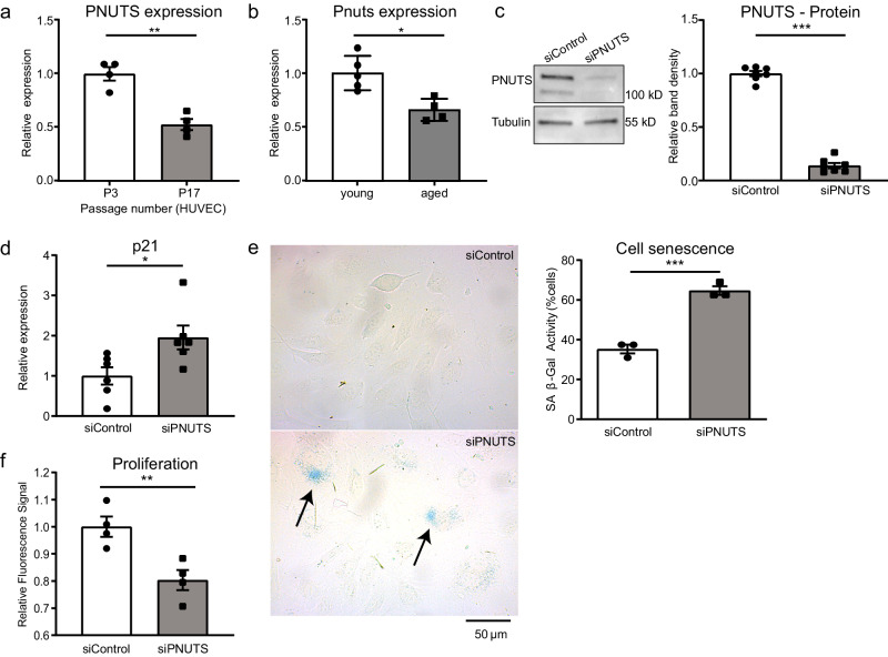
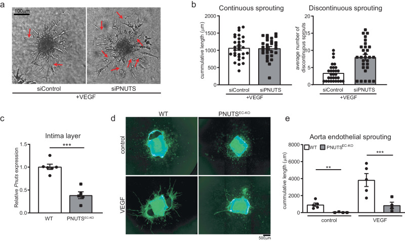
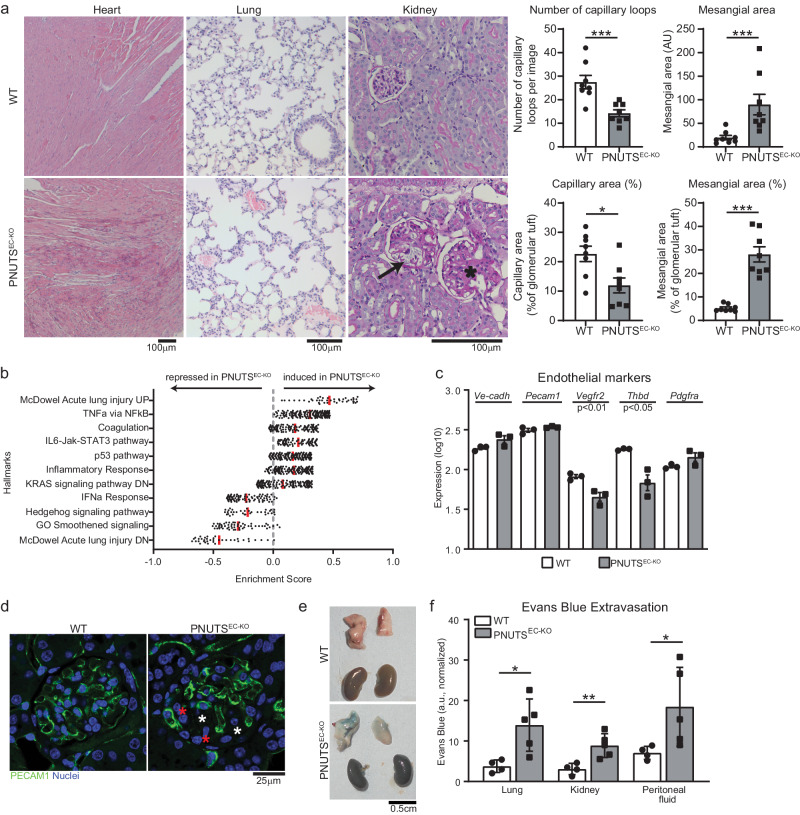
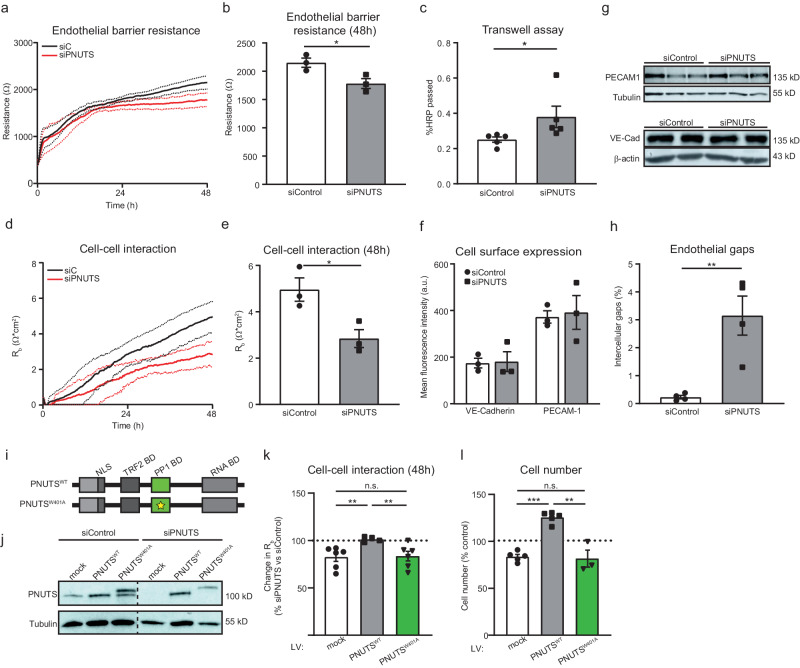
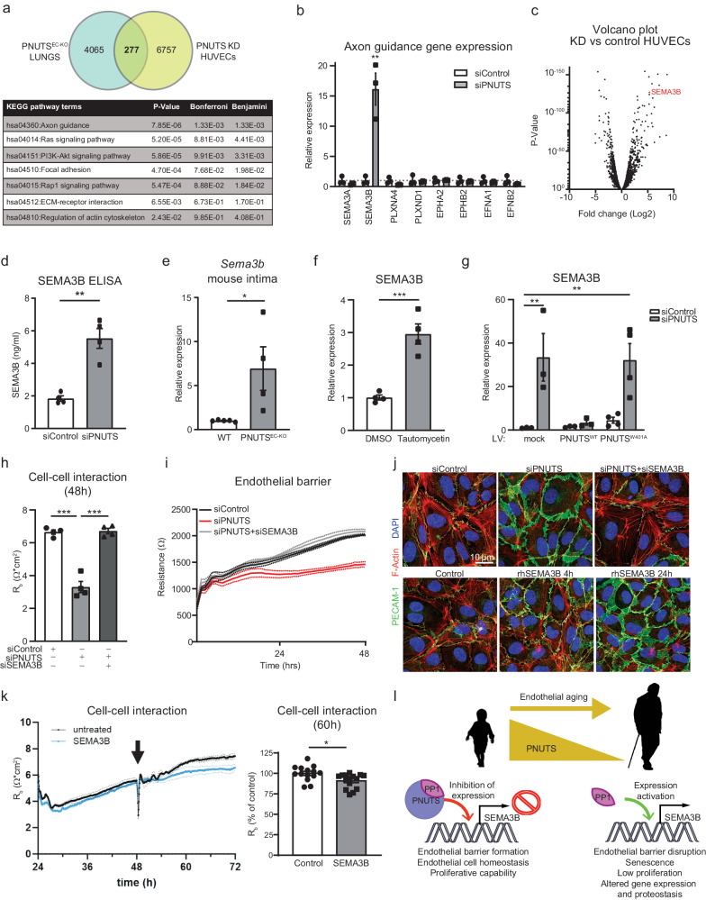
Age-related diseases pose great challenges to health care systems worldwide. During aging, endothelial senescence increases the risk for cardiovascular disease. Recently, it was described that Phosphatase 1 Nuclear Targeting Subunit (PNUTS) has a central role in cardiomyocyte aging and homeostasis. Here, we determine the role of PNUTS in endothelial cell aging. We confirm that PNUTS is repressed in senescent endothelial cells (ECs). Moreover, PNUTS silencing elicits several of the hallmarks of endothelial aging: senescence, reduced angiogenesis and loss of barrier function. Findings are validate in vivo using endothelial-specific inducible PNUTS-deficient mice (Cdh5-CreERT2;PNUTSfl/fl), termed PNUTSEC-KO. Two weeks after PNUTS deletion, PNUTSEC-KO mice present severe multiorgan failure and vascular leakage. Transcriptomic analysis of PNUTS-silenced HUVECs and lungs of PNUTSEC-KO mice reveal that the PNUTS-PP1 axis tightly regulates the expression of semaphorin 3B (SEMA3B). Indeed, silencing of SEMA3B completely restores barrier function after PNUTS loss-of-function. These results reveal a pivotal role for PNUTS in endothelial homeostasis through a SEMA3B downstream pathway that provides a potential target against the effects of aging in ECs.


Conflict of interest statement

N.L.-V. and L.S. are currently employed by AstraZeneca. All other authors declare no competing interests.

Figures

Fig. 1
Fig. 1. PNUTS is an aging-regulated protein and regulates senescence in endothelial cells.
a HUVECs were cultured for 3 or 17 passages (P) and PNUTS expression was measured by qRT-PCR (n = 4). b Aortic intima was isolated from young (8–12 weeks old) and aged (18–20 months old) mice and immediately lysed for RNA isolation. Intima RNA was sequenced. cf HUVECs were transfected with siRNAs targeting PNUTS or a control sequence. c PNUTS protein levels were determined by WB at 48 h after transfection, relative to alpha-tubulin. Densitometric quantification is depicted on the right (n = 7). d Changes in p21 expression were assessed by qRT-PCR. Expression values are relative to siControl-treated HUVECs and normalized to RPSA mRNA (n = 6). e The percentage of senescent HUVECs was analyzed by staining for senescence-associated β-Galactosidase (SA-(β-Gal) 72 h after transfection. Images were taken (4 fields per sample) and the percentage of senescent cells (blue) over the total number of cells in each field was calculated in n = 3 independent experiments, 2–3 biological replicates per group and experiment). f Cell proliferation was assayed by testing the incorporation of EdU after transfection (n = 4). *p < 0.05, **p < 0.01, ***p < 0.001. Error bars depict the standard error of the mean (SEM).
Fig. 2
Fig. 2. PNUTS is necessary for normal angiogenic sprouting.
a In vitro sprouting was analyzed under VEGF (50 ng/ml) stimulation. Representative images are shown, red arrows indicate discontinuous sprouts. b Quantification of cumulative sprout length (left) and number of discontinuous sprouts (right) (n = 30 spheroids per group in 3 independent experiments were measured). c Endothelial Pnuts expression of PNUTSEC-KO and WT mice was assessed by RT-qPCR in intima samples 15 days after initiating tamoxifen treatment and normalized to Rplp0 mRNA (n = 3). d In vivo angiogenesis was tested by aortic ring assay in WT and PNUTSEC-KO mice in basal conditions or upon 48 h of VEGF (10 ng/ml) stimulation. e Quantification of aortic ring sprouting. n = 4–5 mice per group. *p < 0.05, **p < 0.01, ***p < 0.001. Error bars depict the standard error of the mean (SEM).
Fig. 3
Fig. 3. Induction of endothelial-specific PNUTS KO in mice provokes vascular leakage and multiorgan failure.
a Histopathological study of different tissues in PNUTSEC-KO mice compared to WT mice: left, heart samples (haematoxylin-eosin) presenting edema; middle, lung samples (haematoxylin-eosin) presenting thrombi; right, kidney samples (PAS staining) showing glomerulosclerosis (asterisk) and capillary dilatation (black arrow) in renal glomeruli. Quantifications of the kidney sections show the absolute and relative contribution of capillaries and mesangial tissue in glomeruli (n = 8 per group). b RNA-seq was performed with lung tissue of WT and PNUTSEC-KO mice and was analysed for differentially regulated pathways using Gene Set Enrichment Analysis. Enrichment scores of the indicated pathways are plotted on the x axis (n = 3). c The expression levels of the endothelial markers Ve-cadherin, Pecam1, Vegfr2, Thbd and Pdgfra in WT and PNUTSEC-KO lung samples were confirmed by RT-qPCR and normalized to Rplp0 mRNA (n = 3). d The presence of ECs in the glomerular capillary network was investigated by PECAM1 immunostaining in kidneys of WT and PNUTSEC-KO mice. Red and white asterisks indicate potential mesangial proliferation and glomerulosclerotic areas, respectively. e, f An Evans Blue (EB) extravasation assay was performed to measure the vascular extravasation in different organs. e Representative lung and kidney images 1 h after intravenous administration of EB at 25 mg/kg. f Colorimetric measurement of extravasated EB into lung, kidney and peritoneal fluid (n = 4–5 mice per group). *p < 0.05, **p < 0.01, ***p < 0.001. Error bars depict the standard error of the mean (SEM).
Fig. 4
Fig. 4. PNUTS is necessary for endothelial barrier function independently of cell junction gene expression.
ah HUVECs were treated with siRNA (si) targeting PNUTS or a control sequence. a-b) HUVEC barrier resistance was assessed by ECIS at 4000 Hz for 48 h, presented as average resistance ± SEM of n = 3 experiments, 8 biological replicates per group and experiment. c HUVECs were seeded in transwells and HRP passage through the endothelial monolayer was assessed by absorption measurements (450 nm) and shown as percentage of total HRP (n  =  5). d, e Cell-cell interaction was assessed by modeling of data obtained by ECIS measured as Rb (Ω*cm2); presented as average resistance ± SEM of n = 3 experiments, 8 biological replicates per group and experiment. f Cell surface presence of PECAM1 and VE-cadherin was assessed by flow cytometry 48 h after siRNA transfection (n = 3). g Total PECAM1 and VE-cadherin levels were assessed by Western Blotting (WB). Tubulin and β-actin expression was used as loading control. h Cells were grown into confluence and immuno-stained for VE-cadherin, PECAM1 and F-actin. DAPI was used to stain nuclei. The presence of intercellular gaps in endothelial monolayers was measured by quantifying the intercellular areas versus the total area in 4 fields per image, 3 images per experiment, n = 4. i Schematic representation of the PNUTS lentiviral vectors used for the barrier rescue experiments. Both vectors included silent mutations in the seed sequence of siPNUTS. HUVECs were transduced with the indicated constructs for 8–10 days and later transfected with siControl or siPNUTS to silence endogenous expression of PNUTS, before subjecting them to ECIS and cell counting (4–6 independent experiments, 4 biological replicates per group and experiment). j PNUTS expression in total cell lysates of HUVECs treated with indicated vectors and siRNAs was analyzed by WB, Tubulin was used as a loading control. Representative WB is shown. k Change in cell-cell interaction measurement from ECIS 48 h after cell seeding, measured as variation of Rb of siPNUTS- vs siControl-treated cells. l Cell proliferation of ECIS-assayed HUVECs, measured as % number of cells relative to control. *p < 0.05, **p < 0.01, ***p < 0.001. Error bars depict the standard error of the mean (SEM).
Fig. 5
Fig. 5. PNUTS KD induces transcriptomic changes in HUVECs.
a The two subsets of RNAseq data we obtained, from endothelial-depleted PNUTS mouse lungs and PNUTS knockdown (KD) HUVECs, were compared to find common targets of PNUTS depletion. The Venn diagram depicts the finding of 277 common transcripts, which were functionally analysed using KEGG pathways analysis, shown in the table below. b Changes in axon guidance gene expression after PNUTS silencing was assessed by RT-qPCR. Expression values are relative siControl-treated HUVECs and normalized to RPSA mRNA (n = 6). c Volcano plot showing the distribution of gene expression in PNUTS KD versus control HUVECs. SEMA3B is marked in red. d Supernatant SEMA3B concentration was determined by ELISA 72 h after PNUTS silencing (n = 4). e Expression levels of Sema3b mRNA in intima samples of PNUTSEC-KO mice was assessed by RT-qPCR, relative to WT samples and normalized to Rplp0 mRNA. f HUVECs were treated with vehicle or Tautomycetin (166 nM) for 48 h and mRNA was analyzed by RT-qPCR for expression of SEMA3B, normalized to RPSA mRNA (n = 3). g Expression of SEMA3B was measured in mRNA samples of HUVECs assayed in Fig. 4j–l, relative to control cells and normalized to RPSA mRNA. h-i HUVECs were co-transfected with siPNUTS and/or siSEMA3B were subjected to ECIS for 48 h (n = 4 independent experiments, 4 biological replicates per group and experiment). h Cell-cell interaction was modeled. i Endothelial resistance was measured at 4000 Hz. j Top panel: siSEMA3B rescues the effect of PNUTS silencing on adherence junctions (shown as PECAM1 IF staining). Bottom panel: PECAM1 IF staining shows time course of change in adherens junctions upon stimulation with recombinant human SEMA3B. k Cell-cell interaction was modeled using ECIS. The arrow indicates the 48 h time point at which recombinant SEMA3B was added to the medium (or not; untreated). Quantification was performed at 60 h (n = 4 biological replicates and 3–4 technical replicates). l Graphic summary of the proposed mechanism. In young individuals, PNUTS interacts and promotes activity of PP1, which represses the expression of SEMA3B. Endothelial cells are in homeostasis and maintain their barrier function. During aging, PNUTS is repressed in endothelial cells. The absence of PNUTS inhibits PP1 function at the SEMA3B promoter activating SEMA3B expression. SEMA3B exerts repulsive signals between endothelial cells, promoting intercellular gaps and disrupting the barrier. This provokes a series of critical changes in the cells leading to cellular senescence. *p < 0.05, **p < 0.01, ***p < 0.001. Error bars depict the standard error of the mean (SEM).

References

    1. Lopez-Otin C, Blasco MA, Partridge L, Serrano M, Kroemer G. The hallmarks of aging. Cell. 2013;153:1194–1217. doi: 10.1016/j.cell.2013.05.039. - DOI - PMC - PubMed
    1. Christensen K, Doblhammer G, Rau R, Vaupel JW. Ageing populations: The challenges ahead. Lancet. 2009;374:1196–1208. doi: 10.1016/S0140-6736(09)61460-4. - DOI - PMC - PubMed
    1. North BJ, Sinclair DA. The intersection between aging and cardiovascular disease. Circ. Res. 2012;110:1097–1108. doi: 10.1161/CIRCRESAHA.111.246876. - DOI - PMC - PubMed
    1. D’Agostino RB, Sr., et al. General cardiovascular risk profile for use in primary care: The Framingham Heart Study. Circulation. 2008;117:743–753. doi: 10.1161/CIRCULATIONAHA.107.699579. - DOI - PubMed
    1. Vita JA. Endothelial function. Circulation. 2011;124:e906–e912. doi: 10.1161/CIRCULATIONAHA.111.078824. - DOI - PubMed

Publication types