. 2024 May 7;22(1):431.
doi: 10.1186/s12967-024-05150-6.

Multiomics analysis identifies oxidative phosphorylation as a cancer vulnerability arising from myristoylation inhibition

Affiliations

Multiomics analysis identifies oxidative phosphorylation as a cancer vulnerability arising from myristoylation inhibition

Erwan Beauchamp et al. J Transl Med. .

Abstract

Background: In humans, two ubiquitously expressed N-myristoyltransferases, NMT1 and NMT2, catalyze myristate transfer to proteins to facilitate membrane targeting and signaling. We investigated the expression of NMTs in numerous cancers and found that NMT2 levels are dysregulated by epigenetic suppression, particularly so in hematologic malignancies. This suggests that pharmacological inhibition of the remaining NMT1 could allow for the selective killing of these cells, sparing normal cells with both NMTs.

Methods and results: Transcriptomic analysis of 1200 NMT inhibitor (NMTI)-treated cancer cell lines revealed that NMTI sensitivity relates not only to NMT2 loss or NMT1 dependency, but also correlates with a myristoylation inhibition sensitivity signature comprising 54 genes (MISS-54) enriched in hematologic cancers as well as testis, brain, lung, ovary, and colon cancers. Because non-myristoylated proteins are degraded by a glycine-specific N-degron, differential proteomics revealed the major impact of abrogating NMT1 genetically using CRISPR/Cas9 in cancer cells was surprisingly to reduce mitochondrial respiratory complex I proteins rather than cell signaling proteins, some of which were also reduced, albeit to a lesser extent. Cancer cell treatments with the first-in-class NMTI PCLX-001 (zelenirstat), which is undergoing human phase 1/2a trials in advanced lymphoma and solid tumors, recapitulated these effects. The most downregulated myristoylated mitochondrial protein was NDUFAF4, a complex I assembly factor. Knockout of NDUFAF4 or in vitro cell treatment with zelenirstat resulted in loss of complex I, oxidative phosphorylation and respiration, which impacted metabolomes.

Conclusions: Targeting of both, oxidative phosphorylation and cell signaling partly explains the lethal effects of zelenirstat in select cancer types. While the prognostic value of the sensitivity score MISS-54 remains to be validated in patients, our findings continue to warrant the clinical development of zelenirstat as cancer treatment.

Keywords: Cancer; Complex I; N-myristoylation; N-myristoyltransferase; NDUFAF4; NMT inhibitor (NMTI); Oxidative phosphorylation; PCLX-001 (zelenirstat); Respiration.


Conflict of interest statement

As co-founders and/or shareholders of Pacylex Pharmaceuticals Inc. (www.pacylex.com), which owns the rights to patent applications PCT/2012/000696 and PCT/2013/050821, L.G.B., J.R.M., E.B., J.M.G., R.P. and M.Y. declare potential competing interests.

Figures

Fig. 1
Fig. 1
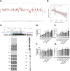
NMT2 levels, but not NMT1 levels, are decreased in numerous hematologic cancer cell lines and tumors. NMT1 (black) and NMT2 (gray) transcript counts in cancer cell lines (A) (data extracted from Depmap 22Q4) and tumors (C) (data extracted from TCGA). NMT2 mRNA expression sorted by cancer cell line (B) and tumor origin (D) (min-to-max box plot; one-way ANOVA comparison to all samples; ***p < 0.0001). NMT2 protein levels in a collection of local lymphocytic cell lines and solid lymphomas. NMT2 levels, assessed by western blot of 35 μg of cell lysate proteins in immortalized “normal” human B-cell line IM9, neoplastic B-cell lymphoma cell lines, leukemic T-cell lines (E), and lysates of various types of human solid lymphomas (F). Immunohistochemical staining for NMT1 (G) and NMT2 (H) in normal lymph nodes, Burkitt’s lymphoma (BL), and diffuse large B-cell lymphoma (DLBCL). Upper row: N1 and N2, normal lymph nodes; Neg, negative control in which primary antibody was omitted. Middle row: Three cases of BL (BL1–3). Lower row: Three cases of DLBCL (LCL1–3). Scale bars: 75 μ. Dependency scores (median non-essential KO effect is 0 and median essential KO effect is −1) obtained after NMT CRISPR knockout in 1078 cancer cell lines (I) NMT1 dependency was significantly correlated with NMT2 expression (r = 0.600; p < 0.0001) (J). Progression-free survival (PFS) Kaplan–Meier analysis of 470 DLBCL patients (GSE31312) with high (red) versus low (blue) NMT1 (K) and NMT2 (L) expression (high and low populations are calculated based on median expression). The P value was determined using the log-rank test
Fig. 2
Fig. 2
The NMT2 locus is methylated on CpG islands in lymphoma cell lines and tumors. Aggregation methylation beta values at the NMT2 locus in tumors and normal tissues showed increased methylation in breast, colon, kidney, and lung tumors compared to those in associated normal tissues (A). Aggregation methylation beta values at the NMT2 locus in tumors correlate with NMT2 expression in DLBCL (B). Structure of the NMT2 genomic region and bisulfite sequencing results for various lymphocytic cell lines and five DLBCL patient tumors used in this study (C). All corresponding DLBCL tumors contained only residual levels of NMT2 by IHC (data not shown). The number of NMT2 mRNA copies after acute 24 h treatments with DAC in the presence or absence of SAHA (1 μM) in BL2 (D) and IM9 (F). NMT2 protein levels normalized to GAPDH in BL2 (E) are shown as the percentage of normalized NMT2 levels in IM9 (G) in the corresponding treatments, as assessed by western blotting values are mean ± s.e.m. of three independent experiments. One representative western blot from three independent experiments is shown. Samples sharing letters are not statistically different (one-way ANOVA)
Fig. 3
Fig. 3
Identification of cancers susceptible to be sensitive to PCLX-001 or PCLX-002 NMTI treatment. Simplified strategy leading to the establishment of a myristoylation inhibition sensitivity signature made of 54 genes (MISS-54) (A) MISS-54 score in tumors sorted by cancer origin (data extracted from TCGA) (B). MISS-54 score (blue color scale; the darker the blue, the more sensitive the tumor) in 11,070 tissue samples (tumor and normal tissues) clustered for their total individual mRNA expression [105] (C). MISS-54 Normalized Enrichment Score (NES) in tumors RNA-seq expression versus their associated normal tissue (D)
Fig. 4
Fig. 4
NMT2 re-expression is toxic to NMT2-deficient cancer cells and NMT KO sensitizes cells to PCLX-001 treatment. NMT2-deficient cells BL2 viability after expressing NMT2 compared to NMT2-normal IM9 cells (p = 0.0095) (A). BL2 cancer cells were grown for 6 months with increasing concentrations of PCLX-001 (from 2 to 20 nM final) to selectively isolate resistant cells. Resistant cell viability curves after treatment with increasing amounts of PCLX-001 for 96 h (n = 3). (B) Corresponding histogram of the calculated EC50 (p = 0.04); (C) Quantification of NMT2 protein level in BL2 resistant cells (D) (p < 0.001; ratios normalized to BL2 normal NMT2 protein level, one western blot representative of four independent experiments is shown). Viability of inducible NMT1 KO HAP1 cells and NMT2 KO/inducible NMT1 KO HAP1 cells incubated with or without doxycycline (1 μg/mL) and with increasing concentration of PCLX-001 for 96 h (E), or pre-incubated with/without doxycycline (1 μg/mL) for 72 h then with increasing concentration of PCLX-001 for 96 h (F)
Fig. 5
Fig. 5
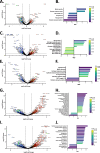
Genetic and pharmacologic myristoylation inhibition drastically changes the proteomes of HAP1 cells. Differential proteomic analysis of inducible NMT1 KO HAP1 cells treated with doxycycline (96 h) (myristoylated proteins bordered in green and complex I proteins shown in orange) (A), NMT2 constitutive KO (C), and inducible NMT1 KO/NMT2 KO (E) with the Hallmark Gene Set Enrichment Analysis of the proteins decreased and increased (False Discovery Rate qVal < 0.25) (B-D-F). Proteomic analysis of inducible parental HAP1 cells treated with PCLX-001 at 0.5 (G) or 5 μM (I) for 72 h (myristoylated proteins bordered in green) with associated Hallmark Gene Set Enrichment Analysis of the proteins decreased and increased in HAP1 cells (False Discovery Rate qVal < 0.25) (H–J)
Fig. 6
Fig. 6
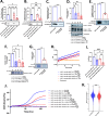
Doxycycline-induced NMT1 KO and PCLX-001 treatment lead to complex I mis-assembly and degradation, reduced activity, and inhibition of cellular oxygen consumption rate. Western blot analysis of SFK member Lyn (A) and NDUFAF4 in HAP1 inducible NMT1 KO and inducible NMT1/NMT2 KO cells treated with or without doxycycline (B) (1 μg/ml) for 72 h or treated with PCLX-001 (1 μM) for 48 h (C). Complex I (NDUFB11), complex V (ATP5A), and complex III (UQCRC2) Blue Native PAGE [106], immunoblotting and quantification for HAP1 inducible NMT1 KO and inducible NMT1/NMT2 KO treated with or without doxycycline (1 μg/mL) for 72 h (D) or with PCLX-001 (1 μM) for 48 h (E). In gel complex I diaphorase activity assay of isolated mitochondria from HAP1 inducible NMT1 KO and inducible NMT1/NMT2 KO treated with/without doxycycline (1 μg/mL) for 72 h (F) or with PCLX-001 001 (1 μM) for 48 h (G). Microplate-based complex I diaphorase activity assay of total cell lysates from HAP1 inducible NMT1 KO and inducible NMT1/NMT2 KO cells treated with or without doxycycline (1 μg/mL) for 72 h (H) and quantification of four independent experiments (I). Total oxygen consumption of HAP1 inducible NMT1 KO and inducible NMT1/NMT2 KO treated with/without doxycycline (1 μg/mL), PCLX-001 (0.5 µM) or IACS10759 (1 µM) was measured over time using a Resipher cell culture monitor (J). MISS-54 scores of tumors with high and low OXPHOS scores (K) [36]
Fig. 7
Fig. 7
PCLX-001 is a dual-action drug that targets pro-survival signaling and OXPHOS/respiration in cancer cells. PCLX-001 (zelenirstat) is the first NMTI tested in humans. By inhibiting both NMTs, PCLX-001 not only induces the degradation of multiple substrates, including proto-oncogenic Src family kinases, essential for receptor tyrosine kinases (RTK) survival signaling [22] but also abrogates the function of NDUFAF4, an essential assembly factor for the respiratory complex I, by promoting its degradation. Loss of complex I activity leads to loss of OXPHOS and respiration of cancer cells. Since OXPHOS is required for metastasis and cancer stem cell survival, the two major causes of death and relapse from cancer, this makes PCLX-001/zelenirstat an attractive dual acting drug prospect for the treatment of cancers

Similar articles

Cited by

References

    1. Yang SH, Shrivastav A, Kosinski C, Sharma RK, Chen MH, Berthiaume LG, Peters LL, Chuang PT, Young SG, Bergo MO. N-myristoyltransferase 1 is essential in early mouse development. J Biol Chem. 2005;280:18990–18995. - PubMed
    1. Deichaite I, Casson LP, Ling HP, Resh MD. In vitro synthesis of pp60v-src: myristylation in a cell-free system. Mol Cell Biol. 1988;8:4295–4301. - PMC - PubMed
    1. Zha J, Weiler S, Oh KJ, Wei MC, Korsmeyer SJ. Posttranslational N-myristoylation of BID as a molecular switch for targeting mitochondria and apoptosis. Science. 2000;290:1761–1765. - PubMed
    1. Taniguchi H. Protein myristoylation in protein-lipid and protein-protein interactions. Biophys Chem. 1999;82:129–137. - PubMed
    1. McCabe JB, Berthiaume LG. N-terminal protein acylation confers localization to cholesterol, sphingolipid-enriched membranes but not to lipid rafts/caveolae. Mol Biol Cell. 2001;12:3601–3617. - PMC - PubMed

Publication types

Substances