. 2024 Jul 3;32(7):2340-2356.
doi: 10.1016/j.ymthe.2024.04.036. Epub 2024 May 6.

Development of an mRNA-based therapeutic vaccine mHTV-03E2 for high-risk HPV-related malignancies

Affiliations

Development of an mRNA-based therapeutic vaccine mHTV-03E2 for high-risk HPV-related malignancies

Jing Wang et al. Mol Ther. .

Abstract

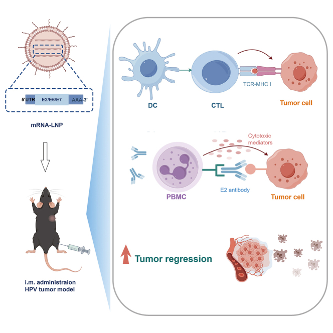
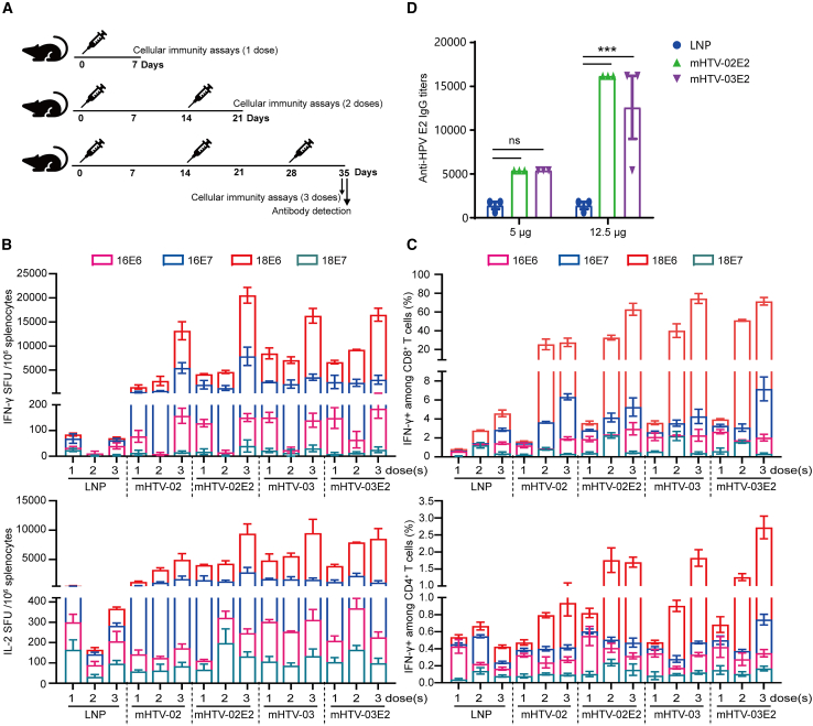
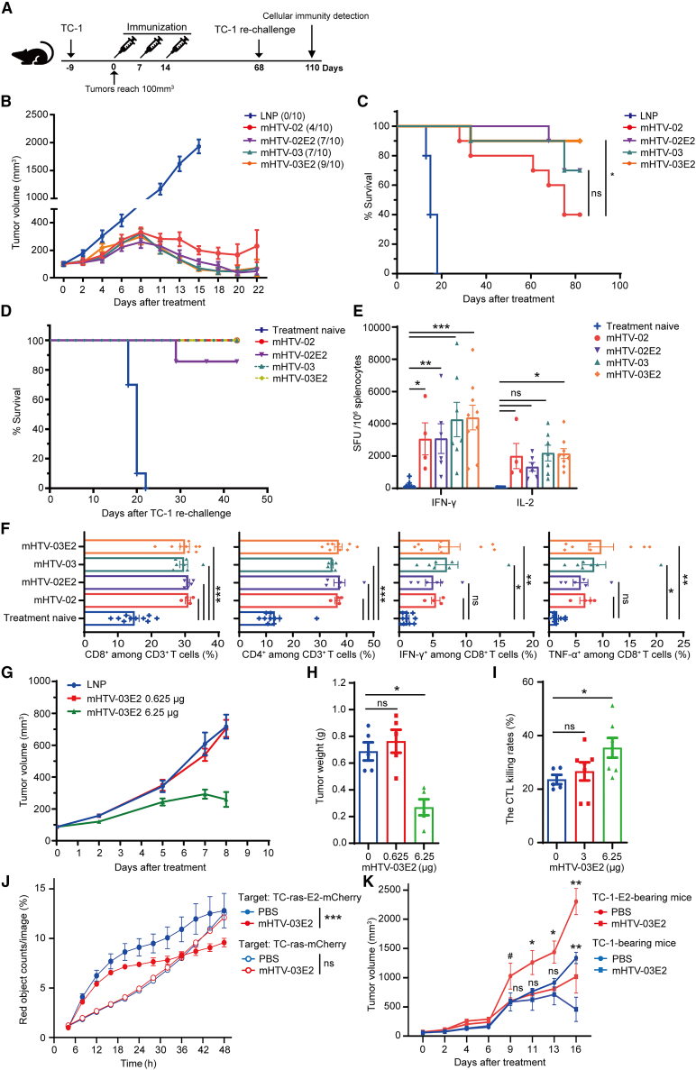
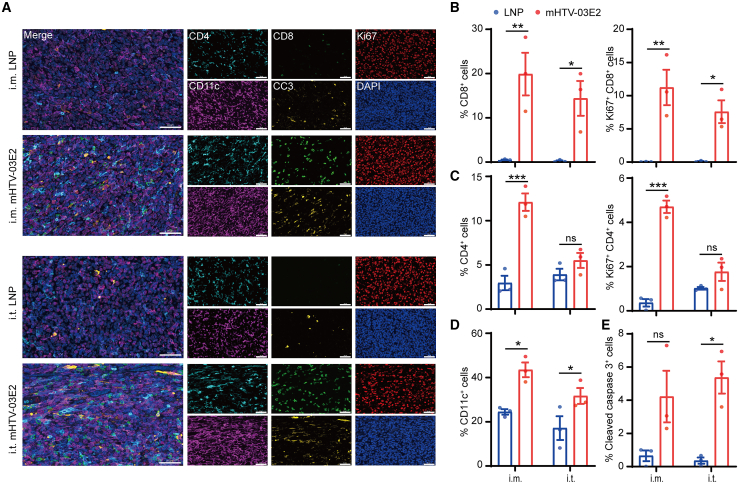
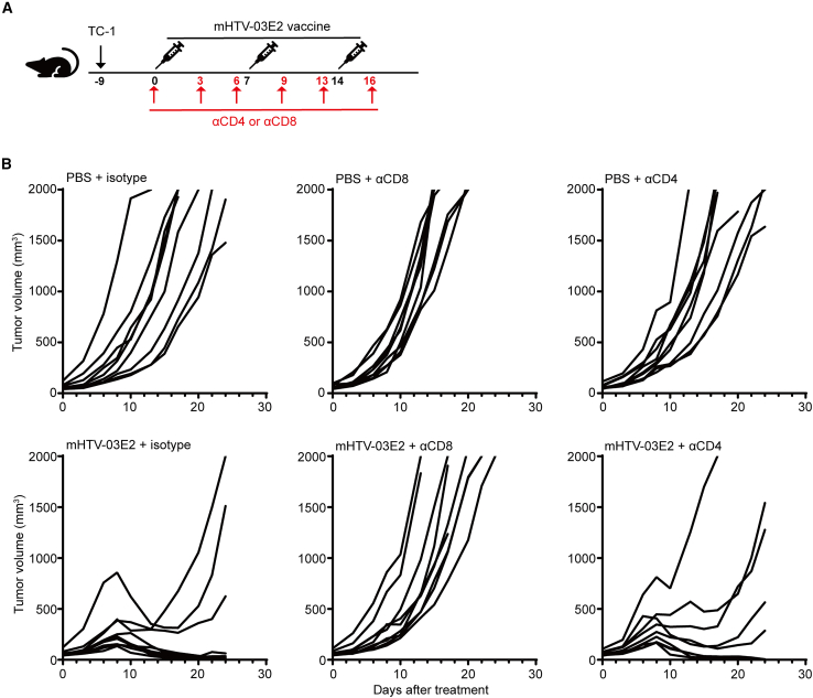
Human papillomavirus (HPV) 16 and 18 infections are related to many human cancers. Despite several preventive vaccines for high-risk (hr) HPVs, there is still an urgent need to develop therapeutic HPV vaccines for targeting pre-existing hrHPV infections and lesions. In this study, we developed a lipid nanoparticle (LNP)-formulated mRNA-based HPV therapeutic vaccine (mHTV)-03E2, simultaneously targeting the E2/E6/E7 of both HPV16 and HPV18. mHTV-03E2 dramatically induced antigen-specific cellular immune responses, leading to significant CD8+ T cell infiltration and cytotoxicity in TC-1 tumors derived from primary lung epithelial cells of C57BL/6 mice expressing HPV E6/E7 antigens, mediated significant tumor regression, and prolonged animal survival, in a dose-dependent manner. We further demonstrated significant T cell immunity against HPV16/18 E6/E7 antigens for up to 4 months post-vaccination in immunological and distant tumor rechallenging experiments, suggesting robust memory T cell immunity against relapse. Finally, mHTV-03E2 synergized with immune checkpoint blockade to inhibit tumor growth and extend animal survival, indicating the potential in combination therapy. We conclude that mHTV-03E2 is an excellent candidate therapeutic mRNA vaccine for treating malignancies caused by HPV16 or HPV18 infections.

Keywords: HPV; animal model; checkpoint blockade; mRNA therapeutic vaccine; tumor.


Conflict of interest statement

Declaration of interests S.C., J.W., W.Z., and Y.D. are co-inventors on pending patent applications related to the HPV-associated mRNA vaccine. H.L., H.Z., H.C., Y.W., T.Z., Y.C., Q.W., J.Z., C.H., Y.D., and W.Z. are employees of RinuaGene Biotechnology Co., Ltd.

Figures

None
Graphical abstract
Figure 1
Figure 1
mHTV mRNA-LNP vaccines induced significant T cell immunity against HPV16/HPV18 E6/E7 antigens in vivo (A) Schematic representation of four mRNA vaccines against HPV16/18. (gray) Signal peptide (SP), (yellow) Flt3L (F), (green) E2 antigen, and reshuffled E6/E7 fragments from (pink) HPV16 and (orange) HPV18 are indicated (blue cap) 5′ cap1, 5′ UTR, 3′ UTR and poly(A) tail are also indicated. (B) The indicated mRNAs were transfected into HEK293T cells, and the antigen expression was detected by western blotting using an anti-HPV18 E7 antibody. Actin was used as a loading control. (C) Scheme of vaccination. Mice were vaccinated two times with mHTV-02, mHTV-02E2, mHTV-03, or mHTV-03E2 (5 μg and 12.5 μg) on days 0 and 14. (D–K) Seven days after the final vaccination, (D) IFN-γ and (E) IL-2 productions were determined by ELISpot assay and (F–I) IFN-γ+ and TNF-α+ in CD8+ or CD4+ cells were detected by flow cytometry in splenocytes stimulated by the HPV16/18 E6 and E7 peptide pool. Mice immunized with empty LNPs were used as negative controls. Data are shown as mean ± SEM and analyzed by two-way ANOVA with multiple comparisons tests (∗p ≤ 0.05, ∗∗p ≤ 0.01, ∗∗∗p ≤ 0.001; ns, not significant).
Figure 2
Figure 2
Immunogenicity of the mHTV vaccines (A) Scheme of vaccination and testing. Mice were vaccinated with mHTV vaccines one, two, or three times at 2-week intervals. (B) Productions of IFN-γ and IL-2 were determined by ELISpot assay and (C) IFN-γ cytokines in CD4+ and CD8+ were detected by flow cytometry 7 days after each vaccination in splenocytes stimulated by the HPV16 E6, HPV16 E7, HPV18 E6 and HPV18 E7 peptide pools, respectively. Numbers 1, 2, and 3 stand for groups with one-, two-, and three-dose vaccine, respectively. Mice immunized with empty LNPs were used as negative controls. (D) The HPV E2-specific IgG antibody titers were determined by ELISA in mice vaccinated with mHTV-02E2 or mHTV-03E2 (5 μg and 12.5 μg), respectively. (B–D) Data are shown as mean ± SEM. Significance was analyzed by two-way ANOVA with multiple comparisons tests in (D) (∗p ≤ 0.05, ∗∗p ≤ 0.01, ∗∗∗p ≤ 0.001; ns, not significant). See also Figures S1–S3.
Figure 3
Figure 3
Therapeutic effects of mHTV vaccines on the TC-1 tumor model (A) Scheme of vaccination. TC-1 tumor-bearing mice were immunized three times at 1-week intervals with mHTV vaccines or LNP via i.m. when the average size of tumors reached about 100 mm3. (B) TC-1 tumor growth and (C) animal survival were monitored and are shown. Survival curves were analyzed by log rank (Mantel-Cox) test. (D–F) Mice with complete regression of tumors upon the treatment of mHTV vaccines were rechallenged with TC-1 cells on day 68. Treatment-naive mice served as the control group. (D) Animal survival was monitored. (E and F) After euthanizing the mice at the endpoints, (E) IFN-γ and IL-2 levels were determined by ELISpot assay, and (F) the proportion of CD4+, CD8+ T cells, IFN-γ, and TNF-α cytokines in CD8+ T cells were measured by flow cytometry in splenocytes stimulated by the HPV16/18 E6 and E7 peptide pools. (G and H) Mice bearing TC-1 tumors (average size of 80 mm3) were immunized twice with mHTV-03E2 (0.625 μg and 6.25 μg) via i.m. (G) Tumor growth curves and (H) tumor weight are shown. (I) The CTL killing rates targeting TC-1-HPV18 were determined in vaccinated mice with different doses of mHTV-03E2. (J) Determination of killing ability of sera samples from mice vaccinated with 12.5 μg of mHTV-03E2 or PBS control against TC-ras-E2-mCherry tumor cell line in cocultures with PBMCs at a 20:1 E:T ratio in the IncuCyte platform. TC-ras-mCherry cells were used as a control cell line. All traces show relative change in live cells by normalizing to the red counts at time point 4 h. (K) TC-1-bearing mice (average size of ∼60 mm3) and TC-1-E2-bearing mice (average size of ∼70 mm3) were immunized three times with mHTV-03E2 (3.125 μg) via i.m. Tumor growth curves are shown. Data are shown as mean ± SEM. Significance was analyzed by (E–I) one-way ANOVA and (J) two-way ANOVA with multiple comparisons tests (∗p ≤ 0.05, ∗∗p ≤ 0.01, ∗∗∗p ≤ 0.001; ns, not significant). (K) Statistical significance between mHTV-03E2 and PBS treatment groups against TC-1-E2 and TC-1 tumors at different time points was determined by unpaired, two-tailed Student’s t tests (#0.05 < p < 0.1, ∗p ≤ 0.05, ∗∗p ≤ 0.01). See also Figures S4 and S5 and Table S2.
Figure 4
Figure 4
Immune cell infiltration induced by mHTV vaccination in mouse tumors (A–D) (A) Mice bearing TC-1 tumors (average size of ∼100 mm3) were immunized twice with mHTV-03E2 via i.m. or i.t. Six days later, (B) the frequencies of lymphocyte subsets, (C) the frequencies of IFN-γ+ GzmB+ CD4+ T and IFN-γ+ GzmB+ CD8+ T cells, and (D) the frequencies of CD4+ and CD8+ Tem cells (CD44+ CD62L) in tumors were measured by flow cytometry. (E–G) (E, left) Mice bearing TC-1 tumors (average size of 80 mm3) were immunized twice with mHTV-03E2 (0.625 μg and 6.25 μg) via i.m. Twenty-four hours later, (E, middle) the frequencies of H-2Kb E6+ and H-2Db E7+ CD8+ T cells, (E, right) the frequencies of naive T (Tn), Tcm and Tem among H-2Db E7+ CD8+ T cells, and (F) the frequencies of myeloid subsets among CD45+ cells were measured by flow cytometry. (G) The ratios of M1/M2 are shown for PBS only and two mRNA vaccine dosages (0.625 μg and 6.25 μg). Data are shown as mean ± SEM. Statistical significance was analyzed by (B–D) unpaired, two-tailed Student’s t test between LNP control and mHTV-02 treatment or (E–G) one-way ANOVA with multiple comparisons tests (∗p ≤ 0.05, ∗∗p ≤ 0.01, ∗∗∗p ≤ 0.001; ns, not significant). See also Figure S6.
Figure 5
Figure 5
Distribution and infiltration characteristics of immune cells in tumor tissues (A) Representative images of multiplex immunohistochemistry staining. Scale bar, 50 μm. (B–E) Quantitative analysis of (B) CD8+ T cells, Ki67+ CD8+ T cells, (C) CD4+ T cells, Ki67+ CD4+ T cells, (D) CD11c+ cells and (E) Cleaved Caspase 3+ cells distributed in the tumors. The percentages of positive cells in the number of cells per whole slide were calculated. Data are shown as mean ± SEM and analyzed by two-way ANOVA with multiple comparisons tests (∗p ≤ 0.05, ∗∗p ≤ 0.01, ∗∗∗p ≤ 0.001; ns, not significant).
Figure 6
Figure 6
CD8+ T cells are required for the mHTV-03E2 induced anti-tumor immunity in the TC-1 tumor model (A) When TC-1 tumors reached an average of 63 mm3, tumor-bearing mice were administered i.m. with mHTV-03E2 (3 μg/mouse) weekly for three doses, and depleting antibody for CD8 or CD4 (10 mg/kg) dosed intraperitoneally twice a week at indicated times (red arrows). PBS plus antibody isotype control was used as negative controls. (B) Tumor growth kinetics for TC-1-bearing mice treated as described in (A). See also Table S3.
Figure 7
Figure 7
Inhibition of tumor growth by mHTV-03E2 vaccine in combination with a PD-L1 antibody (A) When TC-1 tumors reached an average of 63 mm3, tumor-bearing mice were administered with mHTV-03E2 (0.3 μg/mouse for three doses or 3 μg/mouse for a single dose) weekly intramuscularly, and αPD-L1 antibody (10 mg/kg) was dosed intraperitoneally twice a week 3 days after the mHTV administration. PBS and antibody isotype control were used as negative controls. (B) Kinetics of individual tumor growth and (C) animal survival of TC-1-bearing mice treated as described in (A). The data were analyzed by (B) unpaired, two-tailed Student’s t tests or (C) Log rank (Mantel-Cox) test between groups (∗p ≤ 0.05, ∗∗p ≤ 0.01, ∗∗∗p ≤ 0.001). See also Table S4.

References

    1. de Martel C., Plummer M., Vignat J., Franceschi S. Worldwide burden of cancer attributable to HPV by site, country and HPV type. Int. J. Cancer. 2017;141:664–670. - PMC - PubMed
    1. Chabeda A., Yanez R.J.R., Lamprecht R., Meyers A.E., Rybicki E.P., Hitzeroth I.I. Therapeutic vaccines for high-risk HPV-associated diseases. Papillomavirus Res. 2018;5:46–58. - PMC - PubMed
    1. Metz C.K., Skof A.S., Sehouli J., Siedentopf J.P., Gebert P., Weiss F., Alba Alejandre I., Heinrich-Rohr M., Weizsaecker K., Henrich W., et al. Assessment of high-risk human papillomavirus infections and associated cervical dysplasia in HIV-positive pregnant women in Germany: a prospective cross-sectional two-centre study. Arch. Gynecol. Obstet. 2023;308:207–218. - PubMed
    1. Alhamlan F.S., Alfageeh M.B., Al Mushait M.A., Al-Badawi I.A., Al-Ahdal M.N. Human Papillomavirus-Associated Cancers. Adv. Exp. Med. Biol. 2021;1313:1–14. - PubMed
    1. Nyitray A.G., Iannacone M.R. The epidemiology of human papillomaviruses. Curr. Probl. Dermatol. 2014;45:75–91. - PubMed

MeSH terms

LinkOut - more resources