. 2024 Apr 29;8(2):026106.
doi: 10.1063/5.0190561. eCollection 2024 Jun.

Model discovery approach enables noninvasive measurement of intra-tumoral fluid transport in dynamic MRI

Affiliations

Model discovery approach enables noninvasive measurement of intra-tumoral fluid transport in dynamic MRI

Ryan T Woodall et al. APL Bioeng. .

Abstract

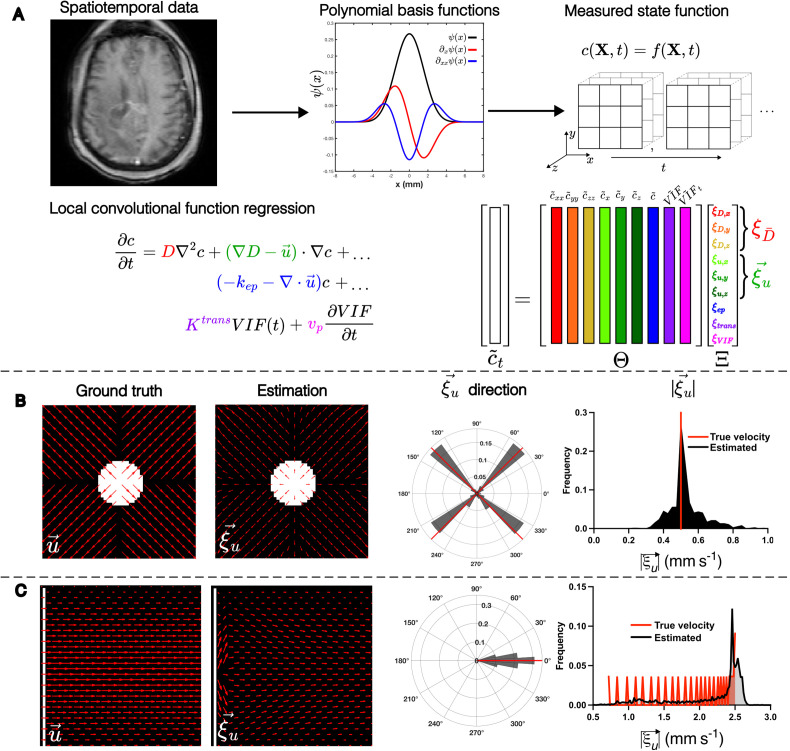
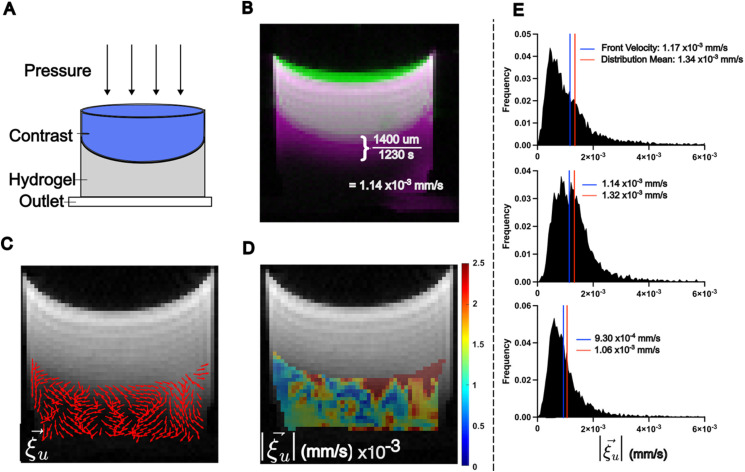
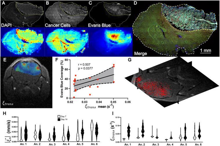
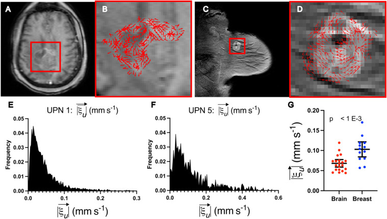
Dynamic contrast-enhanced magnetic resonance imaging (DCE-MRI) is a routine method to noninvasively quantify perfusion dynamics in tissues. The standard practice for analyzing DCE-MRI data is to fit an ordinary differential equation to each voxel. Recent advances in data science provide an opportunity to move beyond existing methods to obtain more accurate measurements of fluid properties. Here, we developed a localized convolutional function regression that enables simultaneous measurement of interstitial fluid velocity, diffusion, and perfusion in 3D. We validated the method computationally and experimentally, demonstrating accurate measurement of fluid dynamics in situ and in vivo. Applying the method to human MRIs, we observed tissue-specific differences in fluid dynamics, with an increased fluid velocity in breast cancer as compared to brain cancer. Overall, our method represents an improved strategy for studying interstitial flows and interstitial transport in tumors and patients. We expect that our method will contribute to the better understanding of cancer progression and therapeutic response.


Conflict of interest statement

The methodologies described herein are disclosed and claimed in a pending patent application co-owned by City of Hope and Virginia Polytechnic Institute and State University listing R.T.W., J.J.C., R.C.R., and J.M.M. as co-inventors. R.T.W., J.J.C., C.A.S., R.C.R., and J.M.M. are co-founders of and hold equity in Cairina Inc.

Figures

FIG. 1.
FIG. 1.
LCFR methodology and validation with in silico phantoms. (a) Methodology of localized convolutional function regression (LCFR), wherein spatiotemporal contrast agent concentration data are convolved with a smooth basis-function and its derivatives, divided into 3 × 3 × 3 (x, y, z) windows. The coefficients of the factored transport PDE are then solved for using linear regression. (b) Validation of LCFR coefficient ξu on a divergent flow field (initial condition, white) with spatially invariant diffusion and the true direction and magnitude of velocity denoted in red bars. (c) Validation of LCFR on a Poiseuille shear flow field with spatially invariant diffusion (initial condition, white) and the true direction and magnitude of velocity denoted in red bars.
FIG. 2.
FIG. 2.
LCFR predicts interstitial fluid velocity in hydrogel phantoms. (a) Experimental setup, wherein a bolus of contrast agent is administered onto a porous hydrogel and drains through due to a hydraulic pressure head. (b) Method of estimating the mean flow velocity of the contrast agent front, using the difference between the initial contrast location (green), and final contrast location (pink), resulting in an estimated contrast agent velocity of 1.14 × 10⁻³ mm/s. (c) The estimated in-plane interstitial flow velocity direction overlaid on the final T1-weighted image. (d) The local 3D magnitude of interstitial flow velocity within the hydrogel. (e) Histograms of three replicate gels, comparing the 3D magnitude of flow velocity within the hydrogel, with the blue line indicating the estimated contrast front velocity, and the red line indicating the mean velocity of the distribution as measured by LCFR.
FIG. 3.
FIG. 3.
LCFR-measured perfusion is correlated with Evans Blue coverage in vivo. (a)–(d) Representative coronal IHC stains through the central tumor slice (top row) and MR resolution-matching intensity projection (bottom row), consisting of DAPI (a), GFP-expressing GL261 cells (b), Evans Blue (c). (d) Merge of all IHC demonstrating tumor heterogeneity. (e) Estimated perfusion field, ξtrans, overlaid on post-contrast T1-weighted image. (f) Scatter plot depicting the correlation and 95% confidence interval of linear regression between mean tumor perfusion as measured by DCE-MRI ( ξtrans) and Evans Blue Coverage (mean tumor stain intensity), for N= 17 histology slices. (g) 3D velocity vector field of estimated interstitial velocity ξu, overlaid on the 3D T1 post-contrast volume. (h) and (i) Violin plots depicting the estimated velocity magnitude (h) and perfusion (i) for six animals imaged 7 and 14 days after tumor implantation.
FIG. 4.
FIG. 4.
LCFR captures differences in fluid transport between breast and brain cancers. (a) and (b) Representative post-contrast T1-weighted image of central slice of residual glioblastoma 2 weeks after resection surgery. (b) Detail of tumor and resection cavity, with overlay of the estimated velocity direction, ξu. (c) and (d) Representative post-contrast T1-weighted image of untreated primary breast cancer lesion. (d) Detail of the enhancing tumor, with overlay of the with overlay of the estimated velocity direction, ξu. (e) Distribution of the in-plane velocity of the entire enhancing glioblastoma and resection cavity (mean = 5.23 × 10⁻¹ ± 5.10 × 10⁻¹). (f) Distribution of the in-plane velocity of the entire enhancing breast tumor (mean = 1.19 × 10⁻¹ ± 1.06 × 10⁻¹). (g) Mean fluid velocities for brain (mean = 6.81 × 10⁻² ± 1.99 × 10⁻² mm/s, N = 20) and breast data (mean = 1.03 × 10⁻¹ ± 3.10 × 10⁻², N = 13), with whiskers indicating mean and 95% confidence interval.

Update of

References

    1. Munson J. M., Bellamkonda R. V., and Swartz M. A., “ Interstitial flow in a 3D microenvironment increases glioma invasion by a CXCR4-dependent mechanism,” Cancer Res. 73, 1536–1546 (2013).10.1158/0008-5472.CAN-12-2838 - DOI - PubMed
    1. Haessler U., Teo J. C. M., Foretay D., Renaud P., and Swartz M. A., “ Migration dynamics of breast cancer cells in a tunable 3D interstitial flow chamber,” Integr. Biol. 4, 401–409 (2012).10.1039/c1ib00128k - DOI - PubMed
    1. Cornelison R. C., Brennan C. E., Kingsmore K. M., and Munson J. M., “ Convective forces increase CXCR4-dependent glioblastoma cell invasion in GL261 murine model,” Sci. Rep. 8, 17057 (2018).10.1038/s41598-018-35141-9 - DOI - PMC - PubMed
    1. Shields J. D., Fleury M. E., Yong C., Tomei A. A., Randolph G. J., and Swartz M. A., “ Autologous chemotaxis as a mechanism of tumor cell homing to lymphatics via interstitial flow and autocrine CCR7 signaling,” Cancer Cell 11, 526–538 (2007).10.1016/j.ccr.2007.04.020 - DOI - PubMed
    1. Ng C. P. and Swartz M. A., “ Fibroblast alignment under interstitial fluid flow using a novel 3-D tissue culture model,” Am. J. Physiol. Circ. Physiol. 284, H1771–H1777 (2003).10.1152/ajpheart.01008.2002 - DOI - PubMed