Review
. 2024 Apr 30;13(4):643-662.
doi: 10.21037/tp-23-602. Epub 2024 Apr 18.

Management advances for congenital diaphragmatic hernia: integrating prenatal and postnatal perspectives

Affiliations
Review

Management advances for congenital diaphragmatic hernia: integrating prenatal and postnatal perspectives

Ahmet A Baschat et al. Transl Pediatr. .

Abstract

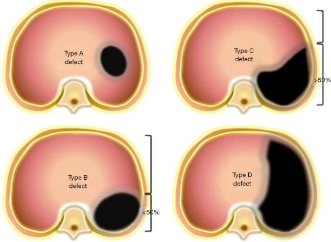
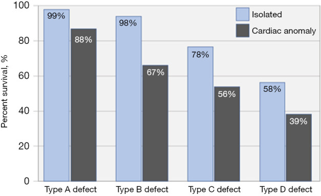
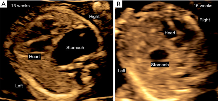
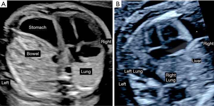
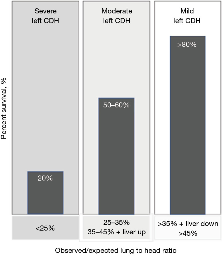
In congenital diaphragmatic hernia (CDH), abdominal organs are displaced into the chest, compress the lungs, and cause mediastinal shift. This contributes to development of pulmonary hypoplasia and hypertension, which is the primary determinant of morbidity and mortality for affected newborns. The severity is determined using prenatal imaging as early as the first trimester and is related to the laterality of the defect, extent of lung compression, and degree of liver herniation. Comprehensive evaluation of fetal CDH includes imaging-based severity assessment, severity assessment, and evaluation for structural or genetic abnormalities to differentiate isolated from complex cases. Prenatal management involves multispecialty counseling, consideration for fetal therapy with fetoscopic endoluminal tracheal occlusion (FETO) for severe cases, monitoring and intervention for associated polyhydramnios or signs of preterm labor if indicated, administration of antenatal corticosteroids in the appropriate setting, and planned delivery to optimize the fetal condition at birth. Integrated programs that provide a smooth transition from prenatal to postnatal care produce better outcomes. Neonatal care involves gentle ventilation to avoid hyperinflation and must account for transitional physiology to avoid exacerbating cardiac dysfunction and decompensation. Infants who have undergone and responded to FETO have greater pulmonary capacity than expected, but cardiac dysfunction seems unaffected. In about 25-30% of CDH neonates extracorporeal life support is utilized, and this provides a survival benefit for patients with the highest predicted mortality, including those who underwent FETO. Surgical repair after initial medical management for the first 24-48 hours of life is preferred since later repair is associated with delayed oral feeding, increased need for tube feeds, and increased post-repair ventilation requirement and supplemental oxygen at discharge. With overall survival rates >70%, contemporary care involves management of chronic morbidities in the context of a multidisciplinary clinic setting.

Keywords: Congenital diaphragmatic hernia (CDH); extracorporeal life support; fetoscopic endoluminal tracheal occlusion (FETO); gentle ventilation; surgical repair.


Conflict of interest statement

Conflicts of Interest: All authors have completed the ICMJE uniform disclosure form (available at https://tp.amegroups.com/article/view/10.21037/tp-23-602/coif). A.A.B. receives royalties from UpToDate and in-kind donation of fetoscopy sheath equipment from Karl Storz. J.L.M. is supported by Janssen Research and Development, LLC; serves as a Data Safety Monitoring Board member for FETO, and on the Executive Board of the North American Fetal Therapy Network. The other authors have no conflicts of interest to declare.

Figures

Figure 1
Figure 1
Schematic representation of diaphragmatic hernia size at surgery. Visual representation of a standardized classification scheme for defect size as observed during hernia repair. “A” defects are entirely surrounded by diaphragmatic muscle, “B” defects have less than 50% of the chest wall devoid of diaphragm, “C” defects have more than 50% of the chest wall lacking diaphragm and in “D” defects the diaphragm is mostly absent.
Figure 2
Figure 2
Survival in relationship to hernia size and presence of a cardiac defect. The bar graphs represent the observed survival in four types of left diaphragmatic hernia, stratified by the presence of an isolated defect or a coexisting cardiac anomaly [based on ref. (11)].
Figure 3
Figure 3
Prenatal ultrasound appearance of diaphragmatic hernia in early gestation. Prenatal ultrasound images obtained in two patients with a left diaphragmatic hernia at 13 weeks’ gestation (A) and 16 weeks’ gestation (B). The first trimester defect was suspected due to the rotation of the cardiac axis to the left and the intrathoracic retrocardiac position of the stomach. The second patient was diagnosed based on the marked mediastinal shift of the heart towards the right of the fetus and the intrathoracic location of the stomach.
Figure 4
Figure 4
Ultrasound appearance of left and right diaphragmatic hernia in the second trimester. Prenatal ultrasound images obtained in a patient with left diaphragmatic hernia at 26 weeks’ gestation (A) and a right diaphragmatic hernia at 22 weeks’ gestation (B). The left diaphragmatic hernia is characterized by a right mediastinal shift of the heart and intrathoracic bowel and stomach herniation. The contralateral right lung is posterior to the right atrium and there is reduction of the observed to estimated lung to head ratio to a moderate hernia severity. In right diaphragmatic hernia the cardiac axis is rotated to the left and the mediastinal shift is less marked despite intrathoracic herniation of the liver. Both lungs measure smaller in size and there is a pericardial effusion (*) which is common in this setting.
Figure 5
Figure 5
Third trimester ultrasound findings in left congenital diaphragmatic hernia. Prenatal ultrasound images obtained in a patient with left diaphragmatic hernia at 37 weeks’ gestation demonstrate a left diaphragmatic hernia with compression and relative hypoplasia of the left ventricle. RV, right ventricle; LV, left ventricle.
Figure 6
Figure 6
Left CDH severity assessed by observed to expected lung to head ratio and liver position. The figure illustrates the prenatal severity assessment of isolated left congenital diaphragmatic hernia based on the ultrasound measured observed lung to head ratio and liver position and the anticipated postnatal survival (37,38). This assessment was utilized to determine eligibility for fetoscopic tracheal occlusion in the randomized TOTAL trials (70,71). CDH, congenital diaphragmatic hernia.
Video 1
Video 1
Fetoscopic balloon insertion and removal.

References

    1. Lally KP. Congenital diaphragmatic hernia. Curr Opin Pediatr 2002;14:486-90. 10.1097/00008480-200208000-00022 - DOI - PubMed
    1. Sola JE, Bronson SN, Cheung MC, et al. Survival disparities in newborns with congenital diaphragmatic hernia: a national perspective. J Pediatr Surg 2010;45:1336-42. 10.1016/j.jpedsurg.2010.02.105 - DOI - PubMed
    1. Gray BW, Fifer CG, Hirsch JC, et al. Contemporary outcomes in infants with congenital heart disease and bochdalek diaphragmatic hernia. Ann Thorac Surg 2013;95:929-34. 10.1016/j.athoracsur.2012.07.010 - DOI - PubMed
    1. Snoek KG, Greenough A, van Rosmalen J, et al. Congenital Diaphragmatic Hernia: 10-Year Evaluation of Survival, Extracorporeal Membrane Oxygenation, and Foetoscopic Endotracheal Occlusion in Four High-Volume Centres. Neonatology 2018;113:63-8. 10.1159/000480451 - DOI - PubMed
    1. Harting MT, Lally KP. The Congenital Diaphragmatic Hernia Study Group registry update. Semin Fetal Neonatal Med 2014;19:370-5. 10.1016/j.siny.2014.09.004 - DOI - PubMed