Review
. 2024 May 3:17:1601-1638.
doi: 10.2147/JPR.S464393. eCollection 2024.

American Society of Pain and Neuroscience Best Practice (ASPN) Guideline for the Treatment of Sacroiliac Disorders

Affiliations
Review

American Society of Pain and Neuroscience Best Practice (ASPN) Guideline for the Treatment of Sacroiliac Disorders

Dawood Sayed et al. J Pain Res. .

Abstract

Clinical management of sacroiliac disease has proven challenging from both diagnostic and therapeutic perspectives. Although it is widely regarded as a common source of low back pain, little consensus exists on the appropriate clinical management of sacroiliac joint pain and dysfunction. Understanding the biomechanics, innervation, and function of this complex load bearing joint is critical to formulating appropriate treatment algorithms for SI joint disorders. ASPN has developed this comprehensive practice guideline to serve as a foundational reference on the appropriate management of SI joint disorders utilizing the best available evidence and serve as a foundational guide for the treatment of adult patients in the United States and globally.

Keywords: best practices; chronic pain; radiofrequency ablation; sacroiliac joint; sacroiliac joint fusion; sacroiliitis.


Conflict of interest statement

All authors were required to disclose conflicts of interest prior to assignment of topics. The senior authors determined the extent of the conflict of interest ensuring balanced inquiry and evaluation for each manuscript section. One of the co-primary authors without conflict were identified for each section and was the adjudication determination official for any issues of potential conflict. All authors were asked to recuse themselves on any recommendation potentially affected by a disclosed conflict. Additionally, authors without conflict vetted all recommendations for bias.DS is a consultant to Abbott, PainTEQ, Saluda, Mainstay, Surgentec, Nevro, and holds stock options with PainTEQ, Neuralace, Mainstay, Vertos, and SPR. TRD is a consultant PainTEQ, CornerLoc, and Spinal Simplicity. VTF receives research funding from Nevro Corporation part of an investigator-initiated study grant that is not related to this manuscript. TEW is a consultant for Medtronic and has received research funding from Medtronic, SPR Therapeutics, Nevro, and Boston Scientific. JSW is a consultant for Abbott, SI Bone, Vertos Medical, Biotronik, Saluda, and AbbVie, receives research funding from Abbott, SI Bone, Saluda, and Medtronic, serves on an advisory board for Abbott, SI Bone, Vertos Medical, and Biotronik, and serves on a speaker board for Abbott, SI Bone, & Abbvie. RSD receives investigator-initiated research grant funding from Nevro Corp and Saol Therapeutics that is paid to his institution. KA is a consultant for Nevro, Saluda, Biotronik, Boston Scientific, and Presidio, reports minor options from PainTEQ. MJD is a consultant for Globus, Camber, LifeSpine, Vyrsa, PainTEQ, Nevro, Abbott, and Biotronik. DTDN is a consultant for SI Bone, Stratus Medical, and Neurovassis. CB is a consultant for Nevro, Abbott, Vertos, Spinal Simplicity, and PainTEQ; reports personal fees from Nevro, Spinal Simplicity, PainTEQ, and Boston Scientific, outside the submitted work. AA is a consultant for Curonix, Medtronic and Avanos. The authors report no other conflicts of interest in this work.

Figures

Figure 1
Figure 1
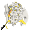
Anatomy of the SIJ. (a) Posterior-oblique view of the sacrum and iliac bones, ligaments, and nervous supply. SIJ is highlighted in pink overlay. (b) Diarthrodial nature of the SIJ space (pink overlay). The joint consists of an anterior one-third consisting of synovium and posterior two-thirds which is primarily ligamentous. (c) Exaggeration of sacral movement in nutation and counternutation. Original medical illustration by Kamil Sochacki, DO.
Figure 2
Figure 2
Leapfrog Radiofrequency Ablation Technique: A bipolar radiofrequency ablation strategy utilizing two probes placed within 10mm of one another along the SIJ. A lesion is made before moving the superior probe inferior to the second probe. A lesion is made, and the process is repeated with the lead probe being positioned inferiorly once again. Original medical illustration by Kamil Sochacki, DO.
Figure 3
Figure 3
Conventional versus Cooled Radiofrequency Ablation. (a) Conventional RFA requires precise placement of the RFA probe within 1–2 mm of the intended target. Conventional RFA probes can reach temperatures of 100°C and insulating properties prevent heat radiofrequency waves from reaching further target tissue. (b) Cooled RFA needles utilize continuously circulating coolant within a hollow exterior shell to modulate temperature at the tip of the probe to around 60°C. This cooling mechanism avoids charring surrounding tissue, allowing for more effective heat transfer beyond the immediate proximity of the probe tip. The result is a significant difference in the overall size, shape, and area of effect of the ablated lesion, as compared to conventional RFA. Original medical illustration by Kamil Sochacki, DO.
Figure 4
Figure 4
RFA with multilesion probe. This technique utilizes a singular probe which is tunneled through the tissue via a single site to generate a true strip lesion at the SIJ. The probe contains three electrodes creating a combination of monopolar and bipolar lesions. Original medical illustration by Kamil Sochacki, DO.
Figure 5
Figure 5
Surgical Techniques: Posterior Allograft Fusion. (a) A large pin (not shown here) is inserted into the joint between the sacrum and the ilium. Next, a tissue dilator and cannula (shown) are inserted to create joint space separation. (b) A rasp is inserted into the cannula to prepare the site for the allograft while decorticating the area. (c) Finally, the allograft which contains the demineralized bone matrix is inserted into the decorticated site to allow for healing and stabilization of the joint. Original medical illustration by Kamil Sochacki, DO.
Figure 6
Figure 6
Surgical Techniques: Lateral and Posterior-Oblique Sacral Fusions. (a) The lateral fusion involves the placement of devices across the SIJ from lateral to medial which fixate the ilium and sacrum together. At least two titanium implants are placed through the ilium, or wing bone of the pelvis, across the SIJ, and into the sacrum, the large bone at the base of the spine, to immediately reduce the motion of the joint. This technique involves disruption of musculature. (b) The posterior-oblique approach involves placing implants in a medial to lateral trajectory. The insertion point is the posterior-superior iliac spine which spares dissection of the musculature and minimizes potential injury to other structures. Original medical illustration by Kamil Sochacki, DO.

References

    1. Deer TR, Grider JS, Pope JE, et al. Best practices for minimally invasive lumbar spinal stenosis treatment 2.0 (MIST): consensus guidance from the American Society of Pain and Neuroscience (ASPN). J Pain Res. 2022;15:1325–1354. doi:10.2147/JPR.S355285 - DOI - PMC - PubMed
    1. Harris RP, Helfand M, Woolf SH, et al. Current methods of the U.S. preventive services task force: a review of the process. Am J Preventive Med. 2001;20(3 SUPPL):21–35. doi:10.1016/S0749-3797(01)00261-6 - DOI - PubMed
    1. Foley BS, Buschbacher RM. Sacroiliac joint pain: anatomy, biomechanics, diagnosis, and treatment. Am J Phys Med Rehabil. 2006;85(12):997–1006. doi:10.1097/01.phm.0000247633.68694.c1 - DOI - PubMed
    1. Cohen SP. Sacroiliac joint pain: a comprehensive review of anatomy, diagnosis, and treatment. Anesth Analg. 2005;101(5):1440–1453. doi:10.1213/01.ANE.0000180831.60169.EA - DOI - PubMed
    1. Alderink GJ. The sacroiliac joint: review of anatomy, mechanics, and function. J Orthop Sports Phys Ther. 1991;13(2):71–84. doi:10.2519/jospt.1991.13.2.71 - DOI - PubMed