Multicenter Study
. 2024 Sep 5;26(9):1723-1737.
doi: 10.1093/neuonc/noae080.

Gliomatosis cerebri in children: A poor prognostic phenotype of diffuse gliomas with a distinct molecular profile

Gunther Nussbaumer  1 Martin Benesch  1 Yura Grabovska  2 Alan Mackay  2 David Castel  3   4 Jacques Grill  3   4 Marta M Alonso  5   6   7 Manila Antonelli  8 Simon Bailey  9 Joshua N Baugh  10 Veronica Biassoni  11 Mirjam Blattner-Johnson  12   13 Alberto Broniscer  14 Andrea Carai  15 Giovanna Stefania Colafati  16 Niclas Colditz  17 Selim Corbacioglu  18 Shauna Crampsie  2 Natacha Entz-Werle  19   20 Matthias Eyrich  21 Lea L Friker  22 Michael C Frühwald  23 Maria Luisa Garrè  24 Nicolas U Gerber  25 Felice Giangaspero  8 Maria J Gil-da-Costa  26 Norbert Graf  27 Darren Hargrave  28 Peter Hauser  29 Ulrich Herrlinger  30 Marion Hoffmann  17 Esther Hulleman  10 Elisa Izquierdo  2 Sandra Jacobs  31   32 Michael Karremann  33 Antonis Kattamis  34 Rejin Kebudi  35 Rolf-Dieter Kortmann  36 Robert Kwiecien  37 Maura Massimino  11 Angela Mastronuzzi  38 Evelina Miele  38 Giovanni Morana  39 Claudia M Noack  40 Virve Pentikainen  41 Thomas Perwein  1 Stefan M Pfister  42   43   44   13 Torsten Pietsch  22 Kleoniki Roka  34 Sabrina Rossi  45 Stefan Rutkowski  46 Elisabetta Schiavello  11 Clemens Seidel  36 Jaroslav Štěrba  47 Dominik Sturm  43   12   13 David Sumerauer  48 Anna Tacke  17 Sara Temelso  2 Chiara Valentini  49 Dannis van Vuurden  10 Pascale Varlet  50 Sophie E M Veldhuijzen van Zanten  51   10 Maria Vinci  38 André O von Bueren  52 Monika Warmuth-Metz  53 Pieter Wesseling  54   10 Maria Wiese  15 Johannes E A Wolff  55 Josef Zamecnik  56 Andrés Morales La Madrid  57 Brigitte Bison  58   59 Gerrit H Gielen  22 David T W Jones  12   13 Chris Jones  2 Christof M Kramm  17
Affiliations
Multicenter Study

Gliomatosis cerebri in children: A poor prognostic phenotype of diffuse gliomas with a distinct molecular profile

Gunther Nussbaumer et al. Neuro Oncol. .

Abstract

Background: The term gliomatosis cerebri (GC), a radiology-defined highly infiltrating diffuse glioma, has been abandoned since molecular GC-associated features could not be established.

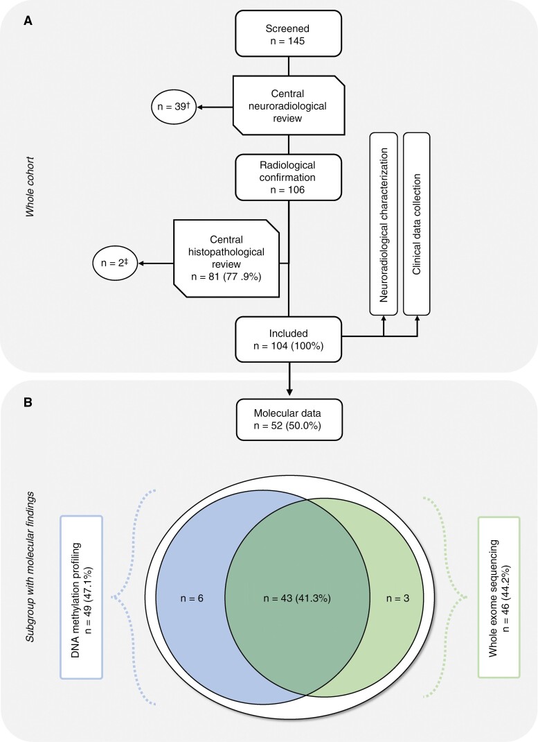
Methods: We conducted a multinational retrospective study of 104 children and adolescents with GC providing comprehensive clinical and (epi-)genetic characterization.

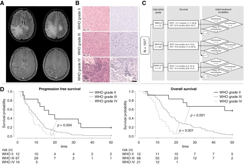
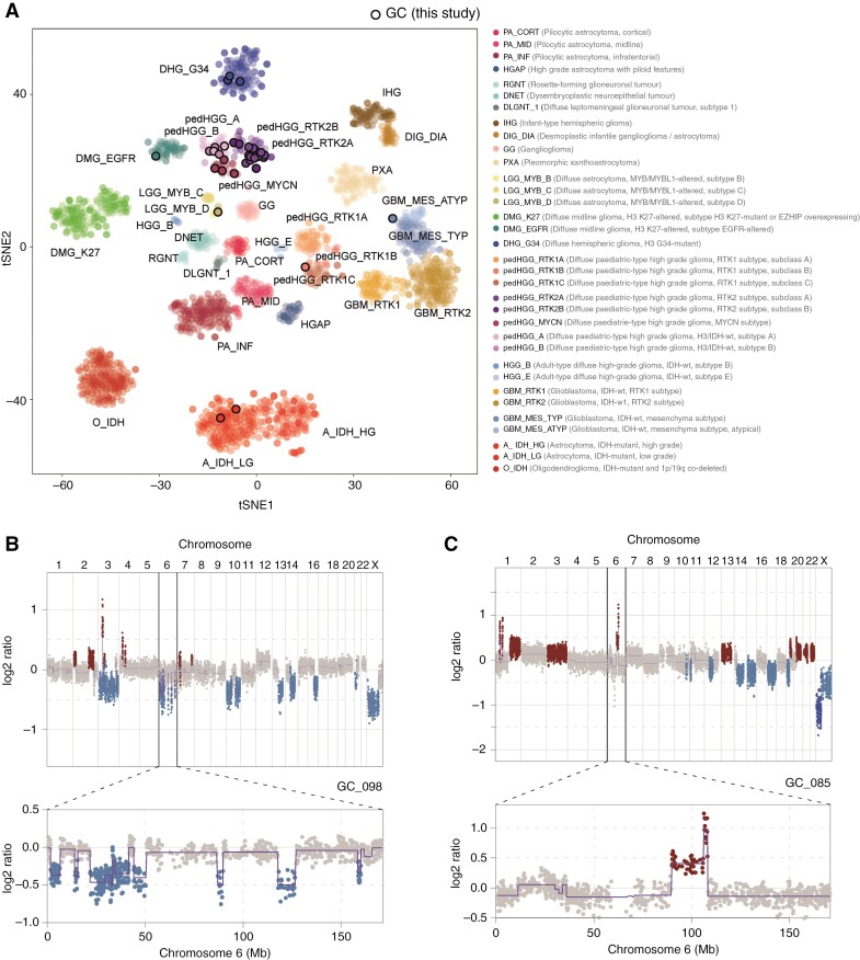
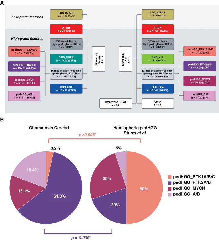
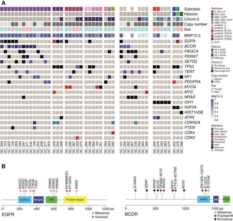
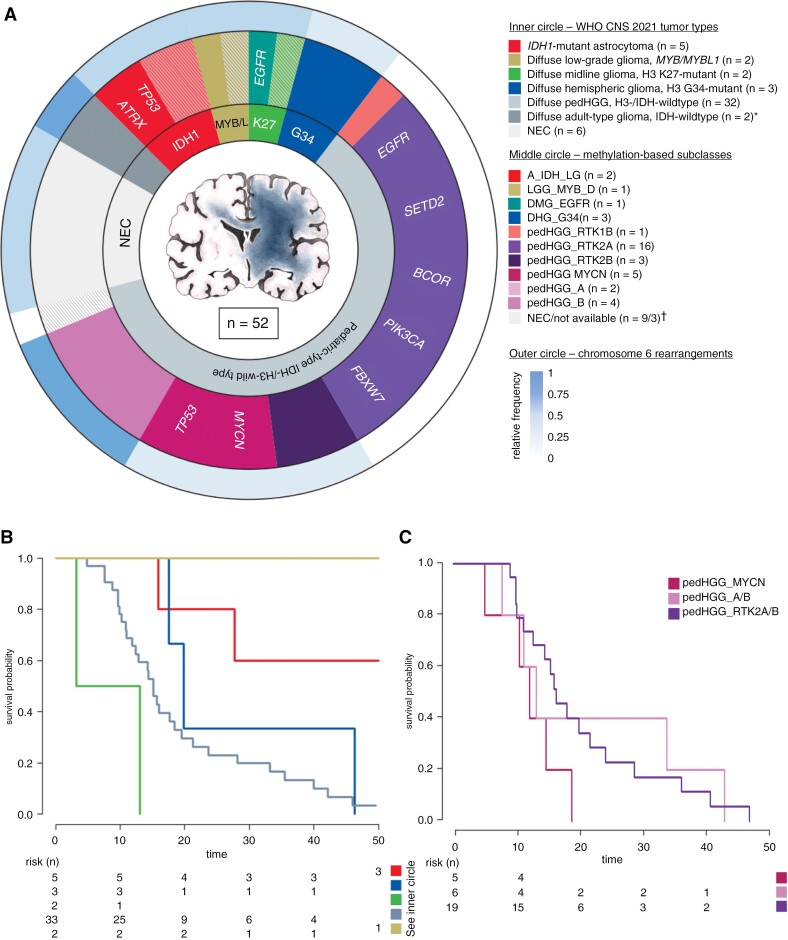
Results: Median overall survival (OS) was 15.5 months (interquartile range, 10.9-27.7) with a 2-year survival rate of 28%. Histopathological grading correlated significantly with median OS: CNS WHO grade II: 47.8 months (25.2-55.7); grade III: 15.9 months (11.4-26.3); grade IV: 10.4 months (8.8-14.4). By DNA methylation profiling (n = 49), most tumors were classified as pediatric-type diffuse high-grade glioma (pedHGG), H3-/IDH-wild-type (n = 31/49, 63.3%) with enriched subclasses pedHGG_RTK2 (n = 19), pedHGG_A/B (n = 6), and pedHGG_MYCN (n = 5), but only one pedHGG_RTK1 case. Within the pedHGG, H3-/IDH-wild-type subgroup, recurrent alterations in EGFR (n = 10) and BCOR (n = 9) were identified. Additionally, we observed structural aberrations in chromosome 6 in 16/49 tumors (32.7%) across tumor types. In the pedHGG, H3-/IDH-wild-type subgroup TP53 alterations had a significant negative effect on OS.

Conclusions: Contrary to previous studies, our representative pediatric GC study provides evidence that GC has a strong predilection to arise on the background of specific molecular features (especially pedHGG_RTK2, pedHGG_A/B, EGFR and BCOR mutations, chromosome 6 rearrangements).

Keywords: H3-wild-type and IDH-wild-type; chromosome 6; gliomatosis cerebri; pedHGG_RTK2; pediatric-type glioma; pediatric-type high-grade glioma.


Conflict of interest statement

D.T.W.J. and S.M.P. are shareholders and cofounders of Heidelberg Epignostix GmbH. All remaining authors have declared that they have no competing interests.

Figures

Figure 1:
Figure 1:
(A) Flowchart of the study cohort. (A) A total of 145 children and adolescents from 14 European countries were screened for suspected GC. 39 cases did not fulfill the neuroradiological criteria and were excluded. Two tumors were excluded as a glial process could not be confirmed unambiguously through central neuropathological review. (B) Composition of the subgroup with available molecular data comprising DNA methylation profiling and whole exome sequencing.
Figure 2:
Figure 2:
Collage of clinical data of the whole cohort. (A) Representative MRI of a pediatric patient with GC. Upper row: Fluid-attenuated inversion recovery (FLAIR): high signal as a sign of diffuse tumor infiltration primarily in the right occipital, parietal, and temporal lobe as well as involvement of the contralateral hemisphere with little mass effect. Lower row: contrast-enhanced T1-weighted images: the occipital part of the tumor shows typical mild multifocal enhancement. (B) Histopathological features of GC. Microscopical examination of three representative GC tumors according to histopathological grading: the left side shows HE staining and the right Ki-67 immunohistochemistry of the respective case: GC_74, WHO grade II; GC_51, WHO grade III; GC_35, WHO grade IV. With higher WHO grade, increasing cellularity and proliferative activity can be detected. Scale bar equate to 100 µm in each case. (C) Composition of the treatment groups according to the WHO grade. 1PFS and OS given as median and the interquartile range in parentheses. Three cases were excluded for this analysis due to absent WHO grading. These patients did not receive an upfront cytotoxic treatment. (D) Kaplan–Meier plot including p-value of the (left) PFS and (right) OS in months according to WHO grading.
Figure 3:
Figure 3:
DNA methylation profiling in pediatric GC. (A) t-Statistic-based stochastic neighbor embedding (t-SNE) projection of a combined methylation dataset according to the MNP12.5 classifier comprising the pediatric GC cases from this study (circled, n = 40/49) plus a reference set of glioma subtypes (n = 2305). The first 2 projections are plotted on the x and y axes, with samples represented by dots colored by the respective subclass as labeled on the figure. (B, C) DNA copy-number plots for the cases GC_098 and GC_085 derived from methylation array data, with log2 ratios plotted (y axis) against genomic location by chromosome (x axis), and colored red for gain, and blue for loss. Above—whole genome; below—chromosome 6 illustrating representing examples of the structural alteration observed in our cohort.
Figure 4:
Figure 4:
Distribution of methylation-based subclasses. (A) Comparison of DNA methylation data according to MNP12.5 of the GC cohort (n = 40) and the published population-based collective by Sturm et al. (n = 80). The subclasses were arranged based on the WHO CNS 2021 classification. In the cohort by Sturm et al., only supratentorial cases with hemispheric location were included. All non-diffuse glioma subclasses, isolated midline location, and with a calibrated score <0.9 were excluded. Infant-type H3-wild-type tumors were not considered in the analysis to approximate the age distribution between the 2 cohorts (median 12 years [1–21] vs. 11.8 years [1.3–18.8]). In the category “others” 22 tumors of “pleomorphic xanthoastrocytoma (-like)” and 2 tumors of the subclass “neuroepithelial tumor, PLAGL1-fused” were present. There was an overlap between these 2 collectives as 2 cases occurred in both the here-reported GC and the control cohort. Therefore, in the subclasses pedHGG_A/B and pedHGG_RTK2A/B, 1 patient is listed once in each of the 2 cohorts. Of note, the only pedHGG_A/B-case in the reference collective was a child from the here-presented GC cohort. (B) Relative frequencies of different subclasses in the diffuse pediatric-type high-grade glioma, H3-wild-type, and IDH-wild-type subgroup of the 2 above-mentioned cohorts. *There was a significant difference in the frequencies of pedHGG_RTK2A/B- and pedHGG_RTK1A/B/C subclasses between the 2 cohorts.
Figure 5:
Figure 5:
Whole-exome sequencing of pediatric GC. (A) Oncoprint representation of an integrated annotation of single-nucleotide variants and DNA copy-number changes for pediatric GC (n = 46). Samples are arranged in columns with genes labeled along rows. Clinicopathological and molecular annotations are provided as bars according to the included key. (B) Lollipop plot of specific variants identified in pediatric GC cases in EGFR (left) and BCOR (right), scaled by number and colored according to the key provided. Abbreviations: cysteine-rich domain (cysteine); receptor L-domain (receptor); growth factor receptor domain 4 (GFR); protein kinase domain (protein kinase); non-ankyrin-repeat domain (NARD); ankyrin repeat domain (ARD); PCGF1-binding domain (PCGF).
Figure 6:
Figure 6:
Summary of the molecular findings in a multilayer circle diagram. (A) In total, molecular data was available of 52 tumors. The inner circle represents different tumor types according to WHO CNS 2021 in synopsis with methylation array data and exome sequencing. The middle circle illustrates the DNA methylation-based subtypes according to the MNP12.5 classifier, which clustered to the corresponding WHO CNS types. In addition, the most frequent genetic alterations of the respective subtypes are given. In both circles the (sub-)types are colored as labeled in the given key. Hatched areas represent cases with inconclusive methylation profiling, but subtype allocation was possible through detection of disease-defining genetic alterations in exome sequencing. *adult-type diffuse gliomas, IDH-wild-type (n = 2) including 1 case each of the subtypes GBM_MES_ATYP and GBM_RTK2. Nine cases were NEC in methylation analyses, and additionally, in 3 cases, no methylation data were available. The outer circle represents the relative frequency of chromosome 6 rearrangements sorted by the respective methylome-based subclass. (B, C) Kaplan–Meier plots of the overall survival in months according to (B) the WHO CNS 2021 and to (C) the methylation-defined subclasses of the pedHGG_H3-/IDH-wt subgroup including pedHGG_A/B. Tumors of the adult-type, IDH-wild-type are not shown in (B).

References

    1. Paugh BS, Qu C, Jones C, et al. Integrated molecular genetic profiling of pediatric high-grade gliomas reveals key differences with the adult disease. J Clin Oncol. 2010;28(18):3061–3068. - PMC - PubMed
    1. Schwartzentruber J, Korshunov A, Liu XY, et al. Driver mutations in histone H3.3 and chromatin remodelling genes in paediatric glioblastoma. Nature. 2012;482(7384):226–231. - PubMed
    1. Zhang J, Wu G, Miller CP, et al. ; St. Jude Children’s Research Hospital–Washington University Pediatric Cancer Genome Project. Whole-genome sequencing identifies genetic alterations in pediatric low-grade gliomas. Nat Genet. 2013;45(6):602–612. - PMC - PubMed
    1. Jones C, Baker SJ.. Unique genetic and epigenetic mechanisms driving signatures of paediatric diffuse high-grade glioma. Nat Rev Cancer. 2014;14(10):651–661. - PMC - PubMed
    1. Jones C, Karajannis MA, Jones DTW, et al. Pediatric high-grade glioma: biologically and clinically in need of new thinking. Neuro-Oncol. 2017;19(2):153–161. - PMC - PubMed

Publication types

MeSH terms

Substances