. 2024 Jun;99(3):290-304.
doi: 10.1007/s12565-024-00775-5. Epub 2024 May 8.

Human shoulder anatomy: new ultrasound, anatomical, and microscopic perspectives

Affiliations

Human shoulder anatomy: new ultrasound, anatomical, and microscopic perspectives

Beatriz Arrillaga et al. Anat Sci Int. 2024 Jun.

Abstract

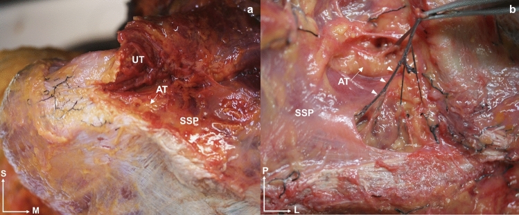
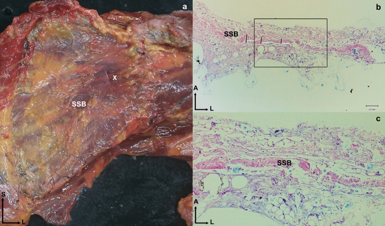
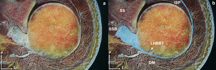
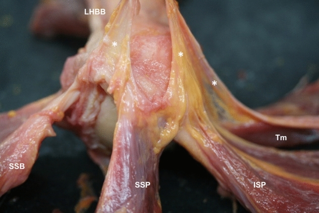
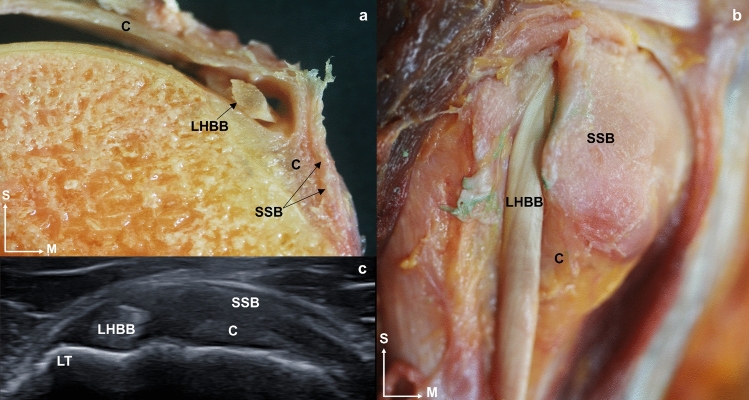
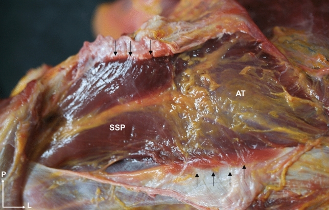
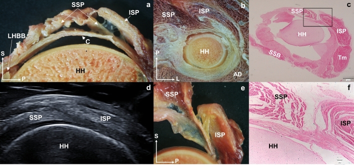
This study aimed to describe the shoulder anatomy, together with the anatomical relationships in adults and early stages of development. The shoulder muscles were studied from ultrasound, anatomical, and microscopic perspectives in a sample of 34 human shoulders. Thickness measurements were taken of the tendons and fasciae of the subscapularis, long head tendon of the biceps brachii, supraspinatus, infraspinatus, and teres minor. Ultrasound and dissection techniques are strongly correlated. However, the measurements obtained from the dissection technique were superior to those obtained from the ultrasound in all cases, except for the thickness of the long head tendon of the biceps brachii, the teres minor tendon, and the fascia thickness of the infraspinatus. In addition, the study of shoulder anatomy revealed no differences between females and males. Relevant findings from dissection included a clear overlap between the infraspinatus and supraspinatus, which shared tendon fibers, and a similar connection between the transverse ligament of the long head tendon of the biceps brachii and the subscapularis, which created a more interconnected shoulder function. The study of the anatomical measurements shows an underestimation of the shoulder measurements in the ultrasound compared with the dissection technique, but a high correlation between the measurements made by the two techniques. We present reference values for the tendon and fascia thicknesses of the rotator cuff, with no differences observed by gender. The relationships between shoulder structures described in the anatomical study imply as well that, in the event of an injury, adjacent tissues may be affected. This extended information may facilitate future optimal clinical explorations.

Keywords: Anatomy; Muscular fascia; Rotator cuff; Tendon; Ultrasound imaging.


Conflict of interest statement

All authors certify that they have no affiliations with or involvement in any organization or entity with a financial or non-financial interest in the subject matter or materials discussed in this manuscript.

Figures

Fig. 1
Fig. 1
Comparison of shoulder tendon thickness between ultrasound and anatomical dissection techniques. Abbreviations US: ultrasound, A: anatomic dissection, SSB: subscapularis, LHBB: long head of the biceps brachii, SSP: supraspinatus, ISP: infraspinatus, Tm: teres minor
Fig. 2
Fig. 2
Anatomical view of the posterior side of the shoulder. a The muscular fascia covers the muscles. b The muscular fascia is removed. It is possible to see the red dye injected by the ultrasound guide only at the infraspinatus and the green dye only at the Tm muscle. c Superior view of the shoulder. The muscular fascia forms an encapsulation of the supraspinatus muscle, and the red dye injected keeps down the muscular fascia. Abbreviations ISP: infraspinatus, Tm: teres minor, SSP: supraspinatus, HH: humeral head, S: superior, L: lateral
Fig. 3
Fig. 3
Images a and b are the same ultrasound cross-sectional view of the left shoulder that shows the fascial septum (white arrows) separating the infraspinatus muscle from the teres minor. b The infraspinatus is highlighted in blue, and the teres minor in yellow. The skin is marked with an asterisk (*). c Superior view of a transverse anatomical section of the left shoulder joint showing the infraspinatus and teres minor muscles and the fascial septum that separates them (white arrows). Abbreviations ISP: infraspinatus, Tm: teres minor, CO: coracobrachialis
Fig. 4
Fig. 4
A superior and anatomical view of a left shoulder shows the separation of the upper trapezius muscular fibers from the supraspinatus muscle, a Adipose tissue (white arrow) is visible between both muscles. b The injected vessels (triangle) pass through fascial tissue. Abbreviations UT: upper trapezius, SSP: supraspinatus, AT: adipose tissue, S: superior, M: medial, P: posterior, L: lateral
Fig. 5
Fig. 5
a Anatomical dissection anterior view of the left shoulder where the subscapularis muscle is visible and surrounded by a thin and own fascia. The white cross (X) shows the location where the histological sample has been taken. b Histological view of the fascia of the subscapularis. The black square can be seen enlarged in image c, where several layers of dense connective tissue can be seen. Abbreviations SSB: subscapularis, S: superior, L: lateral
Fig. 6
Fig. 6
a and b Anatomical view of a transversal cross-section of the left shoulder. The visual depicts a fascial extension (blue color in image b) originating from the tendon of the subscapularis muscle and coursing along the humeral head’s outer edge. It traverses the long head of the biceps brachii tendon before joining the fascia enveloping the infraspinatus muscle. Abbreviations SSB: subscapularis, LHBBT: long head of the biceps brachii tendon, ISP: infraspinatus, SS: scapula spine, DM: deltoid muscle, P: posterior, L: lateral
Fig. 7
Fig. 7
Anatomical dissection of the humeral head revealed a fascial tissue (*), connecting the tendons of the long head biceps brachii, subscapularis, supraspinatus, infraspinatus, and teres minor. Abbreviations LHBB: long head of the biceps brachii, SSB: subscapularis, SSP: supraspinatus, ISP: infraspinatus, Tm: teres minor
Fig. 8
Fig. 8
a A sagittal sectional cut of the humerus where it is possible to see the long head of the biceps brachii tendon inside the capsule and surrounded by the subscapularis tendon. b An anterior view of anatomical dissection where the subscapularis tendon is cut to leave the extracapsular part of the long head of the biceps brachii tendon visible. c An ultrasound image in the transverse axis of the long head of the biceps brachii tendon and in the longitudinal axis of the subscapularis tendon before its insertion into the lesser tubercle of the humerus, and before joining the fascia enveloping the infraspinatus. Abbreviations LHBB: long head of the biceps brachii, C: capsule, SSB: subscapularis, LT: lesser tubercle, ISP: infraspinatus, A: anterior, M: medial, S: superior
Fig. 9
Fig. 9
The superior anatomical dissection view of the shoulder shows the supraspinatus muscle. Its insertion in its own fascia (black arrows), specially in the central zone of the muscle. The presence of adipose tissue within the fascia is also observed. Abbreviations SSP: supraspinatus, AT: adipose tissue, P: posterior, L: lateral
Fig. 10
Fig. 10
Posterior anatomical dissection view of the shoulder. a and b The middle trapezius fibers gives fascial extensions (white arrows) to the superficial and medial part of the infraspinatus fascia and acts as a “fascial tendon”, reinforcing the superior and medial area. a The fascial expansions are continuous and converges with the rhomboid major (black arrows). c The middle trapezius and its extension are already dissected, and the directions of the infraspinatus fascia can be observed. Abbreviations MT: middle trapezius, ISP: infraspinatus, RM: rhomboid major, S: superior, L: lateral
Fig. 11
Fig. 11
Posterior anatomical dissection view of the shoulder that shows the posterior portion of the posterior deltoid muscle, that is lifted to expose the most superior part of the infraspinatus. The muscle fibers of the posterior deltoid also insert into the fascia of the infraspinatus itself (white arrows). Abbreviations PD: posterior deltoid, ISP: infraspinatus, S: superior, L: lateral
Fig. 12
Fig. 12
a Sagittal section of the shoulder section where the tendon of the supraspinatus muscle enters below the tendon of the infraspinatus muscle before reaching its insertion. b Superior view of a transverse anatomical section of the left shoulder where the tendon fibers of the infraspinatus crosses with the supraspinatus muscle. c Histological cross-section of a 7-month-old fetus that shows the arrangement of the shoulder muscles with the supraspinatus and infraspinatus disposition (the black square is shown enlarged in image f). d Ultrasound image at the transverse axis of the supraspinatus and infraspinatus tendons where it is observed how the supraspinatus tendon is introduced under the infraspinatus tendon. e Sagittal section of the shoulder section where the tendon of the supraspinatus muscle enters below the tendon of the infraspinatus muscle before reaching its insertion. f Enlarged image of image c square where it is possible to see how the supraspinatus and infraspinatus fibers intersect. Abbreviations SSP: supraspinatus, ISP: infraspinatus, AD: anterior deltoid, HH: humeral head, S: superior, A: anterior, P: posterior, L: lateral
Fig. 13
Fig. 13
The black lines correspond to the measurement points of the fascia thicknesses. a and b Histological section of the supraspinatus fascia. a Medial area of the supraspinatus fascia. b Lateral area of the supraspinatus fascia. c and d Histological section of the infraspinatus fascia. C Medial area of the infraspinatus fascia. d Lateral area of the infraspinatus fascia. e and f Histological section of the teres minor fascia. e Medial area of the teres minor fascia. f Lateral area of the teres minor fascia. Abbreviations SSP: supraspinatus, ISP: infraspinatus, Tm: teres minor

References

    1. Aly A-R, Rajasekaran S, Ashworth N. Ultrasound-guided shoulder girdle injections are more accurate and more effective than landmark-guided injections: a systematic review and meta-analysis. Br J Sports Med. 2015;49:1042–1049. doi: 10.1136/bjsports-2014-093573. - DOI - PubMed
    1. Bacle G, Gregoire J-M, Patat F, et al. Anatomy and relations of the infraspinatus and the teres minor muscles: a fresh cadaver dissection study. Surg Radiol Anat SRA. 2017;39:119–126. doi: 10.1007/s00276-016-1707-9. - DOI - PubMed
    1. Bağcier B, Geler Külcü GK, Yorulmaz Y, Altunok ÇA. Intra- and inter-rater reliability of ultrasound measurements of supraspinatus tendon thickness, acromiohumeral distance, and occupation ratio in patients with shoulder impingement syndrome. Arch Rheumatol. 2020;35:385. doi: 10.46497/ArchRheumatol.2020.7515. - DOI - PMC - PubMed
    1. Benjamin M. The fascia of the limbs and back–a review. J Anat. 2009;214:1–18. doi: 10.1111/j.1469-7580.2008.01011.x. - DOI - PMC - PubMed
    1. Burkhart SS, Esch JC, Jolson RS. The rotator crescent and rotator cable: an anatomic description of the shoulder’s “suspension bridge”. Arthrosc J Arthrosc Relat Surg. 1993;9:611–616. doi: 10.1016/S0749-8063(05)80496-7. - DOI - PubMed