. 2024 May 20;7(5):3358-3374.
doi: 10.1021/acsabm.4c00296. Epub 2024 May 8.

Fluorescent Chiral Quantum Dots to Unveil Origin-Dependent Exosome Uptake and Cargo Release

Affiliations

Fluorescent Chiral Quantum Dots to Unveil Origin-Dependent Exosome Uptake and Cargo Release

Gaeun Kim et al. ACS Appl Bio Mater. .

Abstract

Exosomes are promising nanocarriers for drug delivery. Yet, it is challenging to apply exosomes in clinical use due to the limited understanding of their physiological functions. While cellular uptake of exosomes is generally known through endocytosis and/or membrane fusion, the mechanisms of origin-dependent cellular uptake and subsequent cargo release of exosomes into recipient cells are still unclear. Herein, we investigated the intricate mechanisms of exosome entry into recipient cells and intracellular cargo release. In this study, we utilized chiral graphene quantum dots (GQDs) as representatives of exosomal cargo, taking advantage of the superior permeability of chiral GQDs into lipid membranes as well as their excellent optical properties for tracking analysis. We observed that the preferential cellular uptake of exosomes derived from the same cell-of-origin (intraspecies exosomes) is higher than that of exosomes derived from different cell-of-origin (cross-species exosomes). This uptake enhancement was attributed to receptor-ligand interaction-mediated endocytosis, as we identified the expression of specific ligands on exosomes that favorably interact with their parental cells and confirmed the higher lysosomal entrapment of intraspecies exosomes (intraspecies endocytic uptake). On the other hand, we found that the uptake of cross-species exosomes primarily occurred through membrane fusion, followed by direct cargo release into the cytosol (cross-species direct fusion uptake). We revealed the underlying mechanisms involved in the cellular uptake and subsequent cargo release of exosomes depending on their cell-of-origin and recipient cell types. Overall, this study envisions valuable insights into further advancements in effective drug delivery using exosomes, as well as a comprehensive understanding of cellular communication, including disease pathogenesis.

Keywords: cell-of-origin; chirality; endocytosis; exosomes; graphene quantum dots; membrane fusion.


Conflict of interest statement

The authors declare no competing financial interest.

Figures

Figure 1.
Figure 1.
Characterization of exosomes from three different cell lines: HepG2, 3T3, and HeLa cells. (a) TEM images of exosomes and size distribution of exosomes based on the TEM images. (b) Size distribution and particle number of exosomes measured by NTA. (c) Surface potential of exosomes analyzed by a Zetasizer. (d) Western blot analysis of exosomal markers expression.
Figure 2.
Figure 2.
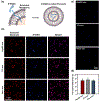
d-GQDs as representative of cargo of exosomes. (a) Schematic illustration of the d-GQDs permeation into exosomes. (b) CLSM images of d-GQDs-loaded (blue) and PKH26-labeled (red) exosomes. (c) Permeation of d-GQDs (blue) into exosomes observed by CLSM. (d) Permeation efficiency quantified by counting d-GQD-loaded exosomes over the total number of exosomes. The samples were prepared by loading 12 μM d-GQDs with exosomes (1 × 10⁹ particles/mL), followed by washing with 1× PBS under the support of 100 kDa centrifugal filter tubes.
Figure 3.
Figure 3.
Cellular uptake profiles of d-GQDs-loaded exosomes derived from three different cell lines into (a) HepG2 cells, (b) 3T3 cells, and (c) HeLa cells (mean ± s.d.). ns: not significant. *p < 0.05; **p < 0.01; ***p < 0.001; ****p < 0.0001. Each channel has red for the cell cytosol area and blue for d-GQDs. Three cell lines were incubated for 6 h in a cell culture medium (Control) with a concentration of 0.2 × 10³ exo/cell. The samples were prepared by loading 12 μM d-GQDs with exosomes (1 × 10⁹ particles/mL).
Figure 4.
Figure 4.
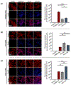
Endocytic uptake profiles of exosomes. (a) CLSM images of three cell lines incubated with exosomes derived from different cell lines. Each channel represents: red for the lysosomes and blue for d-GQDs. The quantification of colocalization between d-GQDs and lysosomes was analyzed in (b) HepG2 cells, (c) 3T3 cells, and (d) HeLa cells (mean ± s.e.). ns: not significant. *p < 0.05; **p < 0.01; ***p < 0.001; ****p < 0.0001. Three cell lines were incubated for 4 h in a cell culture medium (Control) with a concentration of 0.2 × 10³ exo/cell. The samples were prepared by loading 12 μM d-GQDs with exosomes (1 × 10⁹ particles/mL).
Figure 5.
Figure 5.
Identification of the surface proteins on exosomes that facilitate the receptor–ligand interaction-mediated endocytosis. (a) MS-based proteomics. Protein accession numbers were retrieved from the UniProtKB/Swiss-Prot. Black boxes with [+] correspond to the proteins that were detected, while the white boxes with [−] indicate those that were undetected. TGF: transforming growth factor. GalNAc: N-acetylgalactosamine. NRG: neuregulin. CHC: Clathrin heavy chain. HSP: heat shock protein. (b) Western blot analysis of TGF-β1, GalNAc, and NRG1 expression for three types of exosomes. Relative expression levels of (c) TGF-β1, (d) GalNAc, and (e) NRG1 (mean ± s.d.). Data were normalized to the expression level of β-actin. ns: not significant. *p < 0.05; **p < 0.01. ***p < 0.001. ****p < 0.0001.
Figure 6.
Figure 6.
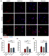
Membrane fusion of exosomes. CLSM images of (a) HepG2 cells, (b) 3T3 cells, and (c) HeLa cells after 1 h of incubation. Each channel represents: blue for nuclei, green for cellular membrane, red for exosomal membrane, and gray for FRET. (d–f) PCC quantitatively analyzed based on merged images. (g–i) FRET evaluation by measuring FI (ex, 484 nm; em, 565 nm) in a cell suspension (mean ± s.d.). ns: not significant. *p < 0.05; **p < 0.01; ***p < 0.001; ****p < 0.0001. Three cell lines were incubated in a cell culture medium (Control) with a concentration of 0.4 × 10³ exo/cell.
Figure 7.
Figure 7.
d-GQDs release and retention from exosomes after 6 h of incubation with HepG2 cells (top line), 3T3 cells (middle line), and HeLa cells (bottom line). Each channel represents: green for the cellular membrane, red for the exosomal membrane, and blue for d-GQDs. The arrows indicate d-GQDs, representing the exosomal cargo released from the exosomes. Three cell lines were incubated with a cell culture medium (Control) with a concentration of 0.2 × 10³ exo/cell. The samples were prepared by loading 12 μM d-GQDs with exosomes (1 × 10⁹ particles/mL).
Figure 8.
Figure 8.
Schematic illustration for the cellular uptake mechanisms of exosomes, depending on their cell-of-origin, followed by cargo release based on the specific cellular uptake mechanisms. (a) Intraspecies endocytic uptake: Exosomes from the same cell-of-origin demonstrated a greater tendency to undergo cellular uptake by parental recipient cells through endocytosis mediated by receptor–ligand interactions, resulting in the entrapment of cargo within lysosomes. (b) Cross-species direct fusion uptake: Exosomes derived from different cells of origin were taken up less by nonparental recipient cells but primarily through direct membrane fusion, resulting in the direct release of cargo into the cytosol.

Update of

Similar articles

Cited by

References

    1. Doyle LM; Wang MZ Overview of Extracellular Vesicles, Their Origin, Composition, Purpose, and Methods for Exosome Isolation and Analysis. Cells 2019, 8 (7), 727. - PMC - PubMed
    1. Johnston J; Stone T; Wang Y Biomaterial-enabled 3D cell culture technologies for extracellular vesicle manufacturing. Biomater. Sci 2023, 11 (12), 4055. - PubMed
    1. Shafiei M; Ansari MN; Razak SI; Khan MU Comprehensive Review on the Applications of Exosomes and Liposomes in Regenerative Medicine and Tissue Engineering. Polymers 2021, 13 (15), 2529. - PMC - PubMed
    1. Saeedi S; Israel S; Nagy C; Turecki G The emerging role of exosomes in mental disorders. Translational Psychiatry 2019, 9 (1). DOI: 10.1038/s41398-019-0459-9. - DOI - PMC - PubMed
    1. Sarko DK; McKinney CE Exosomes: Origins and Therapeutic Potential for Neurodegenerative Disease. Front. Neurosci 2017, 11. DOI: 10.3389/fnins.2017.00082. - DOI - PMC - PubMed

LinkOut - more resources