. 2024 Apr 30;30(5):gaae018.
doi: 10.1093/molehr/gaae018.

Paxillin regulates androgen receptor expression associated with granulosa cell focal adhesions

Affiliations

Paxillin regulates androgen receptor expression associated with granulosa cell focal adhesions

Adelaide E Weidner et al. Mol Hum Reprod. .

Abstract

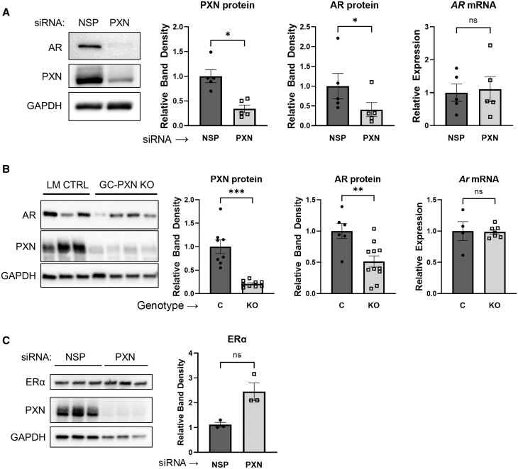
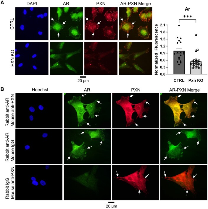
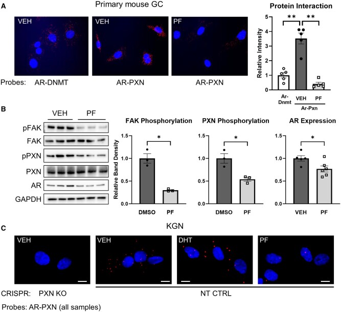
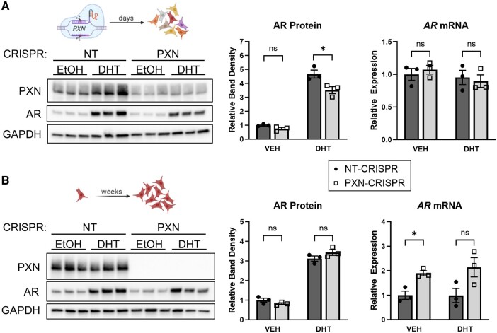
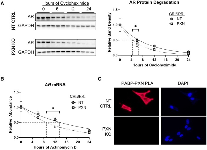
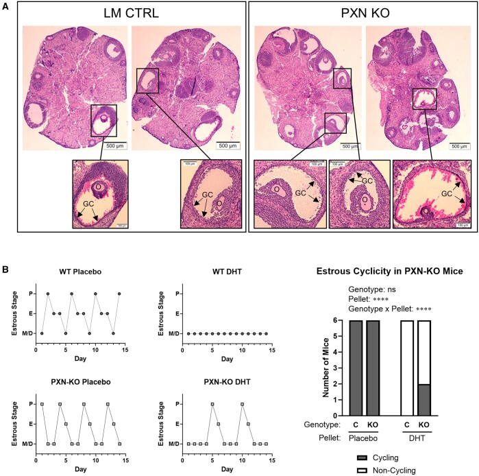
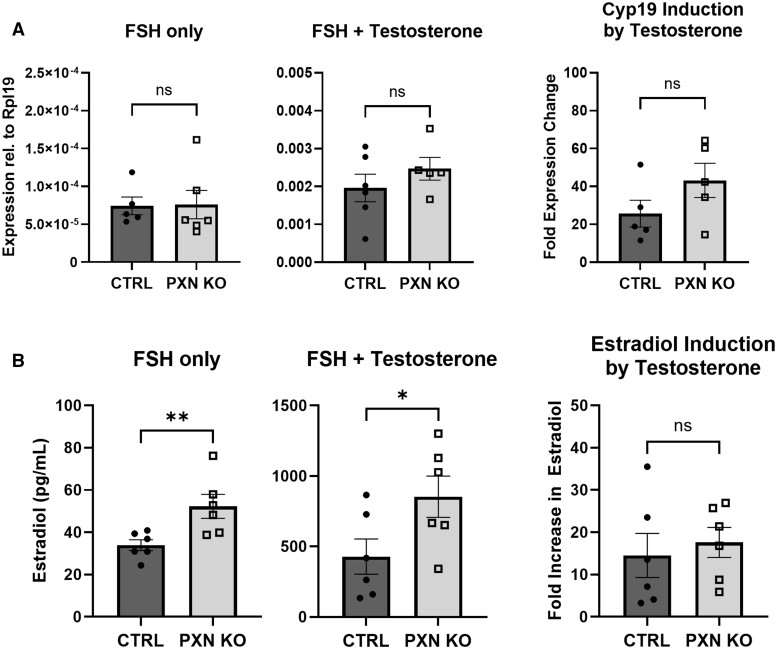
Paxillin is a ubiquitously expressed adaptor protein integral to focal adhesions, cell motility, and apoptosis. Paxillin has also recently been implicated as a mediator of nongenomic androgen receptor (AR) signaling in prostate cancer and other cells. We sought to investigate the relationship between paxillin and AR in granulosa cells (GCs), where androgen actions, apoptosis, and focal adhesions are of known importance, but where the role of paxillin is understudied. We recently showed that paxillin knockout in mouse GCs increases fertility in older mice. Here, we demonstrate that paxillin knockdown in human granulosa-derived KGN cells, as well as knockout in mouse primary GCs, results in reduced AR protein but not reduced mRNA expression. Further, we find that both AR protein and mRNA half-lives are reduced by approximately one-third in the absence of paxillin, but that cells adapt to chronic loss of paxillin by upregulating AR gene expression. Using co-immunofluorescence and proximity ligation assays, we show that paxillin and AR co-localize at the plasma membrane in GCs in a focal adhesion kinase-dependent way, and that disruption of focal adhesions leads to reduced AR protein level. Our findings suggest that paxillin recruits AR to the GC membrane, where it may be sequestered from proteasomal degradation and poised for nongenomic signaling, as reported in other tissues. To investigate the physiological significance of this in disorders of androgen excess, we tested the effect of GC-specific paxillin knockout in a mouse model of polycystic ovary syndrome (PCOS) induced by chronic postnatal dihydrotestosterone (DHT) exposure. While none of the control mice had estrous cycles, 33% of paxillin knockout mice were cycling, indicating that paxillin deletion may offer partial protection from the negative effects of androgen excess by reducing AR expression. Paxillin-knockout GCs from mice with DHT-induced PCOS also produced more estradiol than GCs from littermate controls. Thus, paxillin may be a novel target in the management of androgen-related disorders in women, such as PCOS.

Keywords: PCOS; androgen receptor; focal adhesion; granulosa; ovary; paxillin; polycystic ovary syndrome.


Conflict of interest statement

The authors disclose no conflicts of interest.

Figures

Figure 1.
Figure 1.
Paxillin enhances androgen receptor protein content independently of its mRNA expression in KGN cells and in mouse granulosa cells. (A) Representative western blot, band densitometry, and qPCR of KGN cells after transfection with siRNA targeting non-specific (NSP) control or paxillin (PXN). Data are mean and SEM of five independent experiments. Statistical significance was determined by paired Mann–Whitney test. (B) Representative western blot, band densitometry and qPCR in mouse primary GCs from GC-specific paxillin knockout mice (GC-PXN KO or KO), or littermate controls (LM CTRL or C), n = 3–11 per group. Cells were cultured for 4 days before analysis. Data are mean and SEM of n = 3–11 mice per group. Statistical significance was determined by Mann–Whitney test. (C) ERα protein content in PXN-knockdown KGN cells transfected as in (A) and band densitometry of ERα relative to GAPDH. Statistical significance was determined by Mann–Whitney test. For all comparisons, ns: not significant; *P > 0.05; **P < 0.01; ***P < 0.001. AR: androgen receptor, PXN: paxillin, NSP: nonspecific control, LM CTRL: littermate control, GC-PXN KO: granulosa-specific paxillin knockout, ERα: estrogen receptor alpha. Uncropped blots are shown in Supplementary Fig. S3.
Figure 2.
Figure 2.
Androgen receptor and paxillin co-localize at the granulosa cell membrane. (A) Mouse primary GCs were isolated from GC-specific paxillin knockout mice (PXN KO) or littermate controls (CTRL) and cultured on coverslips before fixation and co-immunofluorescence with antibodies against AR and PXN. AR fluorescence was quantified in 18–23 cells from each sample using Image J and analyzed by Mann–Whitney test. ***P < 0.001. (B) KGN cells were grown on coverslips, fixed, and co-immunoprobed with the primary antibodies listed on the left. All samples were then probed with both anti-mouse and anti-rabbit secondary antibodies. White arrows indicate membrane-localized AR and PXN in all pictures. DAPI: 4',6-diamidino-2-phenylindole, AR: androgen receptor, PXN: paxillin, CTRL: littermate control, PXN KO: granulosa-specific paxillin knockout.
Figure 3.
Figure 3.
Androgen receptor and paxillin interaction requires intact granulosa cell focal adhesions. (A) Proximity ligation assay (PLA) in primary mouse GCs treated for 24 h with 5 μM PF-573228 (PF) or DMSO (VEH) and probed with antibodies against AR and PXN or AR and DNMT as negative control. Protein proximity signal produces red fluorescence, merged with DAPI indicating cell nuclei. Right: red fluorescence was quantified in five randomly selected cells in each sample using Image J and analyzed with Mann–Whitney tests. Magnification: ×40. **P < 0.01. (B) Representative western blot and band densitometry of primary mouse GCs (n = 3–6 mice per group) treated for 24 h with 5 μM PF-573228 (PF) or DMSO (VEH) and analyzed by Mann–Whitney tests. *P < 0.05. (C) PLA assay in KGN cells edited with CRISPR to delete paxillin (PXN KO) or non-targeting control (NT CTRL) and treated with vehicle (VEH), 25 nM dihydrotestosterone (DHT) or 5 μM PF-573228 (PF). All samples were probed with PXN and AR antibodies. Protein proximity signal produces red fluorescence, merged with DAPI indicating cell nuclei. Scale bar: 10 μm. AR: androgen receptor, PXN: paxillin, DNMT: DNA methyltransferase 1, GC: granulosa cells, VEH: vehicle, PF: PF-573228, FAK: focal adhesion kinase, CRISPR: clustered regularly interspaced short palindromic repeats, PXN KO: paxillin knockout, NT CTRL: non-targeting control. Uncropped blots are shown in Supplementary Fig. S5.
Figure 4.
Figure 4.
Androgen receptor expression is rescued after chronic paxillin loss in CRISPR-Cas9-generated paxillin-null KGN clones. (A): KGN cells were transfected with paxillin-targeting or non-targeting control sgRNA and CRISPR-Cas9 lentivirus (PXN-CRISPR or NT-CRISPR, respectively). Successfully transfected cells were selected using GFP-positive flow cytometry 2 days later, cultured for 5 additional days, then treated with 0.1% ethanol (VEH) or 25 nM dihydrotestosterone (DHT) for 24 h before western blotting and qPCR. Band densitometry was analyzed by Mann–Whitney tests. (B) Clonal populations were obtained from single CRISPR-edited KGN cells by expansion in culture over a period of several weeks, then treated and analyzed the same way as in (A). *P < 0.05, ns: not significant. Pictures drawn in Biorender. CRISPR: clustered regularly interspaced short palindromic repeats, PXN: paxillin, NT: non-targeting control, EtOH: ethanol, DHT: dihydrotestosterone, AR: androgen receptor, VEH: vehicle, GFP: green fluorescent protein. Uncropped blots are shown in Supplementary Fig. S6.
Figure 5.
Figure 5.
Paxillin prolongs androgen receptor mRNA and protein half-lives. (A) CRISPR-Cas9-generated clonal paxillin-knockout (PXN KO) and non-targeting control (NT CTRL) KGN cells were treated with 50 μM cycloheximide for indicated times before western blotting. Band densitometry was fitted to a one-phase decay with least squares fit, analyzed by ANOVA and protein half-life was calculated by GraphPad Prism. (B) AR mRNA was measured by qPCR from cells generated as in (A) and treated with 5 μM actinomycin D for up to 24 h, then analyzed the same way as in (A) to determine the mRNA half-life. (C) PLA assay of PABP and PXN and DAPI staining in PXN KO and NT CTRL samples. Red fluorescent signal represents PABP and PXN proximity. Magnification: ×40. NT CTRL: non-targeting control, PXN KO: paxillin knockout, AR: androgen receptor, CRISPR: clustered regularly interspaced short palindromic repeats, PABP: poly-A binding protein, PLA: proximity ligation assay, DAPI: DAPI: 4',6-diamidino-2-phenylindole. Uncropped blots are shown in Supplementary Fig. S7.
Figure 6.
Figure 6.
Reproductive effects of granulosa cell-specific paxillin knockout in a mouse PCOS model. GC-specific paxillin knockout mice (PXN-KO) and littermate controls (WT) were injected with s.c. pellets containing placebo or 2.5 mg DHT on postnatal Day 19. (A) Representative ovary sections stained with hematoxylin and eosin from adult PXN-KO and WT mice treated with DHT as above. High-magnification insets show cystic follicles characterized by thin GC layers (labeled GC) and degenerating oocytes (labeled O). No corpora lutea were observed in any of the ovary sections. (B) Estrous stage was determined daily at 76–90 days after pellet insertion via visual cytology assessment of vaginal lavage. Left, representative estrous cycles. P: proestrus; E: estrus; M/D: metestrus or diestrus. Right, number of mice that experienced at least one estrous cycle (‘Cycling’, defined as proestrus followed immediately by estrus) during the 14-day cycling study. Data were analyzed by Fisher’s exact test (C versus KO and placebo versus DHT) and by Mantel–Haenszel Chi-square test (interaction of pellet treatment and genotype). ns: not significant; ****P < 0.0001. LM CTRL: littermate control, PXN KO: paxillin knockout, GC: granulosa cells, O: oocyte, WT: wild-type, P: proestrus, E: estrus, M/D: metestrus/diestrus, C: control, KO: knockout, DHT: dihydrotestosterone.
Figure 7.
Figure 7.
Granulosa cell-specific paxillin knockout increases estradiol production in mice with dihydrotestosterone-induced PCOS. (A): Cyp19 gene expression was measured by qPCR in primary GCs from littermate control (CTRL) and GC paxillin knockout (PXN KO) mice cultured for 4 days and treated as indicated (FSH: 100 ng/ml, Testosterone: 100nM) during the last 2 days of culture. (B) Estradiol concentration was measured in the culture media collected from cells described in (A). All comparisons were analyzed by Mann–Whitney test; ns: not significant; *P < 0.05; **P < 0.01. Rpl19: ribosomal protein L19, CTRL: control, PXN KO: paxillin knockout.

Similar articles

Cited by

References

    1. Aflatounian A, Edwards MC, Rodriguez Paris V, Bertoldo MJ, Desai R, Gilchrist RB, Ledger WL, Handelsman DJ, Walters KA.. Androgen signaling pathways driving reproductive and metabolic phenotypes in a PCOS mouse model. J Endocrinol 2020;245:381–395. - PubMed
    1. Alpha KM, Xu W, Turner CE.. Paxillin family of focal adhesion adaptor proteins and regulation of cancer cell invasion. Int Rev Cell Mol Biol 2020;355:1–52. - PMC - PubMed
    1. Astapova O, Minor BMN, Hammes SR.. Physiological and pathological androgen actions in the ovary. Endocrinology 2019;160:1166–1174. - PMC - PubMed
    1. Astapova O, Seger C, Hammes SR.. Ligand binding prolongs androgen receptor protein half-life by reducing its degradation. J Endocr Soc 2021;5:bvab035. - PMC - PubMed
    1. Azziz R, Carmina E, Chen Z, Dunaif A, Laven JS, Legro RS, Lizneva D, Natterson-Horowtiz B, Teede HJ, Yildiz BO.. Polycystic ovary syndrome. Nat Rev Dis Primers 2016;2:16057. - PubMed