. 2025 Apr:70:339-353.
doi: 10.1016/j.jare.2024.05.006. Epub 2024 May 7.

Crosstalk between endothelial cells and dermal papilla entails hair regeneration and angiogenesis during aging

Affiliations

Crosstalk between endothelial cells and dermal papilla entails hair regeneration and angiogenesis during aging

Siyi Zhou et al. J Adv Res. 2025 Apr.

Abstract

Introduction: Tissues maintain their function through interaction with microenvironment. During aging, both hair follicles and blood vessels (BV) in skin undergo degenerative changes. However, it is elusive whether the changes are due to intrinsic aging changes in hair follicles or blood vessels respectively, or their interactions.

Objective: To explore how hair follicles and blood vessels interact to regulate angiogenesis and hair regeneration during aging.

Methods: Single-cell RNA-sequencing (scRNA-seq) analyses were used to identify the declined ability of dermal papilla (DP) and endothelial cells (ECs) during aging. CellChat and CellCall were performed to investigate interaction between DP and ECs. Single-cell metabolism (scMetabolism) analysis and iPATH were applied to analyze downstream metabolites in DP and ECs. Hair-plucking model and mouse cell organoid model were used for functional studies.

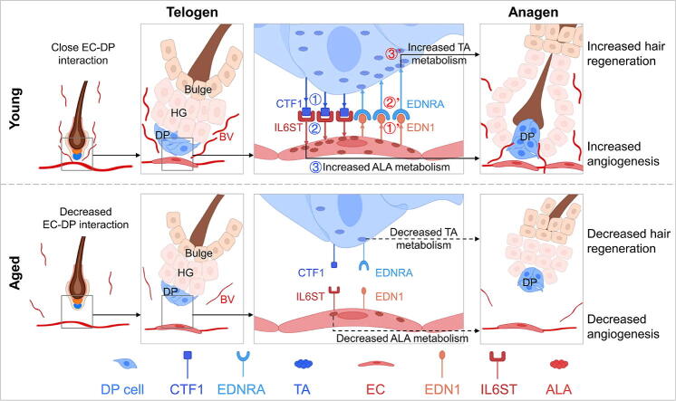
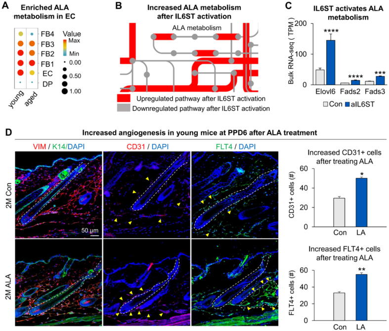
Results: During aging, distance and interaction between DP and ECs are decreased. DP interacts with ECs, with decreased EDN1-EDNRA signaling from ECs to DP and CTF1-IL6ST signaling from DP to ECs during aging. ECs-secreted EDN1 binds to DP-expressed EDNRA which enhances Taurine (TA) metabolism to promote hair regeneration. DP-emitted CTF1 binds to ECs-expressed IL6ST which activates alpha-linolenic acid (ALA) metabolism to promote angiogenesis. Activated EDN1-EDNRA-TA signaling promotes hair regeneration in aged mouse skin and in organoid cultures, and increased CTF1-IL6ST-ALA signaling also promotes angiogenesis in aged mouse skin and organoid cultures.

Conclusions: Our finding reveals reciprocal interactions between ECs and DP. ECs releases EDN1 sensed by DP to activate TA metabolism which induces hair regeneration, while DP emits CTF1 signal received by ECs to enhance ALA metabolism which promotes angiogenesis. Our study provides new insights into mutualistic cellular crosstalk between hair follicles and blood vessels, and identifies novel signaling contributing to the interactions of hair follicles and blood vessels in normal and aged skin.

Keywords: Aging; Angiogenesis; Cell interaction; Hair regeneration; Metabolism; Mutualism.


Conflict of interest statement

Declaration of competing interest The authors declare that they have no known competing financial interests or personal relationships that could have appeared to influence the work reported in this paper.

Figures

None
Graphical abstract
Fig. 1
Fig. 1
Interactions between hair follicles and BV during skin aging. A. Schematic of hair regeneration during aging. B. TSNE plots of DP and EC clusters in young and aged skin by unbiased clustering. C. AUCell score of angiogenesis between young and aged EC. D. PCNA and CD31 immunostaining show angiogenesis in young and aged human scalp skin. Scale bars, 100 μm. Statistical of EC and PCNA+ EC surrounding hair follicles and EC’s location. N ≥ 3, **p < 0.01, *p < 0.05. E. PCNA and CD31 immunostaining show angiogenesis in young and aged mice dorsal skin. Scale bars, 50 μm. Statistical of EC and PCNA+ EC surrounding hair follicles and EC’s location. N ≥ 3, **p < 0.01, *p < 0.05, and ns no significant change. F. Schematic of angiogenesis during hair regeneration in young (left) and aged (right) skin. G. Cellchat analysis of DP and EC interactions between young and aged skin. H. PCNA / CD31 immunostaining shows decreased hair regeneration after inhibiting CDH5. Scale bars, 50 μm.
Fig. 2
Fig. 2
ScRNA-seq analysis of EC-DP reveals decreased expression of EDN1-EDNRA and CTF1-IL6ST in old skin. A. Chord diagram shows decreased Edn1-Ednra / Ednrb signal in aged skin (left); Vlnplot shows mRNA expression of Edn1, Ednra and Ednrb (right). B. Heatmap shows decreased Ctf1-Il6st / Lifr signal in aged skin (left); Vlnplot shows mRNA expression of Ctf1, Il6st and Lifr (right). C. Featureplot shows expression of Edn1, Ednra, Ctf1and Il6st in young and aged skin. D. Statistics of qRT-PCR show decreased expression of target genes in aged skin. N ≥ 3, *p < 0.05. E. Target genes immunostaining shows decreased expression in aged skin. F. Schematics show target gene expression patterns in young and aged skin.
Fig. 3
Fig. 3
EDN1-EDNRA signaling from EC to DP promotes hair regeneration. A. BMP4, SMAD1, β-catenin, and PCNA immunostaining shows hair regeneration after EDN1 recombinant protein or EDNRA inhibitor treatment in young mice; Statistics of average FI of BMP4 (panel 1 bottom), SMAD1+ cells (panel 2 bottom), NIR of β-catenin (panel 3 bottom) and PCNA+ cells (panel 4 bottom). Scale bars, 50 μm. N ≥ 3, **p < 0.01. B. BMP4, SMAD1, β-catenin and PCNA immunostaining shows hair regeneration after EDN1 recombinant protein or EDNRA inhibitor treatment in old mice; Statistics of average FI of BMP4 (panel 1 bottom), SMAD1+ cells (panel 2 bottom), NIR of β-catenin (panel 3 bottom) and PCNA+ cells (panel 4 bottom). Scale bars, 50 μm. N ≥ 3, **p < 0.01, *p < 0.05. C. RNA-seq compares the control group and the iEDNRA group. KEGG analysis shows Mitophagy and AMPK signaling pathway was downregulated in the iEDNRA group. D. RNA-seq compares the control group and the DML group. KEGG analysis shows cell cycle and DNA replication signaling pathways were enrichment in the DML group.
Fig. 4
Fig. 4
TA and HTA metabolism could promote hair regeneration. A. Dotplot of scMetabolism shows TA and HTA metabolism enriched in young DP. B. Featureplot shows expression of TA and HTA metabolic genes in young and aged skin. C. Statistics of bulk RNA-seq shows TA and HTA metabolic genes decreased after inhibiting EDNRA. N ≥ 3, ****p < 0.0001, ***p < 0.001. D. BMP4, SMAD1, β-catenin, and LEF1 immunostaining shows hair regeneration after TA and HTA treatment in young mice; Statistics of average FI of BMP4 (panel 1 bottom), SMAD1+ cells (panel 2 bottom), β-catenin+ cells (panel 3 bottom) and LEF1+ cells (panel 4 bottom). Scale bars, 50 μm. N ≥ 3, **p < 0.01.
Fig. 5
Fig. 5
EDN1-EDNRA and TA also facilitate HF regeneration in mouse cell organoids. A. Featureplot shows expression of Edn1 and Ednra in newborn and adult mouse cell organoids. B. EDN1 and EDNRA immunostaining shows decreased expression in adult mouse cell organoids at D4; Statistics of EDN1+ cells and EDNRA+ cells in newborn and adult mouse cell organoids. N ≥ 3, **p < 0.01. C. BMP4, β-catenin and LEF1 immunostaining and statistics show expression of hair regenerative genes increased after EDN1 recombinant protein and TA treatment in newborn mouse cell organoid; Phase-contrast microscope and statistics show hair regeneration after grafting newborn mouse cell organoid cultures treated with EDNRA inhibitor or TA. D. BMP4, β-catenin and LEF1 immunostaining and statistics show expression of hair regenerative genes increased after EDN1 recombinant protein and TA treatment in adult mouse cell organoid; Phase-contrast microscope and statistics show hair regeneration after grafting adult mouse cell organoid cultures treated with EDN1 recombinant protein or TA. Scale bars, 50 μm. N ≥ 3, **p < 0.01, *p < 0.05, and ns no significant change.
Fig. 6
Fig. 6
CTF1-IL6ST signaling from DP to EC promotes angiogenesis. A. Statistics of qRT-PCR show increased expression of Flt4 after activating IL6ST in young (left) and aged (right) mice. N ≥ 3, *p < 0.05, and ns no significant change. B. PCNA / CD31 and FLT4 immunostaining show angiogenesis after activating or inhibiting IL6ST in young (left) and aged (right) mice. Scale bars, 50 μm. Statistical of PCNA+ EC and FLT4+ EC surrounding hair follicles and EC’s location. N ≥ 3, *p < 0.05, and ns no significant change. C. Schematic shows angiogenesis after activating or inhibiting IL6ST.
Fig. 7
Fig. 7
ALA metabolism promotes angiogenesis. A. Dotplot of scMetabolism shows ALA metabolism enriched in young EC. B. iPATH shows ALA metabolism increased after activating IL6ST. C. Statistics of bulk RNA-seq shows Fads2 increased after activating IL6ST. N ≥ 3, ****p < 0.0001, ***p < 0.001, and ns no significant change. D. VIMTIN / K14, CD31 and FLT4 immunostaining shows angiogenesis after ALA treatment in young mice; Statistics of CD31+ cells and FLT4+ cells. Scale bars, 50 μm. N ≥ 3, **p < 0.01, *p < 0.05.
Fig. 8
Fig. 8
CTF1-IL6ST and ALA also facilitate angiogenesis in mouse cell organoids. A. CTF1 immunostaining shows decreased expression in adult mouse cell organoid at D4; Statistics of CTF1+ cells in newborn and adult mouse cell organoid. N ≥ 3, **p < 0.01. B. IL6ST immunostaining shows decreased expression in adult mouse cell organoid; Statistics of IL6ST + cells in newborn and adult mouse cell organoid. N ≥ 3, *p < 0.05. C. CD31 and FLT4 immunostaining shows EC changes in newborn mouse cell organoids after activating IL6ST; Statistics of EC and FLT4+ cells in newborn mouse cell organoids after activating IL6ST. N ≥ 3, *p < 0.05. D. CD31 and FLT4 immunostaining shows EC changes in adult mouse cell organoids after activating IL6ST; Statistics of EC and FLT4+ cells in adult mouse cell organoids after activating IL6ST. N ≥ 3, *p < 0.05. E. CD31 and FLT4 immunostaining shows EC changes in newborn mouse cell organoid after ALA treatment; Statistics of EC and FLT4+ cells in newborn mouse cell organoid after ALA treatment. N ≥ 3, *p < 0.05, and ns no significant change. F. CD31 and FLT4 immunostaining shows EC changes in adult mouse cell organoids after ALA treatment; Statistics of EC and FLT4+ cells in adult mouse cell organoids after ALA treatment. Scale bars, 50 μm. N ≥ 3, *p < 0.05. G. Schematic shows EC changes after activating IL6ST or treating ALA. H. PCNA / CD31 immunostaining shows angiogenesis in post-transplantation nude mice after activating or inhibiting IL6ST in NB mouse cell organoid; Statistics of PCNA+ ECs and FLT4+ cells in post-transplantation nude mice skin (right). N ≥ 3, **p < 0.01, *p < 0.05.
Fig. 9
Fig. 9
Graphical summary. EC releases EDN1 sensed by DP to activate TA metabolism which induces hair regeneration, while DP emits the CTF1 signal received by EC to enhance ALA metabolism which promotes angiogenesis in young skin. Decreased interactions between EC and DP lead to attenuated hair regeneration and angiogenesis in aged skin.

References

    1. Lei M., Chuong C.M. STEM CELLS. Aging, alopecia, and stem cells. Science. 2016;351:559–560. doi: 10.1126/science.aaf1635. - DOI - PubMed
    1. Ji S., Zhu Z., Sun X., Fu X. Functional hair follicle regeneration: an updated review. Signal Transduct Target Ther. 2021;6:66. doi: 10.1038/s41392-020-00441-y. - DOI - PMC - PubMed
    1. Wu W., Yang J., Tao H., Lei M. Environmental Regulation of Skin Pigmentation and Hair Regeneration. Stem Cells Dev. 2022;31:91–96. doi: 10.1089/scd.2022.29011.wwu. - DOI - PubMed
    1. Lei M., Lin S.J., Chuong C.M. Editorial: Hair Follicle Stem Cell Regeneration in Aging. Front Cell Dev Biol. 2021;9 doi: 10.3389/fcell.2021.799268. - DOI - PMC - PubMed
    1. Chen C.C., Chuong C.M. Multi-layered environmental regulation on the homeostasis of stem cells: the saga of hair growth and alopecia. J Dermatol Sci. 2012;66:3–11. doi: 10.1016/j.jdermsci.2012.02.007. - DOI - PMC - PubMed

LinkOut - more resources