. 2024 May 8;15(5):321.
doi: 10.1038/s41419-024-06700-y.

RAD18 O-GlcNAcylation promotes translesion DNA synthesis and homologous recombination repair

Affiliations

RAD18 O-GlcNAcylation promotes translesion DNA synthesis and homologous recombination repair

Xiaolu Ma et al. Cell Death Dis. .

Abstract

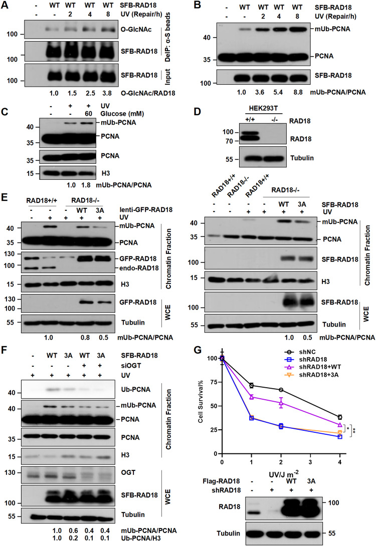
RAD18, an important ubiquitin E3 ligase, plays a dual role in translesion DNA synthesis (TLS) and homologous recombination (HR) repair. However, whether and how the regulatory mechanism of O-linked N-acetylglucosamine (O-GlcNAc) modification governing RAD18 and its function during these processes remains unknown. Here, we report that human RAD18, can undergo O-GlcNAcylation at Ser130/Ser164/Thr468, which is important for optimal RAD18 accumulation at DNA damage sites. Mechanistically, abrogation of RAD18 O-GlcNAcylation limits CDC7-dependent RAD18 Ser434 phosphorylation, which in turn significantly reduces damage-induced PCNA monoubiquitination, impairs Polη focus formation and enhances UV sensitivity. Moreover, the ubiquitin and RAD51C binding ability of RAD18 at DNA double-strand breaks (DSBs) is O-GlcNAcylation-dependent. O-GlcNAcylated RAD18 promotes the binding of RAD51 to damaged DNA during HR and decreases CPT hypersensitivity. Our findings demonstrate a novel role of RAD18 O-GlcNAcylation in TLS and HR regulation, establishing a new rationale to improve chemotherapeutic treatment.


Conflict of interest statement

The authors declare no competing interests.

Figures

Fig. 1
Fig. 1. RAD18 binds OGT and is subject to O-GlcNAcylation predominantly at Ser130/Ser164/Thr468.
A HEK293T cells expressing Flag-RAD18 and Myc-OGT were irradiated with UV (15 J m−2) followed by immunoprecipitation with anti-Flag M2 beads. The cell lysates were detected with anti-Myc and anti-Flag antibodies. The experiments were carried out in triplicate. B HEK293T cells transfected with SFB-RAD18 or empty vector were incubated with Thiamet-G (TMG) and glucose. The cell lysates were denatured and immunoprecipitated with anti-S beads followed by immunoblotting with O-GlcNAc and Flag antibodies. C HEK293T cells transfected with SFB-RAD18 were treated with Thiamet-G and different concentrations of glucose. The cell lysates were immunoprecipitated and analyzed as in (B). D Schematic representation of WT- and 3A-RAD18. HEK293T cells (E) or cells incubated with glucose and TMG (F) were transfected with the indicated SFB-RAD18 constructs, followed by immunoprecipitation as in (B). G Expression of His-RAD18 (WT or 3A) and GST-OGT or GST co-transformed into E. coli Transetta (DE3) cells after IPTG (0.4 mM) induction were examined via SDS-PAGE followed by Coomassie blue staining. H His-RAD18 proteins were purified and analyzed by western blot with anti-O-GlcNAc and anti-His antibodies.
Fig. 2
Fig. 2. O-GlcNAclylation modulates RAD18 recruitment at DNA damage sites.
A Representative image for the dynamic recruitment of WT or 3A GFP-RAD18 to laser-induced DSBs. Data were presented as mean ± SEM from 15 cells. Scale bar, 10 μm. B Quantification of the time course of WT or 3A GFP-RAD18 recruitment after laser microirradiation. C Representative images of GFP-RAD18 foci stained with DAPI after UV irradiation. Scale bars: 2 μm. The protein levels of RAD18 in RAD18+/+ and RAD18-/- U2OS cells were detected by immunoblotting. D Quantification of the percentage of RAD18-/- U2OS cells transfected with WT or 3A GFP-RAD18 constructs with more than 30 RAD18 foci after CPT, Bleomycin and UV exposure by counting at least 200 cells in each experiment. Data represent means ± SEM from three independent experiments. E Chromatin fractions of HEK293T cells expressing WT or 3A SFB-RAD18 were extracted followed by immunoblotting with Flag and H2B antibodies. The whole cell extract (WCE) was harvested and immunoblotted with Flag and β-actin antibodies.
Fig. 3
Fig. 3. RAD18 3A mutation impairs PCNA monoubiquitination and cell survival after UV irradiation.
A HEK293T cells transfected with SFB-RAD18 were irradiated with UV (15 J m−2) and harvested at different time points later. The cell lysates were denatured and immunoprecipitated with anti-S beads followed by immunoblotting with O-GlcNAc and Flag antibodies. Chromatin fractions of HEK293T cells transfected with SFB-RAD18 (B) or incubated with glucose or not (C) were extracted after UV (15 J m−2) irradiation followed by immunoblotting with indicated antibodies. RAD18 O-GlcNAcylation promotes PCNA monoubiquitination. RAD18+/+ or RAD18-/- HEK293T (D) and U2OS (E) or OGT-depleted RAD18-/- HEK293T (F) cells were transfected with WT or 3A RAD18, and irradiated with UV (15 J m−2). The chromatin fractions were harvested and immunoblotted using indicated antibodies. G Clonogenic survival assays in RAD18-knockdown U2OS cells transfected with empty vector, WT or 3A Flag-RAD18 after UV irradiation. Cells were irradiated with indicated doses of UV and further incubated in medium supplemented with 0.4 mM caffeine for 7–10 days. Surviving fraction was expressed as a percentage of mock-treated cells. The representative curve is shown. Error bar: s.d., n = 3. The protein levels of RAD18 were detected by immunoblotting.
Fig. 4
Fig. 4. O-GlcNAclylation promotes CDC7-mediated RAD18 phosphorylation at Ser434.
A WT or 3A SFB-RAD18 and GFP-Polη co-transfected HEK293T cells were treated with glucose (60 mM) and TMG (5 μM). Chromatin fraction was immunoprecipitated followed by immunoblotting with GFP and Flag antibodies after UV (15 J m−2) irradiation. *non-specific band. OGT knockdown HEK293T cells were transfected with WT or 3A SFB-RAD18 and GFP-Polη, followed by incubation with glucose (60 mM) and TMG (5 μM) or not. The whole cell lysates (B) and triton-insoluble fraction (C) were immunoprecipitated and immunoblotted with indicated antibodies post UV (15 J m−2) exposure. *non-specific band. D OGT and O-GlcNAcylation levels in (B, C). E SFB-RAD18 (WT or 3A) were transfected into RAD18-/- HEK293T cells followed by UV (15 J m−2) irradiation. The lysates were immunoprecipitated and analyzed via western blot by RAD18-S434p and Flag antibodies. F Flag-Polη was transfected into WT or 3A GFP-RAD18-complemented RAD18-depleted U2OS cells followed by UV (15 J m−2) irradiation. Quantification of the percentage of GFP-RAD18-positive cells with more than 30 Polη foci after UV (15 J m−2) exposure by counting at least 200 cells in each experiment. Data represent means ± SEM from three independent experiments. The protein levels of RAD18 were detected by immunoblotting. G WT or 3A SFB-RAD18 and HA-CDC7 were transfected into HEK293T cells. The lysates were immunoprecipitated using anti-HA beads and analyzed via western blot using Flag and HA antibodies. H His-RAD18 co-expressed with GST or GST-OGT was incubated with HEK293T cell lysates expressing HA-CDC7. The bound proteins were resolved by SDS-PAGE and analyzed by immunoblotting with HA antibody and staining with Ponceau S. I HEK293T cells transfected with HA-OGT and SFB-RAD18 (WT, S434A or 3A) or empty vector were lysed in a denatured condition and immunoprecipitated with anti-Flag beads followed by immunoblotting with O-GlcNAc and Flag antibodies.
Fig. 5
Fig. 5. RAD18 O-GlcNAcylation is essential for its binding with both ubiquitin and RAD51C.
A RAD18 O-GlcNAcylation promotes HR repair. DR-GFP U2OS cells overexpressing Mcherry empty vector, WT or 3A Mcherry-Flag-RAD18 were treated with siRAD18 oligo followed by I-SceI endonuclease transfection. HR repair efficiency (GFP+Mcherry+%) was analyzed by FACS. The lower panels show immunoblots indicating the RAD18 levels in different conditions. B RAD18 O-GlcNAcylation promotes cell survival post-CPT treatment. Clonogenic survival assays in RAD18-knockdown U2OS cells transfected with empty vector, WT or 3A Flag-RAD18 after CPT treatment. Cells were treated with indicated doses of CPT and further incubated for 7–10 days. Surviving fraction was expressed as a percentage of mock-treated cells. The representative curve is shown. Error bar: s.d., n = 3. C Representative images for colony assay in (B). D 3A mutation inhibits the binding ability of RAD18 with ubiquitin. Purified GST and GST-Ubb were incubated with HEK293T cell lysates expressing SFB-RAD18 (WT or 3A) followed by immunoblotting with Flag antibody. Ponceau staining shows the amounts of GST and GST-Ubb used in the pulldown assay. E RAD18-depleted HEK293T cells were transfected with WT or 3A GFP-RAD18 and SFB-RAD51C. The cell lysates were denatured followed by immunoprecipitation with anti-GFP agarose. The immunoprecipitates were immunoblotted with Flag and GFP antibodies. F Purified His-RAD51C was incubated with OGT knockdown HEK293T cell lysates expressing SFB-RAD18 in incubation with glucose (60 mM) and TMG (5 μM) or not. The bound proteins were analyzed as in (D).
Fig. 6
Fig. 6. RAD18 O-GlcNAcylation facilitates RAD51 focus formation at DSBs.
A WT or 3A SFB-RAD18 were transfected into siRAD18-treated U2OS cells followed by CPT treatment. Representative images of cells expressing RAD51 foci and SFB-RAD18 were shown. Scale bars: 5 μm. B The percentage of SFB-RAD18-positive cells with more than 10 RAD51 foci was determined. Data represent means ± SEM from three independent experiments by counting at least 200 cells. The protein levels of RAD18 were detected by immunoblotting. C Proposed model depicting the role of RAD18 O-GlcNAcylation in mediating TLS and HR pathways. Upon UV irradiation or CPT exposure, RAD18 undergoes O-GlcNAcylation by OGT. O-GlcNAcylated RAD18 promotes CDC7-dependent phosphorylation at Ser434 and further enhances PCNA monoubiquitination in TLS. O-GlcNAcylation also promotes the binding ability of RAD18 and ubiquitin chain at CPT-induced DSB sites, and further recruits RAD51C facilitating RAD51 loading during HR repair.

References

    1. Huang J, Huen MS, Kim H, Leung CC, Glover JN, Yu X, et al. RAD18 transmits DNA damage signalling to elicit homologous recombination repair. Nat Cell Biol. 2009;11:592–603. doi: 10.1038/ncb1865. - DOI - PMC - PubMed
    1. Kobayashi S, Kasaishi Y, Nakada S, Takagi T, Era S, Motegi A, et al. Rad18 and Rnf8 facilitate homologous recombination by two distinct mechanisms, promoting Rad51 focus formation and suppressing the toxic effect of nonhomologous end joining. Oncogene. 2015;34:4403–11. doi: 10.1038/onc.2014.371. - DOI - PubMed
    1. Durando M, Tateishi S, Vaziri C. A non-catalytic role of DNA polymerase η in recruiting Rad18 and promoting PCNA monoubiquitination at stalled replication forks. Nucleic acids Res. 2013;41:3079–93. doi: 10.1093/nar/gkt016. - DOI - PMC - PubMed
    1. Liu T, Huang J, Chen J. RAD18 lives a double life: Its implication in DNA double-strand break repair. DNA repair. 2010;9:1241–8. doi: 10.1016/j.dnarep.2010.09.016. - DOI - PubMed
    1. Ma X, Wang C, Zhou B, Cheng Z, Mao Z, Tang TS, et al. DNA polymerase η promotes nonhomologous end joining upon etoposide exposure dependent on the scaffolding protein Kap1. J Biol Chem. 2022;298:101861. doi: 10.1016/j.jbc.2022.101861. - DOI - PMC - PubMed

Publication types

MeSH terms