. 2024 May 8;14(1):10583.
doi: 10.1038/s41598-024-55594-5.

CSChighE-cadherinlow immunohistochemistry panel predicts poor prognosis in oral squamous cell carcinoma

Affiliations

CSChighE-cadherinlow immunohistochemistry panel predicts poor prognosis in oral squamous cell carcinoma

Rafael Carneiro Ortiz et al. Sci Rep. .

Abstract

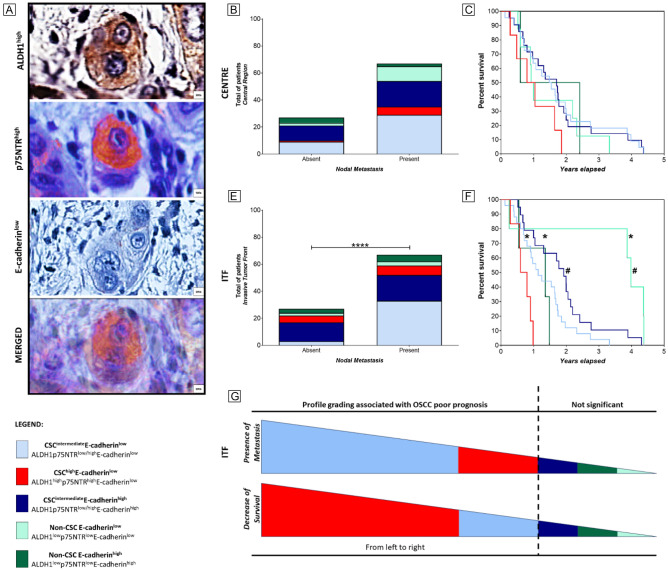
Identifying marker combinations for robust prognostic validation in primary tumour compartments remains challenging. We aimed to assess the prognostic significance of CSC markers (ALDH1, CD44, p75NTR, BMI-1) and E-cadherin biomarkers in OSCC. We analysed 94 primary OSCC and 67 metastatic lymph node samples, including central and invasive tumour fronts (ITF), along with clinicopathological data. We observed an increase in ALDH1+/CD44+/BMI-1- tumour cells in metastatic lesions compared to primary tumours. Multivariate analysis highlighted that elevated p75NTR levels (at ITF) and reduced E-cadherin expression (at the tumour centre) independently predicted metastasis, whilst ALDH1high exhibited independent predictive lower survival at the ITF, surpassing the efficacy of traditional tumour staging. Then, specifically at the ITF, profiles characterized by CSChighE-cadherinlow (ALDH1highp75NTRhighE-cadherinlow) and CSCintermediateE-cadherinlow (ALDH1 or p75NTRhighE-cadherinlow) were significantly associated with worsened overall survival and increased likelihood of metastasis in OSCC patients. In summary, our study revealed diverse tumour cell profiles in OSCC tissues, with varying CSC and E-cadherin marker patterns across primary tumours and metastatic sites. Given the pivotal role of reduced survival rates as an indicator of unfavourable prognosis, the immunohistochemistry profile identified as CSChighE-cadherinlow at the ITF of primary tumours, emerges as a preferred prognostic marker closely linked to adverse outcomes in OSCC.

Keywords: Cancer stem cells; Digital pathology; Epithelial-mesenchymal transition; Immunohistochemistry; Metastasis; Oral cancer.


Conflict of interest statement

The authors declare no competing interests.

Figures

Figure 1
Figure 1
Immunohistochemistry of CSC markers and E-cadherin in OSCC (Centre and ITF) and cervical lymph node metastasis. (A) Central regions of primary tumours display expansive areas with an adherent cell-like structural architecture. (B) The ITF features more invasive cells, often observed individually or in small clusters, and occasionally associated with lymphatic emboli. (C) Lymph node metastases exhibit distinct histological features compared to central regions, especially within extracapsular areas. (D) ALDH1 immunostaining in central tumour areas typically manifests as sporadic cytoplasmic staining, predominantly encircling corneal pearls. (E) ALDH1-positive tumour cells exhibit increased immunostaining within lymphatic emboli. (F) Metastatic tumour cells display notably high levels of ALDH1 staining. (G) Statistical analysis reveals a significant increase in ALDH1-positive cells within metastatic sites compared to both central and ITF regions of primary tumours. (H) CD44 demonstrates homogeneous immunostaining in central regions. (I) Uniform immunostaining within the ITF of primary tumours for CD44. (J) CD44 displays increased immunostaining at metastatic sites. (K) Notably higher CD44 immunostaining levels are observed at metastatic sites compared to central regions of primary tumours. (L) p75NTR presence is predominantly detected in metastatic primary tumours. (M) Limited and scattered expression of p75NTR in central regions. (N) Strong cytoplasmic staining for p75NTR is evident in metastatic sites. (O) No statistical significance in p75NTR expression is observed among the groups. (P) Focal nuclear immunostaining for BMI-1 is predominantly observed in both central regions. (Q) BMI-1 focal nuclear immunostaining is also observed in the ITF of primary tumours. (R) Positive BMI-1 staining is detected in only a few scattered metastatic tumour cells. (S) The majority of BMI-1 immunopositivity significantly decreases in metastatic sites compared to central areas of primary tumours. (T) E-cadherin exhibits strong and consistent membrane intensity in the central regions. (U) Heterogeneous cytoplasmic positivity for E-cadherin is observed in invasive tumour cells within lymphatic emboli. (V) Lymph node metastasis shows strong positivity for E-cadherin. (W) No significant differences in E-cadherin expression are observed among the groups.
Figure 2
Figure 2
CSC model for OSCC poor prognosis. (A) Computerized staining co-localization for CSChighE-cadherinlow (ALDH1highp75NTRhighE-cadherinlow). (B, C) Tumour cell immune profiles at the centre of primary tumours had no impact on prognosis (p > 0.05). (D) CSCintermediate E-cadherinlow, followed by the CSChigh E-cadherinlow immune profile at the ITF of primary OSCC were associated with nodal metastasis (p = 0.0179). (E) CSChigh E-cadherinlow profile was the strongest predictor of poor 5-year overall survival, compared to CSCintermediate E-cadherinlow (p = 0.0082), Non-CSC E-cadherinlow (p = 0.0002), and Non-CSC E-cadherinhigh (p = 0.0254) profiles. The CSCintermediate E-cadherinlow profile also demonstrated lower 5-year overall survival compared to CSCintermediate E-cadherinhigh (p = 0.0374), and Non-CSC E-cadherinlow (p = 0.0019). (F) shows the immune profile grading according to the presence of metastasis and survival decrease; from left to right is the most to the least related profile associated with OSCC's poor prognosis. * Means significant associations between CSChigh E-cadherinlow profile with others, whilst # means significant associations between CSCintermediate E-cadherinlow profile with others.
Figure 3
Figure 3
CSC markers and E-cadherin as a putative panel of poor prognosis in Oral Squamous Cell Carcinoma. (A) Tumour compartments, designed as centre and ITF, exhibited tumour cells with different patterns of CSC and E-cadherin immunopositivity (bottom right square). At the ITF, ALDH1high and p75NTRhigh immunostaining were independent predictors of decreased survival and the presence of metastasis, respectively. The most clinically relevant profile was the CSChighE-cadherinlow (ALDH1highp75NTRhighE-cadherinlow) at the ITF, which was a predictor of metastasis and lower overall survival and, could be used as a panel of biomarkers for poor prognosis in OSCC.

References

    1. Vigneswaran N, Williams MD. Epidemiologic trends in head and neck cancer and aids in diagnosis. Oral Maxillofac. Surg. Clin. 2014;26:123–141. doi: 10.1016/j.coms.2014.01.001. - DOI - PMC - PubMed
    1. Almangush A, et al. Staging and grading of oral squamous cell carcinoma: An update. Oral Oncol. 2020;107:104799. doi: 10.1016/j.oraloncology.2020.104799. - DOI - PubMed
    1. Bose P, Brockton NT, Dort JC. Head and neck cancer: from anatomy to biology. Int. J. Cancer. 2013;133:2013–2023. doi: 10.1002/ijc.28112. - DOI - PubMed
    1. Noguti J, et al. Metastasis from oral cancer: an overview. Cancer Genom. Proteom. 2012;9:329–335. - PubMed
    1. Joseph JP, et al. Hypoxia induced EMT: A review on the mechanism of tumor progression and metastasis in OSCC. Oral Oncol. 2018;80:23–32. doi: 10.1016/j.oraloncology.2018.03.004. - DOI - PubMed

Publication types

MeSH terms